. 2019 Jun:44:311-321.
doi: 10.1016/j.ebiom.2019.05.003. Epub 2019 May 9.

A miR-567-PIK3AP1-PI3K/AKT-c-Myc feedback loop regulates tumour growth and chemoresistance in gastric cancer

Affiliations

A miR-567-PIK3AP1-PI3K/AKT-c-Myc feedback loop regulates tumour growth and chemoresistance in gastric cancer

Feifei Zhang et al. EBioMedicine. 2019 Jun.

Erratum in

Expression of concern in

Abstract

Background: Gastric cancer (GC) ranks the fifth most common cancer, and chemotherapy is one of the most common treatments for GC. However, chemoresistance limits the effectiveness of chemotherapy and leads to treatment failure. This study aims to investigate the biological effect of miR-567 on gastric tumourigenesis and chemoresistance and reveal the possible mechanism.

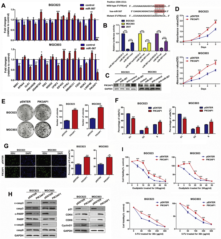
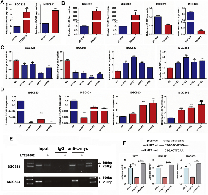
Methods: We measured the expression of miR-567 in 37 paired normal and stomach tumour specimens, as well as GC cell lines by Real-time PCR. The functional effects of miR-567 were validated using in vitro and in vivo assays. Dual-luciferase report assays and Chromatin immunoprecipitation (ChIP) assay were conducted for target evaluation, western blot assay was used to confirm the relationships.

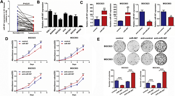
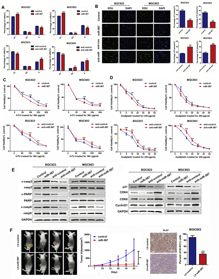
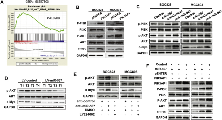
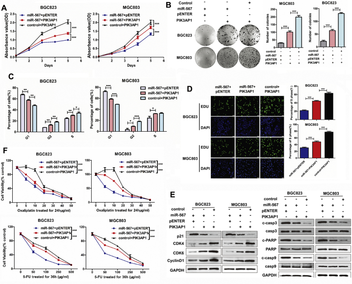
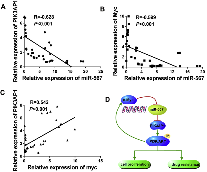
Findings: Our data showed that miR-567 was downregulated in gastric tissues and gastric cancer cells compared with normal tissues and gastric epithelial cells. In vitro, Gain- and lose-of-function assays showed miR-567 not only weakened cells proliferative ability, but also sensitized GC cells to 5-FU and oxaliplatin. In vivo, miR-567 overexpression significantly repressed the tumourigenesis of GC cells compared with the vector control. Mechanistic analysis showed that PIK3AP1 activated AKT phosphorylation in GC. Meanwhile, miR-567 directly targeted PIK3AP1 to inactivate PI3K/AKT/c-Myc pathway and c-Myc inversely regulated miR-567 expression, thus forming a miR-567-PIK3AP1- PI3K/AKT-c-Myc feedback loop explaining the function of miR-567.

Interpretation: Our studies revealed that miR-567 acts as a tumour suppressor gene and suppresses GC tumorigenesis and chemoresistance via a miR-567-PIK3AP1- PI3K/AKT-c-Myc feedback loop. These results suggest that miR-567 may serve as a target for chemoresistance and a potential prognostic biomarker for GC.

Keywords: Chemoresistance; Gastric cancer; Prognostic biomarker; Tumour growth; microRNA-567.


Figures

Fig. 1
Fig. 1
miR-567 is down-regulated in GC tissues and cell lines, and inhibits GC cell proliferation and colony formation. (A) The expression level of miR-567 in GC tissues compared with corresponding non-cancer tissues. (B) Relative expression levels of miR-567 in gastric cancer cell lines and a normal gastric epithelial cell line, GES-1. (C) Real-time PCR was performed to detect the mRNA expression of miR-567 in MGC803 and BGC823 cells, both transfected with miR-567 mimic or anti-miR-567, Student's t-test, mean ± SD, **P < .01; ***P < .001. Cell Counting Kit-8 (CCK-8) assay (D) and colony formation assay (E) of GC cells were performed after transfected with miR-567 mimic or anti-miR-567, Student's t-test, mean ± SD, *P < .05; **P < .01; ***P < .001.
Fig. 2
Fig. 2
miR-567 suppresses GC cell proliferation and increases drug sensitivity in vitro and in vivo. FACS assays (A) and EdU incorporation assays (B) of GC cells were performed after transfected with miR-567 mimic or anti-miR-567, Student's t-test, mean ± SD, *P < .05; **P < .01; ***P < .001. (C&D) Dose-response curves of MGC803 and BGC823 treated with 5-FU for 36 h or oxaliplatin for 24 h, the cells were previously transfected with miR-NC, miR-567, anti-miR-NC or anti-miR-567. Parametric generalized linear model with random effects, Student's t-test, mean ± SD, *P < .05; **P < .01; ***P < .001. (E) Western blot experiments were used to analyse the expression of c-casp3, casp3, c-PARP, PARP, c-casp9, casp9, p21, CDK4, CDK6 and CyclinD1 after miR-567 knockdown and overexpression in MGC803 and BGC823 cells. (F) In vivo image detection of the xenograft tumour growth. Growth curve was measured and drawn, *P < .05. The tumour sections were under IHC staining using antibodies against Ki-67. Quantification of Ki67 staining of the xenograft tumours (right). ***P < .001.
Fig. 3
Fig. 3
PIK3AP1 is the direct target of miR-567 and promotes GC cell proliferation and increases drug sensitivity. (A) Real-time PCR analysis were performed to detect the mRNA expression of candidate genes in MGC803 and BGC823 cells transfected with miR-567 mimic, Student's t-test, mean ± SD. (B) miR-567 and its putative binding sequences in the 3′UTR of PIK3AP1. A mutation was generated in the complementary site that bound to the seed region of miR-567. Luciferase reporter assay was used to determine miR-567 direct targeting the PIK3AP1 3′UTR, Student's t-test, mean ± SD, ***P < .001. (B) Western blot analysis were performed to detect the protein expression of PIK3AP1 in MGC803 and BGC823 cells, both transfected with miR-567 mimic or anti-miR-567. CCK-8 assay (C), colony formation assay (D), FACS assays (E) and EdU incorporation assays (F) of GC cells were performed after transfected with PIK3AP1 or pENTER vector, Student's t-test, mean ± SD, *P < .05; **P < .01. (H) Western blot experiments were used to analyse the expression of pro-apoptosis proteins, cell cycle maker and related proteins in PI3K/AKT pathway after miR-567 knockdown and overexpression in MGC803 and BGC823 cells. (I) Dose-response curves of MGC803 and BGC823 treated with 5-FU for 36 h or oxaliplatin for 24 h, the cells were previously transfected with PIK3AP1 and pENTER. Parametric generalized linear model with random effects, Student's t-test, mean ± SD, *P < .05; **P < .01; ***P < .001.
Fig. 4
Fig. 4
miR-567 negatively regulates with PI3K/AKT-c-Myc signalling in GC. (A) GSEA demonstrated enrichment of PI3K-AKT-mTOR signalling pathway in high PIK3AP1 expression GC group. (B) Western blot experiments were used to analyse the expression of relevant proteins in PI3K/AKT signal pathway after PIK3AP1 overexpression in MGC803 and BGC823 cells. (C) Western blot experiments were used to analyse the expression of relevant proteins in PI3K/AKT signal pathway after miR-567 knockdown and overexpression in MGC803 and BGC823 cells. (D) Western blot experiments were used to analyse the expression of relevant proteins in PI3K/AKT signal pathway in LV-miR-567-transfected cells and LV-miR-NC-transfected cells. (E) The upregulation of p-AKT and c-Myc induced by anti-miR-567 were abrogated after administration of LY294002 in MGC803 and BGC823 cells. (F) The downregulation of p-AKT and c-Myc induced by introduction of PIK3AP1 were abrogated after administration of miR-567 in MGC803 and BGC823 cells.
Fig. 5
Fig. 5
miR-567 suppresses the PI3K/AKT-c-Myc pathway via targeting PIK3AP1. CCK-8 assay (A), colony formation assay (B), FACS assays (C) and EdU incorporation assays (D) showed miR-567-inhibited cell proliferation was counteracted after administration of PIK3AP1, Student's t-test, mean ± SD, *P < .05; **P < .01; ***P < .001. (E) The miR-567-induced downregulation of relevant proteins in cell cycle marker (CDK4, CDK6 and CyclinD1) as well as upregulation of pro-apoptosis proteins and cell cycle marker p21 were abrogated after administration of PIK3AP1 in MGC803 and BGC823 cells. (F) PIK3AP1 overexpression reversed miR-567-induced cell sensitivity to 5-FU compared to control cells. Student's t-test, mean ± SD, ***P < .001.
Fig. 6
Fig. 6
c-Myc inhibits miR-567 by binding to its promoter region. (A–D) Real-time PCR was performed to detect the mRNA expression of miR-567 in MGC803 and BGC823 cells treated with LY294002, PIK3AP1, c-Myc siRNAs or PIK3AP1 siRNAs, Student's t-test, mean ± SD, *P < .05; **P < .01; ***P < .001. (E) PCR gel showing amplification of c-Myc-binding sites after ChIP using antibody against c-Myc. The gel figures were accompanied by the locations of molecular weight markers. (F) Schematic representation of the structure of c-Myc TFBS mutant and c-Myc wild-type (WT) that bound to the promoter regions of miR-567. Relative luciferase activity of the indicated promoter vectors in 293 T, MGC803 and BGC823 cells transfected with c-Myc plasmids, Student's t-test, mean ± SD, **P < .01; ***P < .001.
Fig. 7
Fig. 7
Schematic representation of overall summary. (A) Real-time PCR assay were performed to detect the mRNA expression of miR-567 and PIK3AP1 in GC tissues. (B) Real-time PCR assay were performed to detect the mRNA expression of miR-567 and c-Myc in GC tissues. (C) Real-time PCR assay were performed to detect the mRNA expression of PIK3AP1 and c-Myc in GC tissues. (D) A schematic for an atypical miR-567-PIK3AP1–PI3K/AKT-c-Myc feedback loop.

References

    1. Figueiredo C., Camargo M.C., Leite M., Fuentes-Panana E.M., Rabkin C.S., Machado J.C. Pathogenesis of gastric cancer: genetics and molecular classification. Curr Top Microbiol Immunol. 2017;400:277–304. - PMC - PubMed
    1. Leal M., Wisnieski F., de Oliveira Gigek C. What gastric cancer proteomic studies show about gastric carcinogenesis? Tumour Biol. 2016;37(8):9991–10010. - PubMed
    1. Newton A., Datta J., Loaiza-Bonilla A., Karakousis G., Roses R. Neoadjuvant therapy for gastric cancer: current evidence and future directions. J Gastrointest Oncol. 2015;6(5):534–543. - PMC - PubMed
    1. Wang X., Wang Y., Qiu M. Postoperative chemoradiotherapy in gastric cancer: a phase I study of radiotherapy with dose escalation of oxaliplatin, 5-fluorouracil, and leucovorin (FOLFOX regimen) Med Oncol. 2011;28(Suppl. 1):S274–S279. - PubMed
    1. Hu M., Li K., Maskey N. 15-PGDH expression as a predictive factor response to neoadjuvant chemotherapy in advanced gastric cancer. Int J Clin Exp Pathol. 2015;8(6):6910–6918. - PMC - PubMed

MeSH terms