. 2019 Jul:533:21-33.
doi: 10.1016/j.virol.2019.05.001. Epub 2019 May 2.

Structural basis for catalysis and substrate specificity of a 3C-like cysteine protease from a mosquito mesonivirus

Affiliations

Structural basis for catalysis and substrate specificity of a 3C-like cysteine protease from a mosquito mesonivirus

Manuel Kanitz et al. Virology. 2019 Jul.

Abstract

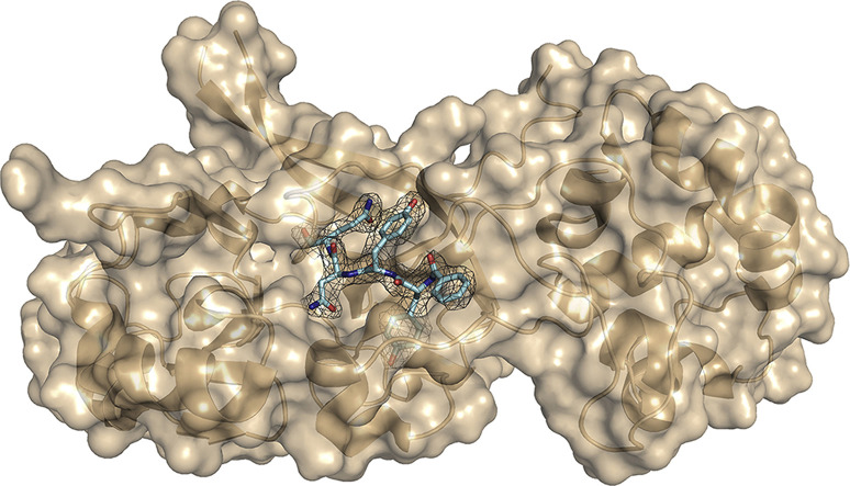
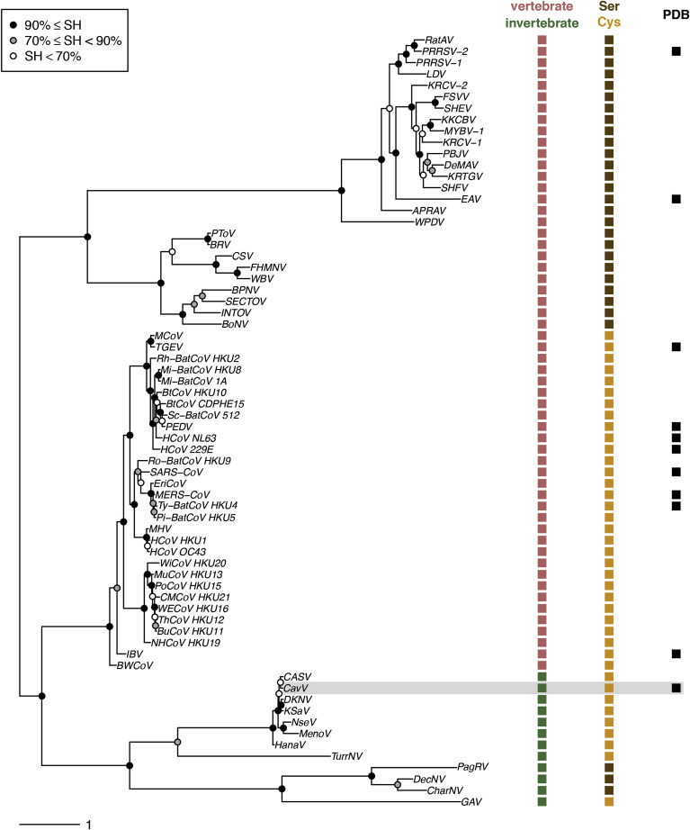
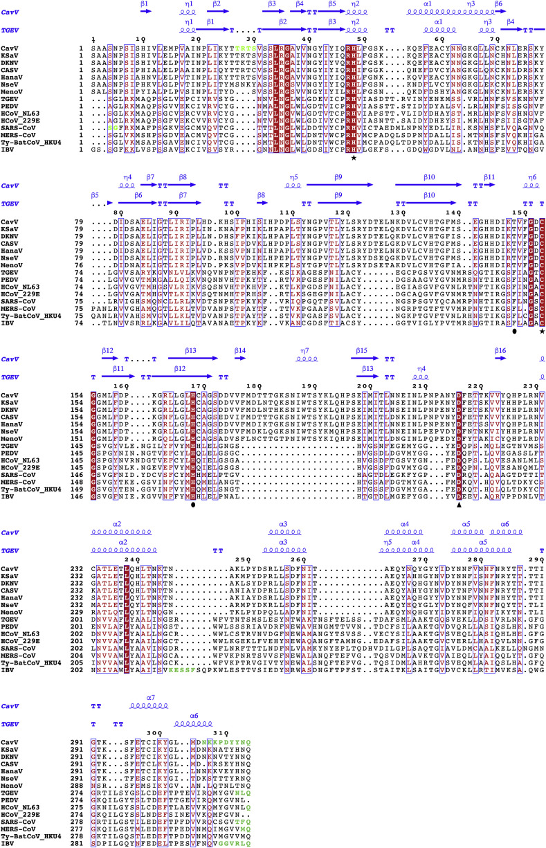
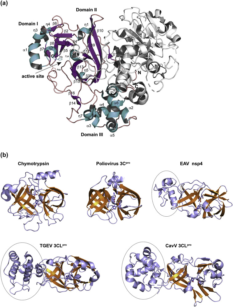
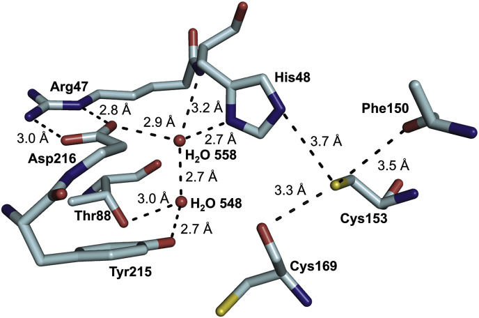
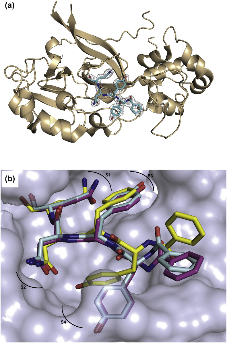
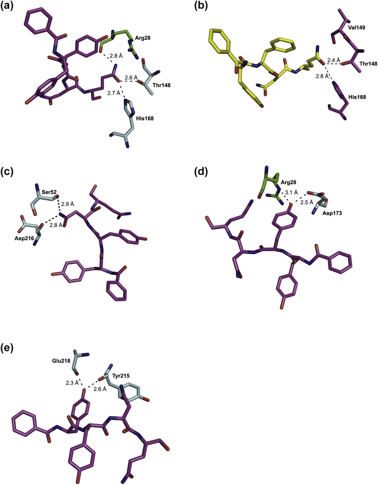
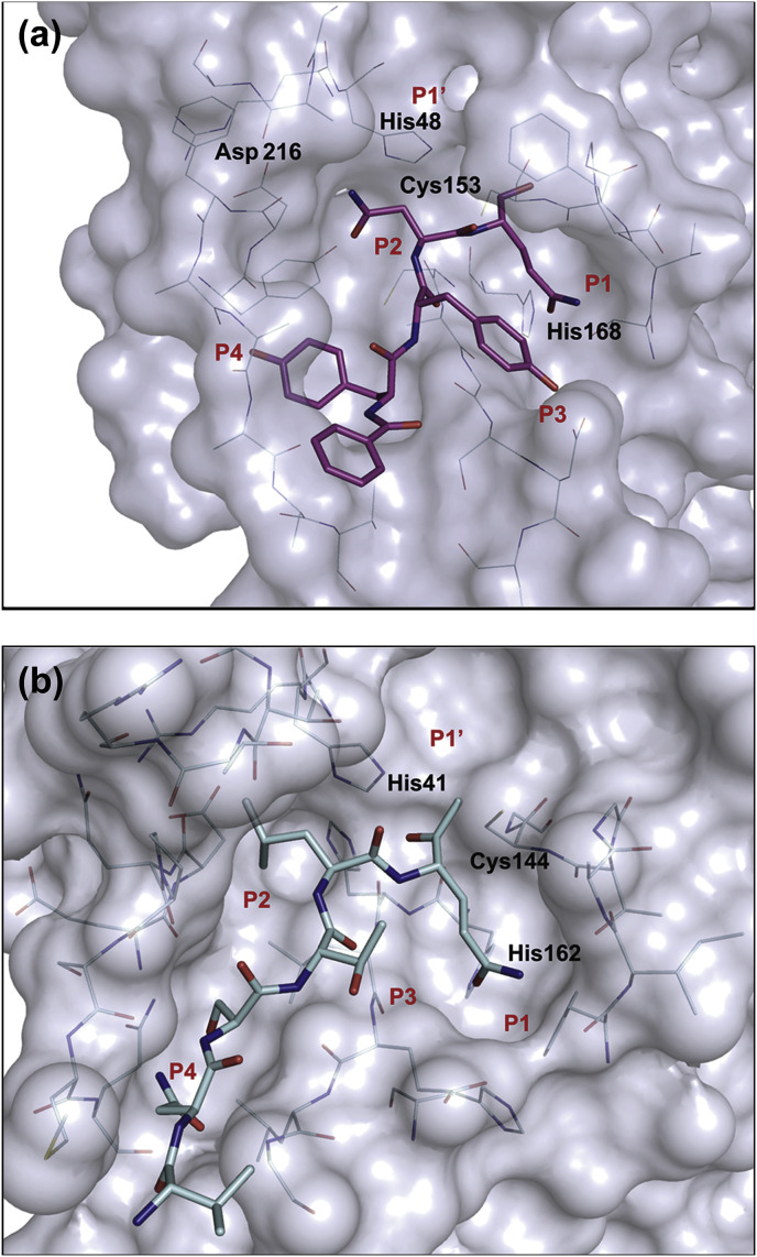
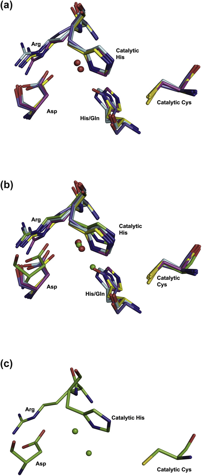
Cavally virus (CavV) is a mosquito-borne plus-strand RNA virus in the family Mesoniviridae (order Nidovirales). We present X-ray structures for the CavV 3C-like protease (3CLpro), as a free enzyme and in complex with a peptide aldehyde inhibitor mimicking the P4-to-P1 residues of a natural substrate. The 3CLpro structure (refined to 1.94 Å) shows that the protein forms dimers. The monomers are comprised of N-terminal domains I and II, which adopt a chymotrypsin-like fold, and a C-terminal α-helical domain III. The catalytic Cys-His dyad is assisted by a complex network of interactions involving a water molecule that mediates polar contacts between the catalytic His and a conserved Asp located in the domain II-III junction and is suitably positioned to stabilize the developing positive charge of the catalytic His in the transition state during catalysis. The study also reveals the structural basis for the distinct P2 Asn-specific substrate-binding pocket of mesonivirus 3CLpros.

Keywords: 3C-like protease; Active site of chymotrypsin-like proteases; Coronavirus; Crystal structure; Invertebrate RNA virus; Mesonivirus.


Figures

Image 1
Graphical abstract
Fig. 1
Fig. 1
Characteristics of nidovirus 3CLpros in a virus phylogeny and host context. Left, RdRp-based phylogeny of representatives of established nidovirus species. Right, presumed primary host of the respective virus species, 3CLpro principal nucleophile residue, and 3CLpro PDB structure availability. Information on CavV is highlighted using a gray background. Information on nidovirus genomes used for phylogeny reconstruction is provided in the Supplemental Information. Box, SH-aLRT branch support is shown using three ranges of values.
Fig. 2
Fig. 2
Structure-based MSA of mesonivirus and coronavirus 3CLpros. Strictly conserved residues are indicated using red background color, partially conserved residues are indicated in red. Secondary structure elements are shown for CavV (PDB ID: 5LAC, chain B) and TGEV (PDB ID: 1LVO, chain A) in blue above the alignment. Residues that were omitted from the respective structure models are indicated in green. Residue numbers given above the alignment refer to the CavV 3CLpro sequence corresponding to the CavV 3CLpro structure (PDB ID: 5LAC, chain B). Crucial CavV 3CLpro residues are indicated below the alignment: catalytic residues – by asterisks; key residues in the S1 subsite of the substrate-binding pocket – by filled circles; the Asp residue that interacts with a conserved water molecule in the active site – by a black triangle. PDB IDs used to produce the structure-based MSA are given in the Supplemental Information.
Fig. 3
Fig. 3
(a) Schematic view of the secondary structure elements of the CavV 3CLpro (PDB ID: 5LAC). Chain B is shown with helices colored in blue, β-strands colored in purple, and loops colored in red; chain A is shown in white. This figure and the following figures were generated using PyMol. (b) Structural homology of nidovirus 3CLpros with chymotrypsin and picornavirus 3C proteases. The double β-barrel that characterizes members of the PA superfamily of proteases is highlighted in orange. Shown are the structures of equine arteritis virus (EAV) nsp4 (PDB ID: 1MBM), transmissible gastroenteritis virus (TGEV) 3CLpro (PDB ID: 1LVO), CavV 3CLpro (PDB ID: 5LAC), chymotrypsin (PDB ID: 5J4Q), and poliovirus 3Cpro (PDB ID: 1L1N). Dotted lines indicate the extra C-terminal helical domains conserved in main proteases of the Nidovirales, represented in this figure by EAV (family Arteriviridae), TGEV (family Coronaviridae), and CavV (family Mesoniviridae). Catalytic residues are shown as sticks.
Fig. 4
Fig. 4
Representation of an extensive H-bond network involving a conserved water molecule and the catalytic Cys153 and His48 in the active site of the CavV 3CLpro structure (PDB ID: 5LAC; chain B). Only amino acids involved in hydrogen bonding are shown as sticks and color-coded by atom type. Dashed lines indicate key hydrogen bonds. For clarity, the backbone carbonyl group of Asp216 and the backbone amide of Arg47 were omitted from this representation.
Fig. 5
Fig. 5
(a) Crystal structure of Bz-YYNQ-H (cyan, color-coded by atom type) in complex with CavV 3CLpro (cartoon, wheat). The electron-density map (2mFo-DFc) for the ligand is displayed at a σ level of 1.0 as gray mesh. (b) Superposition of the peptide aldehyde bound to chain A (blue), chain B (yellow), and chain C (pink), respectively, each represented as sticks and color-coded by atom type. The substrate-binding pocket of chain C (light blue) is shown in surface representation. Subpockets S1 to S4 are indicated.
Fig. 6
Fig. 6
Key interactions of the ligand in the S1 (a), S2 (c), S3 (d), and S4 (e) subpockets, respectively, of monomer C and in the S1 subpocket of monomer B (b) of the CavV 3CLpro (PDB ID: 5LAK). Atoms of the ligand (pink) and protease (chain C in light blue and chain A in green) are represented as sticks and color-coded by atom types. In panel (b), the ligand is shown in yellow and residues of chain B are shown in pink. Only residues involved in hydrogen bonding are shown.
Fig. 7
Fig. 7
Surface representation of mesonivirus and coronavirus 3CLpro substrate-binding sites using (a) the structure of the CavV 3CLpro/inhibitor complex (PDB ID: 5LAK; pink) and (b) the structure of TGEV 3CLpro in a complex with a peptidic inhibitor (Anand et al., 2003) (PDB ID: 1P9U; light blue). Ligands are represented as sticks and color-coded by atom types. Amino acids involved in substrate binding are shown as lines and color-coded by atom types. Catalytic residues and the conserved His residue in the S1 subsite are indicated (see text for details).
Fig. 8
Fig. 8
Superimposition of active-site residues of coronavirus and mesonivirus 3CLpros. Shown are the active sites of the 3CLpros of TGEV (Kim et al., 2012) (PDB ID: 4F49; light blue), IBV (Xue et al., 2008) (PDB ID: 2Q6D; pink), SARS-CoV (Zhu et al., 2011) (PDB ID: 3SNE; yellow), HCoV-HKU1 (Zhao et al., 2008) (PDB ID: 3D23; dark blue), and CavV (PDB ID: 5LAK; green). All structures are represented as sticks and color-coded by atom types. Water molecules of 4F49, 2Q6D, 3SNE and 3D23 are represented as red spheres and water molecules of 5LAK are represented as green spheres. (a) superimposition of 4F49, 2Q6D, 3SNE, and 3D23; (b) superimposition of the coronavirus 3CLpro active sites shown in panel (a) (PDB ID: 4F49, 2Q6D, 3SNE, and 3D23) with the CavV 3CLpro active site (PDB ID: 5LAK); (c) active site of the CavV 3CLpro alone (PDB ID: 5LAK).

References

    1. Adams P.D., Afonine P.V., Bunkoczi G., Chen V.B., Davis I.W., Echols N., Headd J.J., Hung L.W., Kapral G.J., Grosse-Kunstleve R.W., McCoy A.J., Moriarty N.W., Oeffner R., Read R.J., Richardson D.C., Richardson J.S., Terwilliger T.C., Zwart P.H. PHENIX: a comprehensive Python-based system for macromolecular structure solution. Acta Crystallogr D Biol Crystallogr. 2010;66:213–221. - PMC - PubMed
    1. Adams M.J., Lefkowitz E.J., King A.M., Harrach B., Harrison R.L., Knowles N.J., Kropinski A.M., Krupovic M., Kuhn J.H., Mushegian A.R., Nibert M., Sabanadzovic S., Sanfacon H., Siddell S.G., Simmonds P., Varsani A., Zerbini F.M., Gorbalenya A.E., Davison A.J. Ratification vote on taxonomic proposals to the international committee on taxonomy of viruses (2016) Arch. Virol. 2016;161:2921–2949. - PMC - PubMed
    1. Allaire M., Chernaia M.M., Malcolm B.A., James M.N. Picornaviral 3C cysteine proteinases have a fold similar to chymotrypsin-like serine proteinases. Nature. 1994;369:72–76. - PubMed
    1. Anand K., Palm G.J., Mesters J.R., Siddell S.G., Ziebuhr J., Hilgenfeld R. Structure of coronavirus main proteinase reveals combination of a chymotrypsin fold with an extra alpha-helical domain. EMBO J. 2002;21:3213–3224. - PMC - PubMed
    1. Anand K., Ziebuhr J., Wadhwani P., Mesters J.R., Hilgenfeld R. Coronavirus main proteinase (3CLpro) structure: basis for design of anti-SARS drugs. Science. 2003;300:1763–1767. - PubMed

Publication types