. 2019 Aug:76:101930.
doi: 10.1016/j.npep.2019.05.001. Epub 2019 May 6.

Mapping the populations of neurotensin neurons in the male mouse brain

[Article in Spanish]
Affiliations

Mapping the populations of neurotensin neurons in the male mouse brain

[Article in Spanish]
Laura E Schroeder et al. Neuropeptides. 2019 Aug.

Abstract

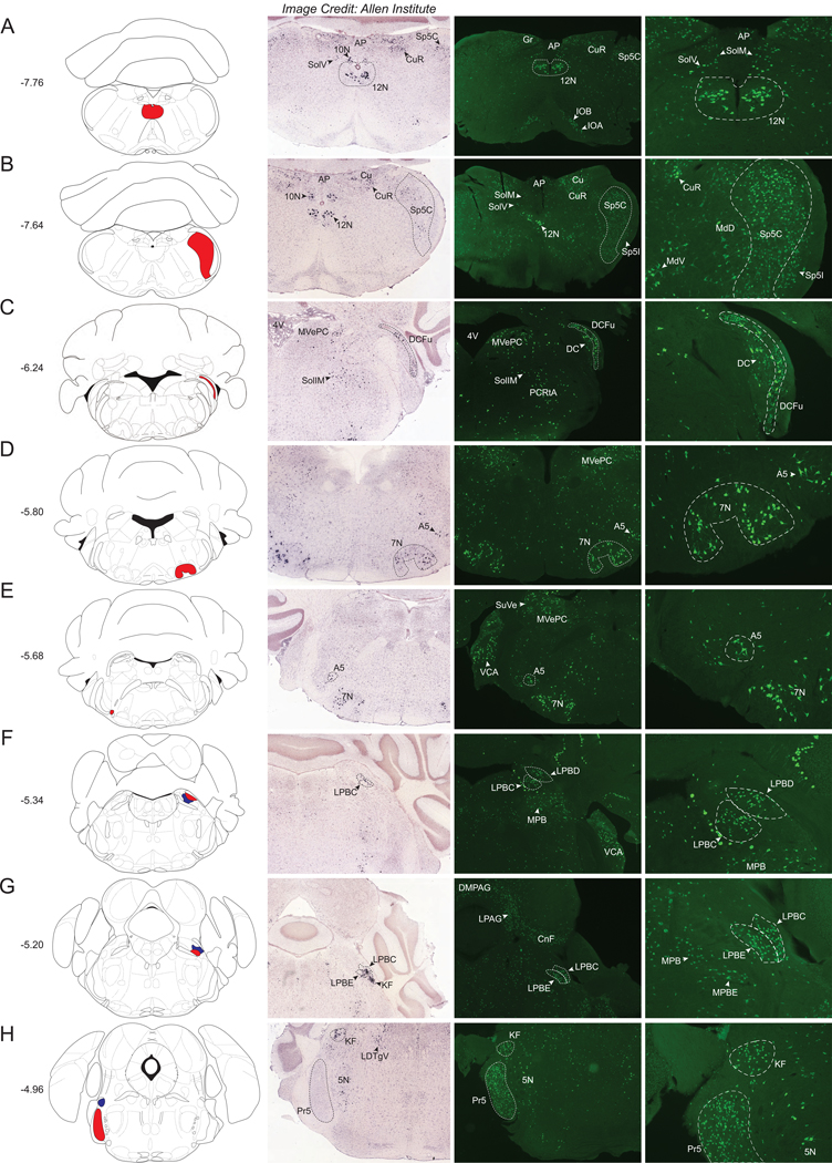
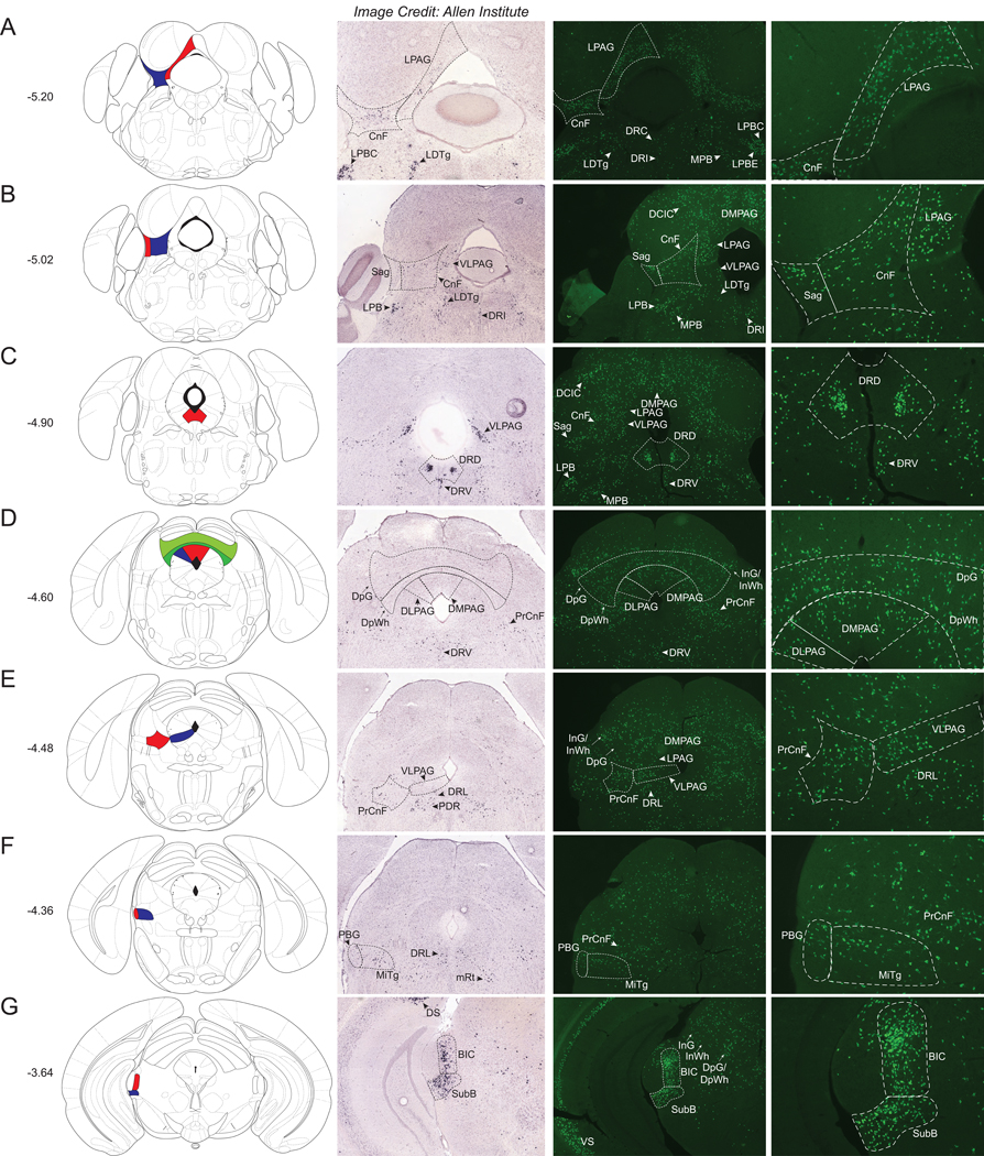
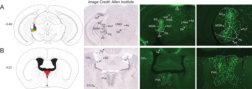
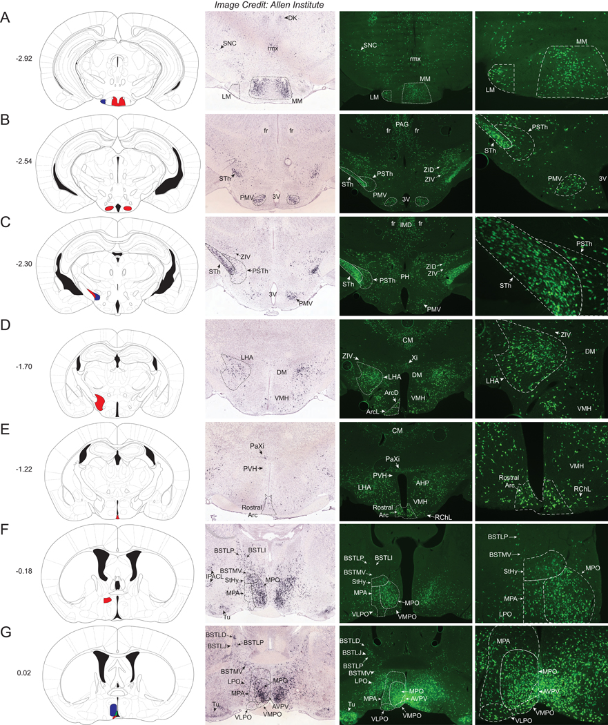
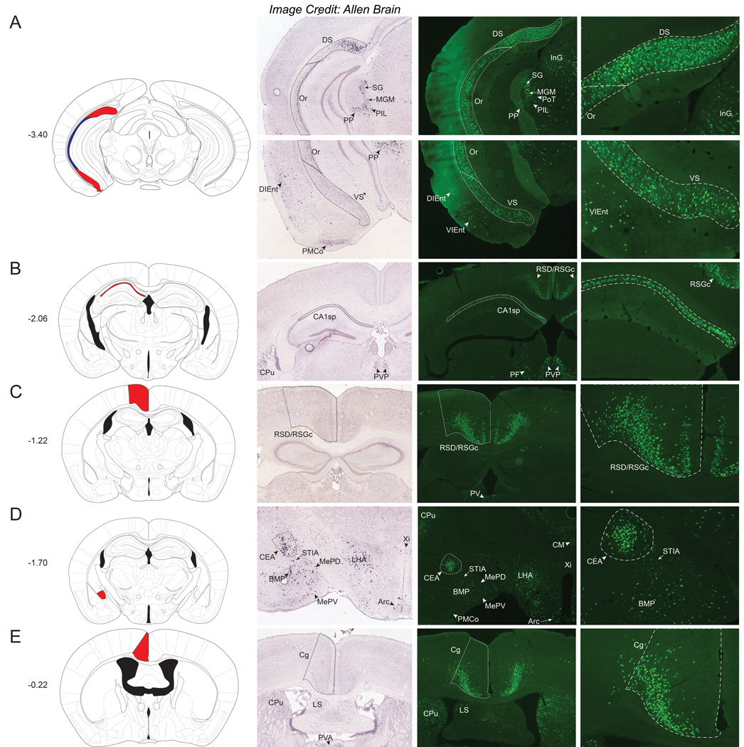
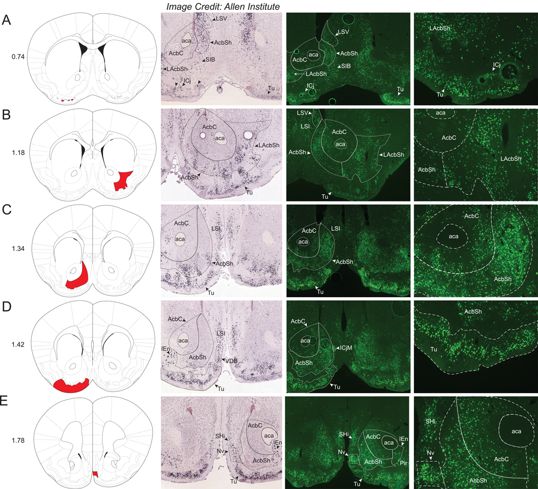
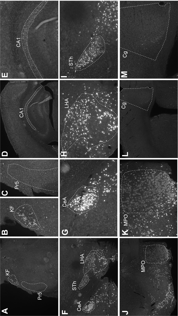
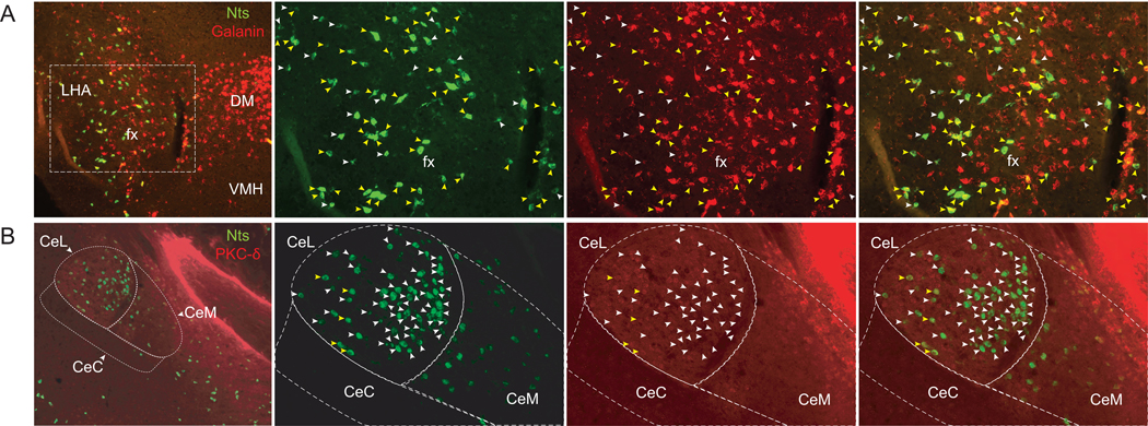
Neurotensin (Nts) is a neuropeptide implicated in the regulation of many facets of physiology, including cardiovascular tone, pain processing, ingestive behaviors, locomotor drive, sleep, addiction and social behaviors. Yet, there is incomplete understanding about how the various populations of Nts neurons distributed throughout the brain mediate such physiology. This knowledge gap largely stemmed from the inability to simultaneously identify Nts cell bodies and manipulate them in vivo. One means of overcoming this obstacle is to study NtsCre mice crossed onto a Cre-inducible green fluorescent reporter line (NtsCre;GFP mice), as these mice permit both visualization and in vivo modulation of specific populations of Nts neurons (using Cre-inducible viral and genetic tools) to reveal their function. Here we provide a comprehensive characterization of the distribution and relative densities of the Nts-GFP populations observed throughout the male NtsCre;GFP mouse brain, which will pave the way for future work to define their physiologic roles. We also compared the distribution of Nts-GFP neurons with Nts-In situ Hybridization (Nts-ISH) data from the adult mouse brain. By comparing these data sets we can distinguish Nts-GFP populations that may only transiently express Nts during development but not in the mature brain, and hence which populations may not be amenable to Cre-mediated manipulation in adult NtsCre;GFP mice. This atlas of Nts-GFP neurons will facilitate future studies using the NtsCre;GFP line to describe the physiological functions of individual Nts populations and how modulating them may be useful to treat disease.

Keywords: Central amygdala; Galanin; Lateral hypothalamus; Nucleus accumbens; Olfactory tubercle; Parabrachial nucleus; Periaqueductal gray; Preoptic area; Thalamus.


Conflict of interest statement

Disclosure Summary: The authors have no conflicts of interest to declare.

Figures

Figure 1.
Figure 1.
Nts-GFP and Nts-ISH in the Hindbrain. From left to right, each row contains a Bregma-numbered atlas image (Franklin and Paxinos, 2013), an image of Nts-ISH at the same Bregma level, courtesy of the Allen Brain Atlas (Lein et al., 2007), a 4x image of Nts-GFP neurons, and a 10x image of Nts-GFP neurons from the same area. Red and blue shaded areas in the atlas image are outlined in the Nts-GFP images. A) Bregma −7.76, Red shading corresponds to the Hypoglossal nucleus (12N), B) Bregma −7.64, Red shading corresponds to the Spinal trigeminal tract, caudal part (Sp5C), C) Bregma −6.24, Red shading corresponds to the Dorsal cochlear nucleus, fusiform layer (DCFu), D) Bregma −5.80, Red shading corresponds to the Facial nucleus (7N), E) Bregma −5.68, Red shading corresponds to the A5 noradrenaline cell group (A5), F) Bregma −5.34, Red shading corresponds to the Lateral parabrachial nucleus, dorsal part (LPBD) whereas blue shading corresponds to the Lateral parabrachial nucleus, central part (LPBC), G) Bregma −5.20, Red shading corresponds to the Lateral parabrachial nucleus, external part (LPBE) while blue shading corresponds to the LPBC, H) Bregma −4.96, Red shading corresponds to the Principal sensory trigeminal nucleus (Pr5) whereas blue shading corresponds to the Koelliker-Fuse nucleus (KF). AP= Area postrema, 10N = dorsal motor nucleus of the vagus, Gr= Gracile nucleus, CuR= Cuneate nucleus, Cu= Cuneate nucleus and fasciculus, Sp5I= Spinal trigeminal tract, interpolar part, IOB= Inferior olive subnucleus B of the medial nucleus, IOA= Inferior olive subnucleus A of the medial nucleus, SolM= Solitary nucleus, medial part, SolV= Solitary nucleus, ventral part, MdV= Medullary reticular nucleus, ventral part, MdD= Medullary reticular nucleus, dorsal part, 4V= 4th ventricle, MVePC= Medial vestibular nucleus, parvicellular part, DC= Dorsal cochlear nucleus, SollM= Solitary nucleus, intermediate part, PCRtA= Parvicellular reticular nucleus, SuVe= Superior vestibular nucleus, VCA= Ventral cochlear nucleus, anterior part, MPB= Medial parabrachial nucleus, DMPAG= Dorsomedial periaqueductal gray, LPAG= Lateral periaqueductal gray, CnF= Cuneiform nucleus, 5N= Motor trigeminal nucleus.
Figure 2.
Figure 2.
Nts-GFP and Nts-ISH in the Midbrain. From left to right, each row contains a Bregma-numbered atlas image (Franklin and Paxinos, 2013), an image of Nts-ISH at the same Bregma level, courtesy of the Allen Brain Atlas (Lein et al., 2007), a 4x image of Nts-GFP neurons, and a 10x image of Nts-GFP neurons from the same area. Red, blue, and green shaded areas in the atlas image are outlined in the Nts-GFP images. A) Bregma −5.20, Red shading corresponds to the Lateral periaqueductal gray (LPAG) while blue shading corresponds to the Cuneiform nucleus (CnF), B) Bregma −5.02, Red shading corresponds to the Sagulum nucleus (Sag) whereas blue shading corresponds to the CnF, C) Bregma −4.90, Red shading corresponds to the Dorsal raphe nucleus, dorsal part (DRD), D) Bregma −4.60, Red shading corresponds to the Dorsomedial periaqueductal gray (DMPAG), blue shading corresponds to the Dorsolateral periaqueductal gray (DLPAG), dark green shading corresponds to the Deep white layer of the superior colliculus (DpWh), and light green shading corresponds to the Deep gray layer of the superior colliculus (DpG), E) Bregma −4.48, Red shading corresponds to the Precuneiform area (PrCnF) whereas blue shading corresponds to the ventrolateral periaqueductal gray (VLPAG), F) Bregma −4.36, Red shading corresponds to the Parabigeminal nucleus (PBG) while blue shading corresponds to the Microcellular Tegmental nucleus (MiTg), G) Bregma −3.64, Red shading corresponds to the Brachium of the Inferior Colliculus (BIC) whereas blue shading corresponds to the Subbrachial nucleus (SubB). LDTg= Laterodorsal tegmental nucleus, LPBC= Lateral parabrachial nucleus, central part, LPBE=Lateral parabrachial nucleus, external part, MPB= Medial parabrachial nucleus, DRC= Dorsal raphe nucleus, caudal part, DRI=Dorsal raphe nucleus, interfascicular part, MPB= Medial parabrachial nucleus, DCIC= Dorsal cortex of the inferior colliculus, DRV= Dorsal raphe nucleus, ventral part, DRL= Dorsal raphe nucleus, lateral part, InG/InWh= Intermediate gray or white layer of the superior colliculus, PDR= Posterodorsal raphe nucleus, mRt=Mesencephalic reticular formation, DS= Dorsal Subiculum, VS=Ventral Subiculum.
Figure 3.
Figure 3.
Nts-GFP and Nts-ISH in the Thalamus. From left to right, each row contains a Bregma-numbered atlas image (Franklin and Paxinos, 2013), an image of Nts-ISH at the same Bregma level, courtesy of the Allen Brain Atlas (Lein et al., 2007), a 4x image of Nts-GFP neurons, and a 10x image of Nts-GFP neurons from the same area. Shaded areas in the atlas image are outlined in the Nts-GFP images. A) Bregma −3.40, Red shading corresponds to the Suprageniculate Thalamic nucleus (SG), blue shading corresponds to the Medial Geniculate nucleus, medial part (MGM), yellow shading corresponds to the Peripeduncular nucleus (PP), green shading corresponds to the Posterior Intralaminar Thalamic nucleus (PIL), and maroon shading corresponds to the Posterior Thalamic nucleus, Triangular (PoT), B) Bregma −0.22, Red shading corresponds to the Paraventricular Thalamic nucleus, Anterior (PVA). DS= Dorsal Subiculum, LPAG=Lateral periaqueductal gray, Aq=Aqueduct, DK= Nucleus of Darkschewitsch, InG/InWh= Intermediate gray or white layer of the superior colliculus, CPu= Caudate Putamen, Cg= Cingulate Cortex, LSD= Lateral Septal Nucleus, dorsal part, BSTLP= Bed nucleus of the stria terminalis, lateral division, posterior part.
Figure 4.
Figure 4.. Nts-GFP and Nts-ISH in the Hypothalamus.
From left to right, each row contains a Bregma-numbered atlas image (Franklin and Paxinos, 2013), an image of Nts-ISH at the same Bregma level, courtesy of the Allen Brain Atlas (Lein et al., 2007), a 4x image of Nts-GFP neurons, and a 10x image of Nts-GFP neurons from the same area. Shaded areas in the atlas image are outlined in the Nts-GFP images. A) Bregma −2.92, Red shading corresponds to the medial mammillary nucleus, medial part (MM) whereas blue shading corresponds to the lateral mammillary nucleus (LM), B) Bregma −2.54, Red shading corresponds to the Premammillary nucleus, ventral (PMV), C) Bregma −2.30, Red shading corresponds to the Subthalamic nucleus (STh) while blue shading corresponds to the Parasubthalamic nucleus (PSTh), D) Bregma −1.70, Red shading corresponds to the Lateral Hypothalamus (LHA), E) Bregma −1.22, Red shading corresponds to the Rostral Arcuate nucleus (Arc), F) Bregma −0.18, Red shading corresponds to the Striohypothalamic nucleus (StHy), G) Bregma +0.02, Red shading corresponds to the Ventromedial Preoptic nucleus (VMPO), blue shading corresponds to the Medial Preoptic nucleus (MPO), and green shading corresponds to the Anteroventral Periventricular nucleus (AVPV). DK= Nucleus of Darkschewitsch, rmx= Retromammillary decussation, SNC= Substantia Nigra Compacta, fr= Fasciculus retroflexus, PSTh= Parasubthalamic nucleus, STh= Subthalamic nucleus, 3V= 3rd ventricle, PAG= Periaqueductal Gray, ZID= Zona Incerta, Dorsal, ZIV= Zona Incerta, Ventral, IMD= Intermediodorsal Thalamic nucleus, PH= Posterior Hypothalamic nucleus, DM= Dorsomedial Hypothalamic nucleus, VMH=Ventromedial Hypothalamic nucleus, CM= Central Medial Thalamic nucleus, Xi= Xiphoid Thalamic nucleus, PaXi= Paraxiphoid nucleus of Thalamus, ArcD= Arcuate hypothalamic nucleus, Dorsal, ArcL= Arcuate hypothalamic nucleus, Lateral, PVH= Paraventricular nucleus of the Hypothalamus, AHP= Anterior Hypothalamic Area, Posterior, RChL= Retrochiasmatic, Lateral, BSTLI= Bed nucleus of the stria terminalis, lateral division, intermediate part, BSTLP= Bed nucleus of the stria terminalis, lateral division, posterior part, BSTMV= Bed nucleus of the stria terminalis, medial division, ventral part, BSTLD= Bed nucleus of the stria terminalis, lateral division, dorsal part, BSTLJ= Bed nucleus of the stria terminalis, lateral division, juxtacapsular part, IPACL= Interstitial nucleus of the posterior limb of the anterior commissure, MPA= Medial Preoptic Area, Tu= Olfactory tubercle, VLPO= Ventrolateral Preoptic nucleus, LPO= Lateral Preoptic nucleus.
Figure 5.
Figure 5.. Nts-GFP and Nts-ISH in the Cortex.
From left to right, each row contains a Bregma-numbered atlas image (Franklin and Paxinos, 2013), an image of Nts-ISH at the same Bregma level, courtesy of the Allen Brain Atlas (Lein et al., 2007), a 4x image of Nts-GFP neurons, and a 10x image of Nts-GFP neurons from the same area. Shaded areas in the atlas image are outlined in the Nts-GFP images. A) Bregma −3.40, Red shading corresponds to the Dorsal and Ventral Subiculum (DS and VS) while blue shading corresponds to the Oriens layer of the hippocampus (Or), B) Bregma −2.06, Red shading corresponds to Field CA1 of the hippocampus (CA1sp) C) Bregma −1.22, Red shading corresponds to the Retrosplenial Dysgranular and Granular Cortex (RSD/RSGc), D) Bregma −1.70, Red shading corresponds to the Central Amygdalar nucleus (CEA), E) Bregma −0.22, Red shading corresponds to the Cingulate Cortex (Cg). SG= Suprageniculate Thalamic nucleus, MGM=Medial Geniculate nucleus, PIL= Posterior Intralaminar Thalamic nucleus, PP= Peripeduncular nucleus, PoT= Posterior Thalamic nucleus, InG= Intermediate gray layer of the superior colliculus, DIEnt= Dorsointermedial Entorhinal Cortex, VIEnt= Ventral Intermediate Entorhinal Cortex, PMCo= Posteromedial cortical amygdalar nucleus, CPu= Caudate Putamen, PVP= Paraventricular Thalamic nucleus, Posterior, PF= Parafascicular Thalamic nucleus, PV= Paraventricular Thalamic nucleus, STIA= ST, intraamygdalar division, BMP= Basomedial Amygdalar nucleus, posterior, MePD= Medial Amygdalar nucleus, posterodorsal, MePV= Medial Amygdalar nucleus, posteroventral, LHA= Lateral Hypothalamus, Arc= Arcuate hypothalamic nucleus, Xi= Xiphoid Thalamic nucleus, CM= Central Medial Thalamic nucleus, LS=Lateral Septal Nucleus.
Figure 6.
Figure 6.. Nts-GFP and Nts-ISH in the Forebrain.
From left to right, each row contains a Bregma-numbered atlas image (Franklin and Paxinos, 2013), an image of Nts-ISH at the same Bregma level, courtesy of the Allen Brain Atlas (Lein et al., 2007), a 4x image of Nts-GFP neurons, and a 10x image of Nts-GFP neurons from the same area. Shaded areas in the atlas image are outlined in the Nts-GFP images. A) Bregma +0.74, Red shading corresponds to the Islands of Cajella (ICj), B) Bregma +1.18, Red shading corresponds to the Nucleus Accumbens, lateral shell (LAcbSh), C) Bregma +1.34, Red shading corresponds to the Nucleus Accumbens, Shell (AcbSh), D) Bregma +1.42, Red shading corresponds to the Olfactory tubercle (Tu), E) Bregma +1.78, Red shading corresponds to the Navicular Postolfactory nucleus (Nv). LSV=Lateral Septal Nucleus, ventral part, LSI=Lateral Septal Nucleus, intermediate part, aca=anterior commissure, AcbC=Nucleus Accumbens, Core, ICjM=Island of Cajella, Major Island, SIB=Substantia innominata, VDB=Nucleus of the Vertical Limb of the Diagonal Band, SHi=Septohippocampal nucleus, Pir=Piriform Cortex.
Figure 7.
Figure 7.. Verification of Nts-ISH in Specific Brain Regions.
RNA Scope ISH for Nts (white) in wild type male mice (n=3). Panels show representative images of findings. A) 4x image of section containing the Pr5 and KF. B) KF (20x) and C) Pr5 (20x). CA1 region of the hippocampus at D) 4x and E) 20x. F) 4x image of section containing the CeA, LHA and STh. 20x images of from this section of the G) CeA, H) LHA and I) STh. Abundant Nts is found in the J) 4x image of the MPO and K) can be seen in numerous MPO cell bodies at 20x. By contrast, note the dearth of Nts in the L) 4x image and K) 20x image of the Cg. Pr5=Principal sensory trigeminal nucleus, KF=Koelliker-Fuse nucleus (KF), CA1=Field CA1 of the hippocampus, CeA=central amygdala, LHA= lateral hypothalamic area, STh= subthalamic nucleus, MPO=medial preoptic area, Cg=cingulate cortex.
Figure 8.
Figure 8.. Heterogeneity of Nts Neurons Within the LHA and CEA.
From left to right, each row contains a 10x image of merged red and green channels, followed by 20X images of green, red, and merged channels. A) RNA Scope dual-fluorescent ISH for Nts (green) and Galanin (Gal, red) in the LHA. Yellow arrows identify neurons expressing Nts and robust levels of Gal. White arrows identify neurons expressing Nts and negligible Gal. B) Section of the CEA from an NtsCre;GFP mouse immunostained for GFP (Nts-GFP, green) and PKC-δ (red). Yellow arrows identify the few neurons co-labeled with Nts-GFP and PKC-δ, which lie primarily within the lateral aspect of the CEA (CeL). White arrows identify CEA neurons that contain Nts but no detectable PKC-δ. LHA= Lateral Hypothalamus, fx=fornix, DM=Dorsomedial Hypothalamic nucleus, VMH=Ventromedial Hypothalamic nucleus, CeL=Central Amygdalar nucleus, lateral, CeC=Central Amygdalar nucleus, central, CeM=Central Amygdalar nucleus, medial.

References

    1. Al-Rodhan NRF, Richelson E, Gilbert JA, McCormick DJ, Kanba KS, Pfenning MA, Nelson A, Larson EW, Yaksh TL, 1991. Structure-antinociceptive activity of neurotensin and some novel analogues in the periaqueductal gray region of the brainstem. Brain Res. 557, 227–235. 10.1016/0006-8993(91)90139-M - DOI - PubMed
    1. Alexander MJ, 1999. Colocalization of Neurotensin Messenger Ribonucleic Acid (mRNA) and Progesterone Receptor mRNA in Rat Arcuate Neurons under Estrogen-Stimulated Conditions1. Endocrinology 140, 4995–5003. - PubMed
    1. Alexander MJ, 1993. Estrogen-regulated synthesis of neurotensin in neurosecretory cells of the hypothalamic arcuate nucleus in the female rat. Endocrinology 133, 1809–1816. - PubMed
    1. Alexander MJ, Kiraly ZJ, Leeman SE, 1991. Sexually dimorphic distribution of neurotensin/neuromedin N mRNA in the rat preoptic area. J. Comp. Neurol. 311, 84–96. 10.1002/cne.903110107 - DOI - PubMed
    1. Aronin N, Carraway RE, Ferris CF, Hammer RA, Leeman SE, 1982. The stability and metabolism of intravenously administered neurotensin in the rat. Peptides 3, 637–642. 10.1016/0196-9781(82)90164-4 - DOI - PubMed