. 2019 May 16;177(5):1243-1251.e12.
doi: 10.1016/j.cell.2019.04.021. Epub 2019 May 9.

Structural Insights into the Process of GPCR-G Protein Complex Formation

Affiliations

Structural Insights into the Process of GPCR-G Protein Complex Formation

Xiangyu Liu et al. Cell. .

Abstract

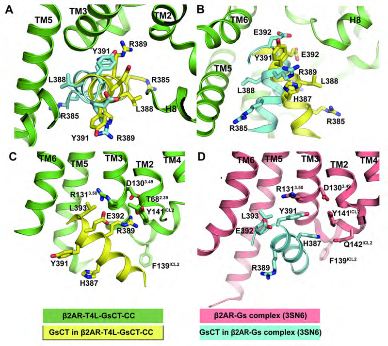
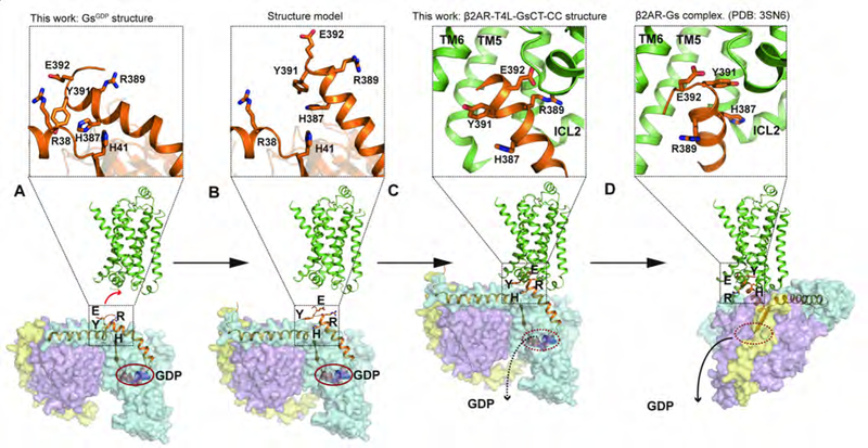
The crystal structure of the β2-adrenergic receptor (β2AR) bound to the G protein adenylyl cyclase stimulatory G protein (Gs) captured the complex in a nucleotide-free state (β2AR-Gsempty). Unfortunately, the β2AR-Gsempty complex does not provide a clear explanation for G protein coupling specificity. Evidence from several sources suggests the existence of a transient complex between the β2AR and GDP-bound Gs protein (β2AR-GsGDP) that may represent an intermediate on the way to the formation of β2AR-Gsempty and may contribute to coupling specificity. Here we present a structure of the β2AR in complex with the carboxyl terminal 14 amino acids from Gαs along with the structure of the GDP-bound Gs heterotrimer. These structures provide evidence for an alternate interaction between the β2AR and Gs that may represent an intermediate that contributes to Gs coupling specificity.

Keywords: G protein; G protein-coupled receptor; coupling specificity; intermediate state; protein engineering.


Conflict of interest statement

DECLARATION OF INTERESTS

B.K.K. is a co-founder of and consultant for ConfometRx, Inc.

Figures

Figure 1:
Figure 1:. Design of the β2AR-T4L-GsCT-CC construct and overall β2AR-T4L-GsCT-CC structure.
(A) Topology map of β2AR-T4L-GsCT-CC construct. (B) Agonist competition studies show β2AR-T4L-GsCT-CC and β2AR-T4L-GsCT exhibit higher affinity for the agonist isoproterenol (ISO) than do β2AR-T4L or β2AR (p < 0.0001) (n=3). The differences between β2AR-T4L-GsCT-CC and β2AR-T4L-GsCT are not statistically significant (p = 0.13). Statistics comparing were performed by extra sum-of-squares F test. Data are represented as mean ± SEM. (C) Overall structure of β2AR-T4L-GsCT-CC. (D) Structure alignment of β2AR-T4L-GsCT-CC structure with active β2AR structure (Protein Data Bank accession number 3SN6). (E) Alignment of β2AR-T4L-GsCT-CC structure with inactive β2AR structure (Protein Data Bank accession number 2RH1). TM6 in β2AR-T4L-GsCT-CC is almost identical to its position in the active structure, and is ~14Å away compared to inactive structure. See also Figure S1, S2, S3 and Table S1.
Figure 2:
Figure 2:. Identification of an alternate interaction between the α5 helix and β2AR
(A and B) Two different views comparing the orientation of GsCT from β2AR-T4L-GsCT-CC structure (yellow) with GsCT from β2AR-Gs complex structure (cyan). (C) E392Gαs interact with R1313.50, R389Gαs interacts with Y141ICL2 and T682.39 in the β2AR-T4L-GsCT-CC structure. (D) Y391Gαs forms cation-π interaction with R1313.50, H387Gαs interacts with Y141ICL2 and Q142ICL2 in β2AR-Gs complex structure (Protein Data Bank accession number 3SN6). See also Figure S4 and S5.
Figure 3:
Figure 3:. The structure of the α5 helix in GsGDP
(A) Alignment of GsGDP with Gi1GDP (Protein Data Bank accession number 1GP2). (B) Alignment of GsGDP with GqGDP (Protein Data Bank accession number 3AH8). The overall structures are similar, but the C terminal end of the α5 helix was not resolved in Gi1GDP or GqGDP. (C) Y391Gαs and H387Gαs on the α5 helix interact with R38Gαs, H41Gαs and R42Gαs on the αN-β1 loop. Cation-pi interaction is shown with a green arrow, hydrogen bonds are shown with green dashes. (D) Conformational changes in the GsCT in going from the GsGDP structure (orange) to the β2AR-Gsempty complex structure (cyan, Protein Data Bank accession number 3SN6) involve disrupting an extensive network of hydrophobic and polar interactions between the α5 helix and the rest of the Ras domain. See also Figure S2 and Table S1.
Figure 4:
Figure 4:. R389Gαs and E392Gαs are essential for efficient complex formation
(A) Fluorescence spectra of monobromobimane–labeled β2AR in presence of 1 µM WT Gs, 1 µM R389A/E392A Gs or 1 µM H387A/Y391A Gs. Each spectrum represents an average of 3 independent spectra. (B) β2AR triggered 3H-GDP release of WT Gs, R389A/E392A Gs or H387A/Y391A Gs (n=3). The statistically significant differences (p < 0.05) between the wild type and mutant are labeled with an asterisk (*). (C) Isoproterenol competition curves of β2AR reconstituted into high-density lipoprotein (HDL) particles, either as receptor alone or in presence of WT Gs, R389A/E392A Gs or H387A/Y391A Gs. The values and statistics of the agonist competition curves are shown in the inset table. Data are given as mean±SEM from 4–6 independent experiments performed in duplicate or triplicate. Statistics comparing WT and mutant Gs were performed by extra sum-of-squares F test. The differences between WT Gs and R389A/E392A Gs are statistically significant (p < 0.0001), so are the differences between WT Gs and H387A/Y391A Gs (p < 0.0001). However, the differences between R389A/E392A Gs and H387A/Y391A Gs are not statistically significant (p = 0.13). See also Figure S6.
Figure 5:
Figure 5:. Interaction between the β2AR and R389Gαs and E392Gαs may initiate GDP release
(A) Interaction between the β2AR and R389Gαs and E392Gαs would affect interactions between H387Gαs, Y391Gαs and the αN-β1 loop. These changes could be propagated to the P loop and β6-α5 loop through β1 strand and α5 helix. (B) H387Gαs interacts with H41Gαs from β1 strand; Q390Gαs interacts with T242Gαs at α2-β4 loop. (C) Effect of Gαs H387A/Q390A mutation on basal GDP release of Gαs. The wild type and mutant are statistically different (p < 0.0001). Statistics comparing the H387A/Q390A with WT Gs was performed by the extra sum-of-squares F test where a model of individually best-fitted values of Koff of the two different datasets was compared to a model where the parameters were shared among the datasets. The data are given as mean±SD of three independent experiments.
Figure 6:
Figure 6:. Proposed process of GPCR – G protein complex formation
(A) Before the formation of β2AR-Gs protein complex, α5 helix of GsGDP is bent with H387Gαs and Y391Gαs interacting with the RAS domain while E392Gαs and R389Gαs are surface exposed. (B and C) An early interaction with the β2AR involves E392Gαs and R389Gαs that requires straightening of the α5 helix, which may trigger GDP release. The β2AR-T4L-GsCT-CC structure may represent this intermediate state. (D) Relative large conformation changes between GPCR and Gs are required to change from the intermediate state shown in panel C to nucleotide-free complex (Protein Data Bank accession number 3SN6). H387Gαs and Y391Gαs interact with β2AR in this state (zoom in window on top of panel D). See also Figure S5.
Figure 7:
Figure 7:. The proposed intermediate complex may contribute to G protein coupling specificity.
Interactions between R389Gαs, E392Gαs and D3.49, R3.50 suggest the interaction pattern may be conserved among most Family A GPCRs. The orientation of the Gs peptide (yellow) requires larger outward movement of TM6 (Green, β2AR). While the smaller movement of TM6 observed in Gi coupled GPCRs is not compatible with this binding mode (Grey, µOR).

References

    1. Abraham MJM,T; Schulz R; Páll S; Smith JC; Hess B; Lindahl E (2015). GROMACS: High performance molecular simulations through multi-level parallelism from laptops to supercomputers. SoftwareX 1–2, 19–25.
    1. Adams PD, Afonine PV, Bunkoczi G, Chen VB, Davis IW, Echols N, Headd JJ, Hung LW, Kapral GJ, Grosse-Kunstleve RW, et al. (2010). PHENIX: a comprehensive Python-based system for macromolecular structure solution. Acta crystallographica Section D, Biological crystallography 66, 213–221. - PMC - PubMed
    1. Baltensperger K, Karoor V, Paul H, Ruoho A, Czech MP, and Malbon CC (1996). The beta-adrenergic receptor is a substrate for the insulin receptor tyrosine kinase. J Biol Chem 271, 1061–1064. - PubMed
    1. Caffrey M, and Cherezov V (2009). Crystallizing membrane proteins using lipidic mesophases. Nat Protoc 4, 706–731. - PMC - PubMed
    1. Carpenter B, Nehme R, Warne T, Leslie AG, and Tate CG (2016). Structure of the adenosine A(2A) receptor bound to an engineered G protein. Nature 536, 104–107. - PMC - PubMed

Publication types

MeSH terms

Substances