. 2019 May 20;29(10):1634-1646.e6.
doi: 10.1016/j.cub.2019.04.032. Epub 2019 May 9.

Origin of a Core Bacterial Gene via Co-option and Detoxification of a Phage Lysin

Affiliations

Origin of a Core Bacterial Gene via Co-option and Detoxification of a Phage Lysin

Amelia M Randich et al. Curr Biol. .

Abstract

Temperate phages constitute a potentially beneficial genetic reservoir for bacterial innovation despite being selfish entities encoding an infection cycle inherently at odds with bacterial fitness. These phages integrate their genomes into the bacterial host during infection, donating new but deleterious genetic material: the phage genome encodes toxic genes, such as lysins, that kill the bacterium during the phage infection cycle. Remarkably, some bacteria have exploited the destructive properties of phage genes for their own benefit by co-opting them as toxins for functions related to bacterial warfare, virulence, and secretion. However, do toxic phage genes ever become raw material for functional innovation? Here, we report on a toxic phage gene whose product has lost its toxicity and has become a domain of a core cellular factor, SpmX, throughout the bacterial order Caulobacterales. Using a combination of phylogenetics, bioinformatics, structural biology, cell biology, and biochemistry, we have investigated the origin and function of SpmX and determined that its occurrence is the result of the detoxification of a phage peptidoglycan hydrolase gene. We show that the retained, attenuated activity of the phage-derived domain plays an important role in proper cell morphology and developmental regulation in representatives of this large bacterial clade. To our knowledge, this is the first observation of a phage gene domestication event in which a toxic phage gene has been co-opted for core cellular function at the root of a large bacterial clade.

Keywords: Alphaproteobacteria; Asticcacaulis; Caulobacter; GH24 lysozyme; bacterial evolution; prophage domestication.


Conflict of interest statement

Declaration of Interests

The authors declare no competing interests.

Figures

Figure 1.
Figure 1.. The SpmX is vertically inherited in Caulobacterales.
(A) Schematic of SpmX architecture, including the conserved muramidase domain (see Figure S1 for alignments), the variable intermediate domain, and two C-terminal transmembrane (TM) segments. Bar indicates amino acid sequence conservation among spmX alleles (see Table S1 for a list of spmX genes used in this study). (B) Phylogenetic trees of representative species from Caulobacterales and other Alphaproteobacteria for concatenated housekeeping gene alignments (left) and for SpmX (right), with branch colors indicating the amino acid identity at position 20 of SpmX (D20L in yellow, D20R in red, and D20G in green). See Table S6 for genome IDs. The concatenated housekeeping tree is fully supported with posterior probability of 1.0 for all clades. Asterisks indicate clades in the SpmX tree with posterior probabilities > 0.95. See Figure S2 for the relationship of the SpmX muramidase domain within the lysozyme superfamily.
Figure 2.
Figure 2.. The SpmX muramidase domain retains the canonical GH motif but contains inactivating mutations in the catalytic cleft.
(A) P22 lysozyme (PDB 2ANX) as a model lysozyme colored with rainbow gradient from blue N-terminus to red C-terminus. The catalytic glutamate appears in fuchsia and the GH beta-hairpin in light blue. (B) HMM logos of GH lysozymes made using WebLogo 3 [52]. Logos were constructed from protein sequences of (i) T4 lysozyme-like genes (n = 94), (ii) representative autolysins/endolysins from the Conserved Domain Database including P22 lysozyme (n = 20) but excluding SpmX genes, (iii) closest BLAST hits from non-SpmX muramidases (n = 60), and (iv) SpmX muramidases (n = 66), and organized in a cladogram to resemble the sequence cluster tree diagram in Figure S2. Amino acids are color-coded according to chemical properties, with uncharged polar residues in green, neutral residues in purple, basic residues in blue, acidic residues in red, and hydrophobic residues in black. The height of each letter is proportional to the relative frequency of a given identity and the height of the stack indicates the sequence conservation at that position. T4L numbering is used for ease of comparison. Asterisks mark positions critical for enzymatic activity and open circles mark positions associated with GH motif stability [26,27]. Refer to Figure S1 for alignments of SpmX muramidases, which are listed in Table S1. See also Table S5 for non-SpmX GH24 gene IDs.
Figure 3.
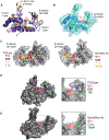
Figure 3.. The structure of SpmX muramidase domain has a wider, more dynamic catalytic cleft than related phage lysins.
(A) Structural alignment of P22 lysozyme (PDB 2ANX, the model used for molecular replacement) in purple, R21 endolysin from P21 (PDB 2HDE, a distantly related GH24 T4L lysozyme) in navy blue, and SpmX-Mur-Ae in gold (PDB 6H9D). The catalytic glutamate is shown in red. Root mean square deviation (rmsd) 1.7 Å and 40% identity over 141 aligned Cα atoms, Dali Z-score 21.5 between P22 lysozyme and SpmX-Mur-Ae. See Table S2 for data collection and refinement statistics. (B) Structural alignment of the three SpmX-Mur-Ae molecules, chains A (green), B (light blue), and C (dark blue), from the asymmetric unit. The surface of chain B is shown in partially transparent light blue. The doubleheaded arrow indicates the tilt of about 16° between the GH beta-hairpins of chains B and A. (C) Overlays of ribbon diagrams and surfaces of P22 lysozyme (2ANX, left) and SpmX-Mur-Ae (6H9D, right) illustrating the conformation of the critical residues E11 (red), D20 (dark blue), R14 (yellow), and Y18 (orange). T4L numbering is used for ease of comparison. These structures have been rotated 180° around the y-axis from their representation in (A, B, D, E). (D) Surface representation of P22 lysozyme (2ANX) with inset showing ribbon diagram and conformation of catalytic cleft with the canonical E11/D20/T26 catalytic triad. (E) Surface representation of SpmX-Mur-Ae (6H9D) with inset showing ribbon diagram and conformation of remodeled catalytic cleft with E11/D20L/T26M.
Figure 4.
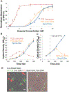
Figure 4.. The D20L mutation attenuates P22 hydrolytic activity.
(A) Remazol brilliant blue assays on C. crescentus sacculi using purified P22 lysozyme, P22 lysozyme D20L mutant, and C. crescentus SpmX muramidase. Active enzymes release peptidoglycan monomers covalently-bound to RBB into the supernatant that are detected by absorbance at 595 nm. Error bars are ± standard deviation for each normalized absorbance (n = 3). Lines are drawn to help guide the eye toward basic trends. Data points are from various days and sacculi preparations, but with internal normalization to Hen Egg White Lysozyme (HEWL). See Figure S3 for peptidoglycan binding activity and Figure S4AB for SpmX mutant activity in RBB assays. (B and C) Growth curves of Lemo21(DE3) E. coli expressing P22 lysozyme (blue), P22 lysozyme D20L mutant (green), and C. crescentus SpmX muramidase (red). Proteins were expressed from pET22b with a N-terminal PelB signal sequence. In (B), strains were grown in 5 mM rhamnose without IPTG for maximal repression of basal expression from the plasmids. In (C), strains were grown without rhamnose and induced with 400 μM IPTG at the indicated time. See Figure S4CDE for enzymatic activity and periplasmic expression of SpmX-Mur and various mutants. (D) Phase/fluorescent overlays show live/dead staining of Lemo21(DE3) cells expressing P22Lyso-D20L and SpmX-Mur-Cc after four hours of induction. Green, membrane permeable SYTO 9 stains DNA in live cells and red, membrane impermeable propidium iodide nucleic acid dyes labels released nucleoids and DNA from lysed bacteria. The rounding of the E. coli in (i) is characteristic of spheroplast formation and lysis by hydrolytic activity on the cell wall. Scale bars are 5 μm.
Figure 5.
Figure 5.. Inactivating the muramidase domain partially delocalizes SpmX in vivo.
Phase and fluorescent images of (A) C. crescentus, (B) A. biprosthecum, and (C) A. excentricus. In the top panel, phase images with derived schematics emphasizing stalks and morphologies are shown for (i) WT and (ii) ΔspmX cells. In Aii, C. crescentus cells exhibiting characteristic ΔspmX divisional defects are marked with asterisks and a cell growing stalks from both poles has its stalks marked with red arrowheads. Phase and fluorescent images of cells expressing (iii) SpmX-eGFP, (iv) SpmX-E11A-eGFP, or (v) SpmX-N105R-eGFP from the native chromosomal locus are shown in the lower panels. In Aiv and Av, cells with divisional defects are marked with white asterisks. In Biii and Biv, cells with one lateral or subpolar stalk are marked with white arrowheads. In Civ, cells with foci at the tips of stalks are marked with white arrowheads. All scale bars are 5 μm. See Figure S5 for quantification of fluorescence and morphology data.
Figure 6.
Figure 6.. Removing or replacing the muramidase domain depletes native SpmX protein levels in vivo.
(A) Phase and fluorescent images of strains in which the native spmX allele was replaced with the following gene fusions in the ΔspmX parent strain (ii): (i) spmX-L20D-sfGFP (iii) WT spmX-sfGFP, (iv) spmX-E11A-sfGFP, (v) MurBs-Δmur-SpmX-sfGFP where MurBs is the muramidase domain from Brevundimonas subvibrioides SpmX, and (vi) spmX-L20R-sfGFP. All scale bars are 5 μm. (B) Western blot comparing the ΔspmX parent strain to SpmX mutants and chimeras inserted at the spmX locus. In all cases, the primary antibody is directed against the C-terminal GFP fusion.

References

    1. Hall JPJ, Brockhurst MA, and Harrison E (2017). Sampling the mobile gene pool: innovation via horizontal gene transfer in bacteria. Phil Trans R Soc B 372, 20160424. - PMC - PubMed
    1. Koonin EV (2016). Viruses and mobile elements as drivers of evolutionary transitions. Phil Trans R Soc B 371, 20150442. - PMC - PubMed
    1. Feiner R, Argov T, Rabinovich L, Sigal N, Borovok I, and Herskovits AA (2015). A new perspective on lysogeny: prophages as active regulatory switches of bacteria. Nat. Rev. Microbiol 13, 641–650. - PubMed
    1. Harrison E, and Brockhurst MA (2017). Ecological and Evolutionary Benefits of Temperate Phage: What Does or Doesn’t Kill You Makes You Stronger. BioEssays 39, 1700112. - PubMed
    1. Howard-Varona C, Hargreaves KR, Abedon ST, and Sullivan MB (2017). Lysogeny in nature: mechanisms, impact and ecology of temperate phages. ISME J. 11, 1511–1520. - PMC - PubMed

Publication types

MeSH terms

LinkOut - more resources