. 2019 Jun 11;12(6):1329-1341.
doi: 10.1016/j.stemcr.2019.04.009. Epub 2019 May 9.

Modeling Motor Neuron Resilience in ALS Using Stem Cells

Affiliations

Modeling Motor Neuron Resilience in ALS Using Stem Cells

Ilary Allodi et al. Stem Cell Reports. .

Abstract

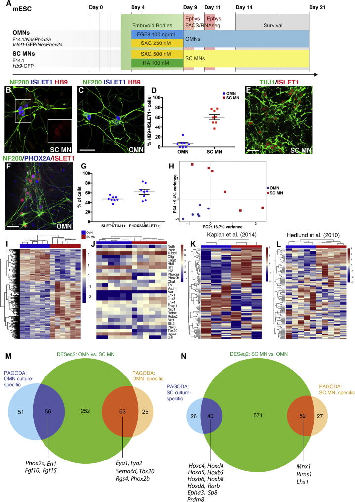
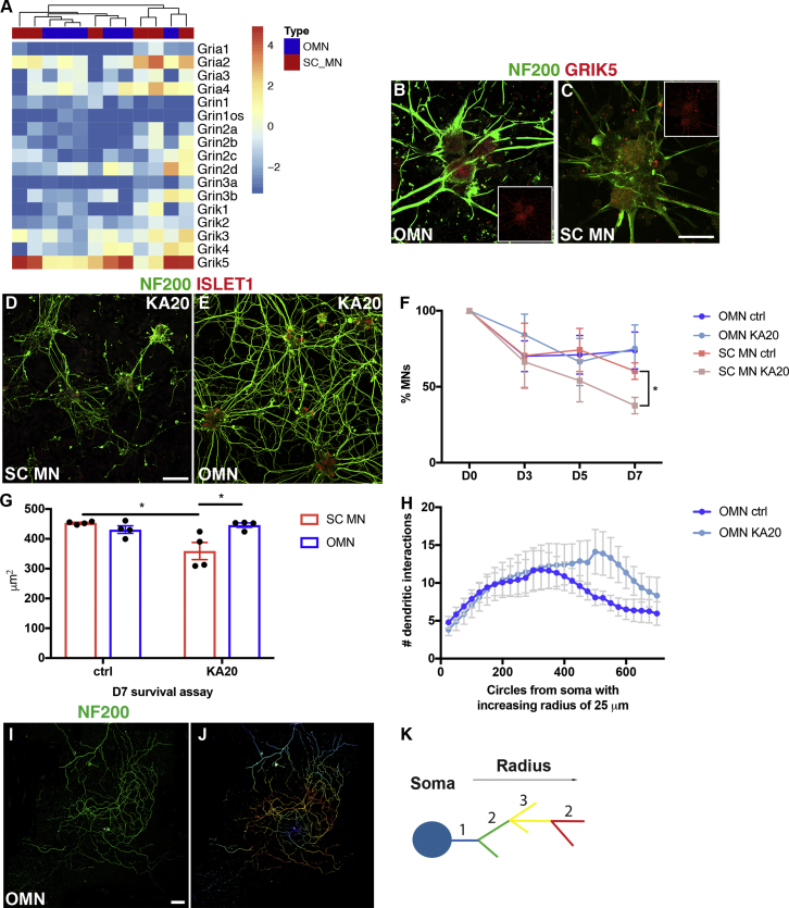
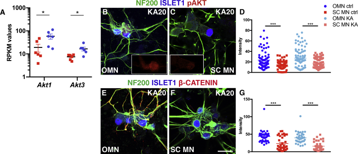
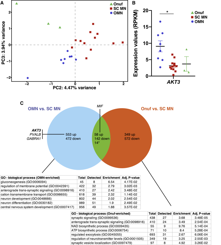
Oculomotor neurons, which regulate eye movement, are resilient to degeneration in the lethal motor neuron disease amyotrophic lateral sclerosis (ALS). It would be highly advantageous if motor neuron resilience could be modeled in vitro. Toward this goal, we generated a high proportion of oculomotor neurons from mouse embryonic stem cells through temporal overexpression of PHOX2A in neuronal progenitors. We demonstrate, using electrophysiology, immunocytochemistry, and RNA sequencing, that in vitro-generated neurons are bona fide oculomotor neurons based on their cellular properties and similarity to their in vivo counterpart in rodent and man. We also show that in vitro-generated oculomotor neurons display a robust activation of survival-promoting Akt signaling and are more resilient to the ALS-like toxicity of kainic acid than spinal motor neurons. Thus, we can generate bona fide oculomotor neurons in vitro that display a resilience similar to that seen in vivo.

Keywords: ALS; LCM sequencing; Onuf’s nucleus; Phox2a; RNA sequencing; amyotrophic lateral sclerosis; neuronal vulnerability and resistance; oculomotor neurons; spinal motor neurons; stem cells.


Figures

Figure 1
Figure 1
PHOX2A Overexpression Drives the Generation of Bona Fide Oculomotor Neurons from Stem Cells (A) Time line depicting in vitro protocols for the generation of resistant oculomotor neuron (OMN) and vulnerable spinal cord motor neuron (SC MN). The mESC column reports the four cell lines used in this study. Patterning was performed for 5 days following EB formation. mRNA-seq coupled with FACS was performed on EB dissociation day (day 9 of the protocol). The survival assay was performed 5 days after EB dissociation (day 14 to 21). (B and C) SC MNs generated from E14.1 ESCs express HB9, ISLET1, and NF200 (B). OMNs generated from E14.1 ESCs overexpressing the transcription factor Phox2a under the Nestin enhancer co-express ISLET1 and NF200 in the absence of HB9 (C). Scale bar, 60 μm. (D) Percentage of HB9+/ISLET1+ cells in SC MN (64.1% ± 5.2%, mean ± SEM, n = 4) and OMN (6.3% ± 1.3%, mean ± SEM, n = 4) cultures, respectively (at least 120 Islet1+ cells counted per condition, bar and whiskers represent means ± SEM). (E) Microphotographs showing ISLET1+/TUJ1+ cells in OMN cultures. (F) OMNs also express the specific marker PHOX2A as indicated by asterisks. Scale bar, 100 μm (applies also to E). (G) Quantification of ISLET1+ over TUJ1+ cells demonstrates that half the neuronal population appears to be ISLET1+ (47.5% ± 5.9%, mean ± SEM, n = 4; total number of TUJ1+ cells counted: 1,325). Quantification of PHOX2A+ over ISLET1+ cells (experiments performed in quadruplicates with two technical replicates each, total number of Islet1+ cells counted: 746) indicates that 62% ± 5.2% (mean ± SEM, n = 4) of the ISLET1+ population is also PHOX2A+. All quantifications were performed 5 days after EB dissociation, and experiments were conducted in quadruplicates including two technical replicates per experiment (bar and whiskers represent means ± SEM). (H) PCA of OMN and SC MN samples based on all genes expressed confirmed cell differential identities. mRNA-seq analysis of OMNs and SC MNs isolated by FACS was performed after 5 days of patterning (day 9 of the protocol). (I) A total of 1,017 DEGs was found between the two different cell types (adjusted p < 0.05, n = 6). Heatmap shows the top 500 most significant DEGs by adjusted p value. (J–L) Heatmap of expected progenitors and motor neuron transcripts but also OMN and SC MN specific transcripts obtained in the two generated motor neuron populations (J and L). Using the top 100 DEGs obtained from our RNA-seq analysis (K), we could separate datasets originating from in vivo microarray studies on early postnatal (J) and adult (L) rodent OMNs and SC MNs. (M and N) Venn diagrams showing gene sets enriched either in brain stem cultures or OMN-specific as revealed by PAGODA analysis (M), and gene sets preferentially found in spinal cord cultures or MN specific (N).
Figure 2
Figure 2
In Vitro-Generated Oculomotor Neurons Are Relatively Resistant to ALS-like Toxicity (A) OMNs and SC MNs express similar mRNA levels of glutamate ionotropic receptor AMPA, NMDA, and kainate type subunits. The heatmap shows log2 RPKM values of these subunits, and no separate clustering of the two cell types is observed. (B and C) Immunohistochemistry performed on generated OMNs (B) and SC MNs (C) at D1 in vitro in control conditions; similar levels of glutamate ionotropic receptor kainate type subunit 5 (GRIK5) are found in both cell types. Scale bar in (C), 60 μm (applies also to B). (D and E) Microphotographs presenting SC MN (D) and OMN (E) response to kainic acid-induced toxicity (20 μM) for a week. Scale bar in (D), 100 μm (applies also to E). (F) Curves represent percentages of MN survival over time in OMN and SC MN cultures. OMNs were visualized as NF200+ISLET1+HB9 cells, while SC MNs as NF200+ISLET1+HB9+ cells. OMNs show increased survival to kainic acid toxicity at D7 (bar and whiskers represent means ± SEM, two-way ANOVA and Tukey's multiple comparison test, F(9, 56) = 2.333, p = 0.0261, SC MNs n = 4, OMNs n = 5) when compared with SC MNs (experiments were performed at least in quadruplicate, with technical replicates and with at least 130 motor neurons counted per condition in each experiment). (G) Analysis of the length of neuronal processes in both oculomotor and spinal motor neuron cultures exposed to kainic acid for 7 days showed that oculomotor neurons were unaffected by kainic acid while spinal motor neurons displayed a shortening of neurites (bar and whiskers represent means ± SEM, two-way ANOVA, p < 0.05, n = 4). (H–K) Sholl analysis was performed on OMN at D7 survival assay in control and KA20 conditions (H) to further assess individual MN arborization complexity during toxicity. (H) Comparison of the average number of neurite intersections of OMN in control and KA20 toxicity conditions with radial step size of 25 μm. OMNs did not show reduction in arborization (bar and whiskers represent means ± SEM, multiple t test, n = 10). (I) Example of OMN at day 7 of KA20 toxicity. Scale bar in (I), 100 μm (applies also to J). (J) Sholl mask was applied to individual OMNs after specifying the radius from the center of the soma of the neuron and created concentric circles every 25 μm of increasing radius. (K) Schematic depicting identification of neurite segments by Sholl analysis. Color code is assigned depending on arbor localization from the soma in an inside-out manner following the given radius. Multiple intersections within the same segment display the same color.
Figure 3
Figure 3
mESC-Derived Oculomotor Neurons Have High Levels of Akt Signaling (A) RPKM values from RNA sequencing of mESC-derived oculomotor and spinal motor neurons enriched by FACS shows preferential expression of Akt1 and Akt3 transcripts in the generated OMNs (bar and whiskers represent means ± SEM, adjusted p < 0.05, n = 6). (B and C) Microphotographs show pAKT levels in OMN (B) and SC MN (C) cultures at D7 toxicity assay assessed by immunohistochemistry, pAKT staining alone in small insets (red channel). (D and F) Fluorescence intensities (D), analyzed in a semi-quantitative manner, indicate higher expression of pAKT in OMNs (F) (bar and whiskers represent means ± SEM, two-way ANOVA and Tukey's multiple comparison test, F(1, 353) = 66.1, ∗∗∗p < 0.0001, n = 4). Scale bar in (F), 60 μm (applies also to B and C). (E and G) β-CATENIN expression is maintained in OMN also during kainate toxicity (E). (G) Semi-quantitative analysis of β-CATENIN over NF200 staining intensities in OMNs and SC MNs in kainate conditions (bar and whiskers represent means ± SEM, t test, t = 6.799, df = 69, ∗∗∗p < 0.0001, n = 3). Scale bar in (E), 60 μm.
Figure 4
Figure 4
Human Oculomotor Neurons Show a Transcriptional Profile, with Elevated AKT3, Unique from Onuf's Nucleus Motor Neurons (A) PCA of all expressed genes separated into Onuf's, oculomotor (OMN), and spinal (cervical and lumbar) (SC_MN) motor neuron groups. (B) RNA-sequencing data showed that AKT3 was elevated in OMNs compared to the other motor neuron groups (adjusted p < 0.05, OMN n = 8, SC_MN n = 12, Onuf's n = 3, bar and whiskers represent means ± SEM). (C) To distinguish the number of DEGs that were shared or unique to each resistant neuron group compared to vulnerable spinal motor neurons, we analyzed DEGs between OMN and SC_MN and between Onuf's and SC_MN. This analysis showed that the majority of DEGs were unique to each resistant motor neuron group, with OMNs showing 553 upregulated and 472 downregulated unique DEGs and Onuf's motor neurons showing 349 upregulated and 572 downregulated unique DEGs. Oculomotor and Onuf's shared 58 upregulated and 142 downregulated DEGs compared with vulnerable spinal motor neurons. 14 DEGs were regulated in the opposite direction in the two motor nuclei.

References

    1. Afgan E., Baker D., van den Beek M., Blankenberg D., Bouvier D., Cech M., Chilton J., Clements D., Coraor N., Eberhard C. The Galaxy platform for accessible, reproducible and collaborative biomedical analyses: 2016 update. Nucleic Acids Res. 2016;44:W3–W10. - PMC - PubMed
    1. Allodi I., Comley L., Nichterwitz S., Nizzardo M., Simone C., Benitez J.A., Cao M., Corti S., Hedlund E. Differential neuronal vulnerability identifies IGF-2 as a protective factor in ALS. Sci. Rep. 2016;6:25960. - PMC - PubMed
    1. Allodi I., Hedlund E. Directed midbrain and spinal cord neurogenesis from pluripotent stem cells to model development and disease in a dish. Front. Neurosci. 2014;8:109. - PMC - PubMed
    1. Bjorke B., Shoja-Taheri F., Kim M., Robinson G.E., Fontelonga T., Kim K.T., Song M.R., Mastick G.S. Contralateral migration of oculomotor neurons is regulated by Slit/Robo signaling. Neural Dev. 2016;11:18. - PMC - PubMed
    1. Boillee S., Yamanaka K., Lobsiger C.S., Copeland N.G., Jenkins N.A., Kassiotis G., Kollias G., Cleveland D.W. Onset and progression in inherited ALS determined by motor neurons and microglia. Science. 2006;312:1389–1392. - PubMed

Publication types