Review
. 2019 Jun 14;294(24):9326-9341.
doi: 10.1074/jbc.REV119.006860. Epub 2019 May 12.

Face-time with TAR: Portraits of an HIV-1 RNA with diverse modes of effector recognition relevant for drug discovery

Affiliations
Review

Face-time with TAR: Portraits of an HIV-1 RNA with diverse modes of effector recognition relevant for drug discovery

Sai Shashank Chavali et al. J Biol Chem. .

Abstract

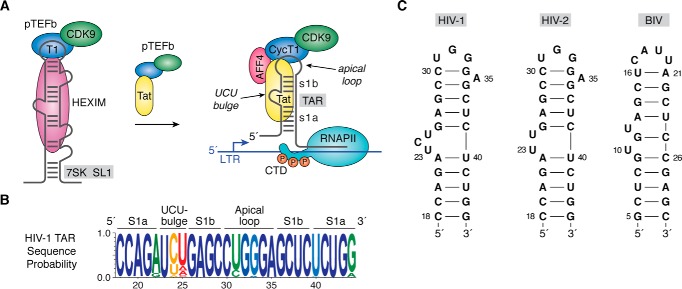
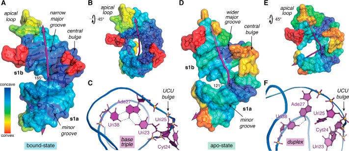
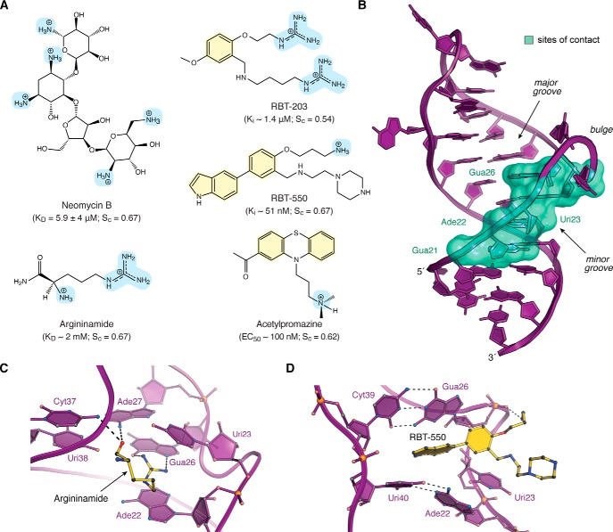
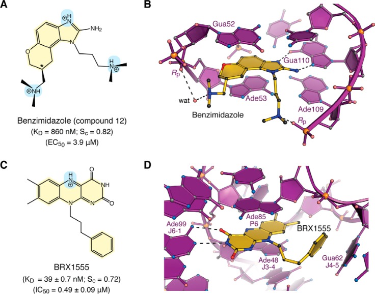
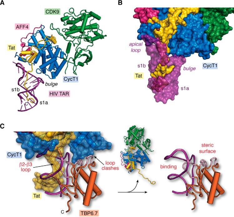
Small molecules and short peptides that potently and selectively bind RNA are rare, making the molecular structures of these complexes highly exceptional. Accordingly, several recent investigations have provided unprecedented structural insights into how peptides and proteins recognize the HIV-1 transactivation response (TAR) element, a 59-nucleotide-long, noncoding RNA segment in the 5' long terminal repeat region of viral transcripts. Here, we offer an integrated perspective on these advances by describing earlier progress on TAR binding to small molecules, and by drawing parallels to recent successes in the identification of compounds that target the hepatitis C virus internal ribosome entry site (IRES) and the flavin-mononucleotide riboswitch. We relate this work to recent progress that pinpoints specific determinants of TAR recognition by: (i) viral Tat proteins, (ii) an innovative lab-evolved TAR-binding protein, and (iii) an ultrahigh-affinity cyclic peptide. New structural details are used to model the TAR-Tat-super-elongation complex (SEC) that is essential for efficient viral transcription and represents a focal point for antiviral drug design. A key prediction is that the Tat transactivation domain makes modest contacts with the TAR apical loop, whereas its arginine-rich motif spans the entire length of the TAR major groove. This expansive interface has significant implications for drug discovery and design, and it further suggests that future lab-evolved proteins could be deployed to discover steric restriction points that block Tat-mediated recruitment of the host SEC to HIV-1 TAR.

Keywords: HIV TAR RNA; RNA structure; RNA virus; RNA-binding protein; RNA–drug interaction; RNA–protein interaction; arginine-sandwich motif; drug development; drug discovery; structural biology; structural model; structure–function; super-elongation complex; viral transactivation.


Conflict of interest statement

The authors declare that they have no conflicts of interest with the contents of this article.

Figures

Figure 1.
Figure 1.
HIV-1 TAR role in transcription, sequence conservation, and secondary structure. A, cartoon diagram of an inactive pTEFb complex comprising CDK9 and CycT1 in the context of HEXIM protein bound to 7SK ncRNA in the host. The arrow indicates that addition of the HIV-1 regulatory protein Tat competes with HEXIM, removing pTEFb from 7SK, which is then escorted by Tat to the TAR RNA element of HIV-1 (141). TAR is essential for transcription and is depicted as a stem loop interrupted by a central bulge that comprises nucleotides 18–44 of the viral transcript (78, 142). Tat interacts directly with TAR and promotes formation of a host SEC comprising pTEFb, scaffold proteins such as AFF4, and other factors (43, 47, 52, 68, 143, 144). CDK9 phosphorylates host RNA polymerase II in its CTD, which releases pausing and stimulates synthesis of full-length viral transcripts (33, 145–147). B, Web-logo showing the sequence conservation of HIV-1 TAR based on circulating forms of the virus compiled as described (77); blue represents the greatest conservation, and red indicates the poor conservation. Elements of the secondary structure including helical stems s1a and s1b are labeled. C, secondary structures of various TAR RNAs. The canonical Cyt30–Gua34 pair of HIV-1 TAR is supported by chemical modification, NMR, sequence conservation, and CycT1-binding requirements (65, 66, 77, 148, 149). A key difference between HIV-1 and HIV-2 TAR is deletion of Cyt24 in the central bulge (150). Details of the BIV TAR secondary structure were derived from Refs. , , . CTD, C-terminal domain.
Figure 2.
Figure 2.
Overview of TAR conformational differences in the ligand-bound and ligand-free states. A, surface map of concave and convex features for the bound-state of HIV-2 TAR (PDB entry 6mce) (76). The helical axis (purple line) deviates subtly from linearity with an angle of 165°. B, view of A rotated +45°; convex features map mainly to the apical loop and bulge. C, ribbon model passing through the phosphate backbone showing the Uri23·Ade27–Uri38 major-groove base triple—a hallmark long-range interaction characteristic of the ligand-bound state. The flanking UCU bulge is depicted. Coordinates were derived from the HIV-1 TAR–TBP6.7 complex (PDB entry 6cmn) (77). D, surface map of concave and convex features for apo-state HIV-1 TAR (PDB entry 1anr) (80). The helical axis bends substantially with an overall angle of 121°. The structure is characterized by more convex surfaces compared with the bound state. E, view of D rotated +45° to emphasize the helical bend. F, ribbon model revealing the Ade27–Uri38 duplex but not the major groove triple. Bases of the flanking UCU bulge penetrate the core contributing to the bend. The helical axis, angle, major-groove width, and depth were calculated by Curves+ (152); when applicable, parameters were computed as the average of the NMR ensemble. Concave and convex properties for each nucleotide of the lowest-energy NMR structures were calculated by Cx (83) and displayed on a Curves+ output file as a heat-map surface using PyMOL (Schrödinger, LLC). Here and elsewhere, perceived hydrogen bonds and related interactions are depicted as broken lines.
Figure 3.
Figure 3.
Chemical structures, interaction properties, and representative modes of small-molecule binding to HIV TAR. A, chemical diagrams for various small molecules that bind TAR and have been characterized structurally by experimental approaches. Positively charged groups are light blue, and aromatic rings are pale yellow. Equilibrium KD values for TAR binding to neomycin and argininamide were derived from NMR (69, 92). Ki values for RBT-203 and RBT-550 were measured for the ability to displace a Tat-derived peptide from TAR, as monitored by FRET (106, 107). The EC50 value of acetylproamizine was estimated based on an EMSA analysis of concentration-dependent disruption of a TAR–Tat–CycT1 complex (64). Here and elsewhere, shape correlation coefficients for RNA–ligand interfaces were calculated by the program Sc on a scale of 0 to 1.0 (102). Calculations in A were applied to the following: TAR–neomycin (PDB entry 1qd3) (92); TAR–argininamide (PDB entry 1akx) (69); TAR–RBT-203 (PDB entry 1uub) (107); TAR–RBT550 (PDB entry 1uts) (106); and TAR–acetylpromazine (PDB entry 1lvj) (64). Sc values are the average derived from the reported NMR ensembles. Solvent-accessible surface areas of RNA–ligand interfaces were calculated by PISA (153). B, ribbon diagram of HIV-1 TAR (PDB entry 6cmn) (77) depicting the locations of nucleotides (green surface) that interact with various small molecules in Table 1. Most ligands bind in the major groove at the interface between s1a and s1b; neomycin binds in the minor groove (92). C, ribbon and ball–and–stick diagram of HIV-2 TAR in complex with argininamide. D, ribbon and ball–and–stick diagram of HIV-1 TAR in complex with RBT-550.
Figure 4.
Figure 4.
Chemical properties and modes of drug binding to representative ncRNAs. A, chemical structure of benzimidazole variant “compound 12”. The KD of binding to HCV domain IIa and EC50 from replicon assays are shown (118). The chiral center is labeled with an asterisk. B, ball–and–stick diagram of HCV domain IIa (purple) bound to compound 12 (yellow) (PDB entry 3tzr) (28). The stereochemistry was not resolved in electron density maps. C, chemical structure of the FMN analogue BRX1555. The KD value of drug binding, the EC50 value from single-round transcription assays, and the IC50 value for bacterial growth inhibition are provided (25). D, ball-and-stick diagram of the Fusobacterium nucleatum FMN riboswitch in complex with BRX1555 (PDB entry 6dn3) (25). The respective ncRNA–inhibitor structures were chosen based on visual inspection of ligand fit to electron-density maps and associated quality-control indicators.
Figure 5.
Figure 5.
Tat organization and molecular recognition of TAR by Tat ARM domains, lab-evolved proteins, and cyclic peptides. A, functional domain organization of the HIV-1 Tat protein (154). The transactivation domain comprises an N-terminal acidic and proline-rich domain, a cysteine-rich domain that binds Zn(II) (i.e. zinc finger or ZnF), a core region, and a basic ARM. Additional domains include a glutamine-rich region and the E2 CTD. B, summary of peptide sequence interactions with TAR RNAs from C–J (blue box). Amino acids of naturally occurring BIV and HIV-1 Tat ARMs are shown; the sequence alignment is based on common recognition modes of RNA targets as follows: Gua11(26) and Gua14(28) of BIV(HIV) TAR are recognized by Arg-73(R52) and Arg-70(R49) of BIV(HIV) Tat (yellow box). Amino acids of lab-evolved proteins and cyclic peptides from structure-based design. The sequence alignment is based on common spatial recognition at Gua26 and Gua28 of HIV-1 TAR by Arg-47 and Arg-49 of TBP6.7 and Arg-3 and Arg-5 of JB181. Specific RNA–peptide interaction types are listed above each amino acid; the symbols are as follows: π indicates cation–π or aromatic stacking; H equals hydrogen-bond recognition of a guanine Hoogsteen edge; Pi indicates salt-bridge formation to the phosphate backbone; b indicates hydrogen-bond recognition to a nucleobase; a pentagon indicates a hydrogen-bond contact to ribose. Symbols of nonstandard amino acids are as follows: B equals l-2,4-diaminobutyric acid; O equals l-ornithine; backward P is d-proline. C, global view depicting BIV TAR (purple ribbon) recognition by the BIV-Tat ARM (yellow worm) (PDB entry 1biv) (75). The Sc value was calculated from the lowest energy NMR core structure (amino acids 67–79). D, close-up view of BIV TAR recognition by BIV Tat at the UU bulge. Despite differences in the central bulge compared with HIV TAR (Fig. 1C), BIV TAR exhibits a major-groove base triple at Uri10·Ade13–Uri24. The Tat peptide undergoes a sharp bend with dihedral angles of ϕi + 1 75°, ψi + 1 −10°, and ϕi + 2 −133°, ψi + 2 63° characteristic of a type V′ turn (130); the peptide has no other β-hairpin characteristics. For clarity, only amino acids engaged in peptide–RNA were included in diagrams. E, global view depicting HIV-2 TAR recognition by the HIV-1 Tat peptide (PDB entry 6mce) (76). The Sc value was calculated from the lowest energy NMR core structure (amino acids 48–54). F, close-up view of HIV-2 TAR recognition at its UU bulge by HIV-1 Tat. Three nucleobases compose an ASM (cyan highlight) that engages Arg-52 of Tat. G, global view depicting HIV-1 TAR recognition by TBP6.7 (PDB entry 6cmn) (77). The lab-evolved β2–β3 loop (yellow) recognizes the TAR major groove. The Sc value was calculated from the co-crystal structure. H, close-up view of HIV-1 TAR recognition in the central UCU bulge by TBP6.7. Arg-47 engages TAR at the ASM, similar to F. I, global view of HIV-1 TAR recognition by the cyclic peptide JB181 (PDB entry 6d2u) (63). The Sc value was calculated from the lowest energy NMR structure. J, close-up view of HIV-1 TAR recognition at the UCU bulge by JB181. The DP13–LP14 turn is shown to emphasize the restrained peptide conformation. CTD, C-terminal domain.
Figure 6.
Figure 6.
Structural model of the core super-elongation complex bound to HIV TAR(1–60). A, hypothetical model of HIV-1 TAR in complex with CycT1–CDK9–AFF4–Tat(1–60). The putative Tat ARM trajectory is based on superposition of HIV-1 TAR in the context of the recent co-crystal structure of the SEC core complex (PDB entry 6cyt) (74) upon HIV-2 TAR in the context of the recent TAR–Tat(44–60) structure (PDB entry 6mce) (76), as depicted in Fig. 5C. Rather than localizing the Tat ARM domain to the s1b stem (74), the model predicts that the ARM runs through TAR's major groove. B, surface model of the hypothetical SEC–TAR–Tat model from A. The surface emphasizes three virus–host protein contact points to TAR. First, the RNA apical loop makes a modest number of interactions with Tat (yellow) and CycT1 (blue). Second, the proximal segment of the Tat ARM (amino acids 47–52) contacts TAR within s1b and the bulged loop. Third, the distal Tat ARM (amino acids 53–57) contacts TAR at s1a. For emphasis, TAR is depicted as a semi-transparent surface to allow Tat visualization in the major groove. Notably, there are no observed contacts between the TAR bulge and CycT1, which is behind the RNA in this orientation. C, two sites of steric blocking are predicted when TBP6.7 is docked onto the SEC–TAR–Tat model of A. Specifically, interference occurs by the β2–β3 loop of TBP6.7 (labeled “binding”) where the Tat ΑRΜ interacts with s1b and the UCU bulge of TAR (Fig. 5, G and H). TBP6.7 loops also interfere with the positioning of CycT1 in the context of the SEC due to steric clashes (labeled “steric surface”). The net result is displacement of the SEC, resulting in the TAR–TBP6.7 complex (right panel). The hypothetical model (left panel) was prepared by superposition of the HIV-1 TAR–TBP6.7 co-crystal structure (PDB entry 6cmn) (77) upon the SEC–TAR–Tat model of A.

References

    1. Cech T. R., and Steitz J. A. (2014) The noncoding RNA revolution–trashing old rules to forge new ones. Cell 157, 77–94 10.1016/j.cell.2014.03.008 - DOI - PubMed
    1. Pennisi E. (2012) Genomics. ENCODE project writes eulogy for junk DNA. Science 337, 1159–1161 10.1126/science.337.6099.1159 - DOI - PubMed
    1. Djebali S., Davis C. A., Merkel A., Dobin A., Lassmann T., Mortazavi A., Tanzer A., Lagarde J., Lin W., Schlesinger F., Xue C., Marinov G. K., Khatun J., Williams B. A., Zaleski C., et al. (2012) Landscape of transcription in human cells. Nature 489, 101–108 10.1038/nature11233 - DOI - PMC - PubMed
    1. Matsui M., and Corey D. R. (2017) Non-coding RNAs as drug targets. Nat. Rev. Drug Discov. 16, 167–179 10.1038/nrd.2016.117 - DOI - PMC - PubMed
    1. Li Z., and Rana T. M. (2014) Therapeutic targeting of microRNAs: current status and future challenges. Nat. Rev. Drug Discov. 13, 622–638 10.1038/nrd4359 - DOI - PubMed

Publication types

MeSH terms

Substances