

Macrophage Imaging of Intracranial Aneurysms
- PMID: 31080227
- PMCID: PMC6635145
- DOI: 10.2176/nmc.st.2019-0034
Macrophage Imaging of Intracranial Aneurysms
Abstract
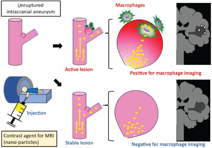
Considered with a poor outcome of subarachnoid hemorrhage due to rupture of intracranial aneurysms (IAs), treatment interventions to prevent rupture of the lesions are mandatory for social health. As treatment option is limited to surgical manipulations, like microsurgical clipping, endovascular coiling or deployment of flow diverter, and these surgical interventions have a potential risk of complications in nature, a proper selection of rupture-prone IAs among ones incidentally found is essential. Today, a rupture risk in each case is estimated by several factors like patient characteristics and morphological ones of each lesion. However, unfortunately, an IA without treatment sometimes unexpectedly ruptures resulting in a devastating outcome or an IA surgically treated is turned out to have a thick wall. To achieve more efficient treatment interventions, the development of a novel diagnostic modality is required. Here, mainly through the accumulation of experimental findings, the crucial contribution of macrophage-mediated chronic inflammatory responses to IA progression have been revealed, making macrophage being a promising target for a diagnosis. If we could non-invasively visualize accumulation of macrophages in lesions, this imaging technique 'macrophage imaging' may enable a qualitative evaluation of IAs to stratify rupture-prone 'dangerous' lesions among many stable ones. Thereby, a development of macrophage imaging makes an indication of surgical interventions being more accurate and also greatly facilitates a development of a novel medical therapy if used as a surrogate marker.
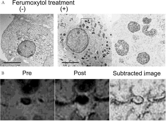
Keywords: chronic inflammation; ferumoxytol; imaging; intracranial aneurysm; macrophage.
Conflict of interest statement
All authors have no conflicts of interest and registered online Self-reported COI Disclosure Statement Forms through the website for The Japan Neurosurgical Society.
Figures
Similar articles
-
Inflammatory changes in the aneurysm wall: a review.J Neurointerv Surg. 2018 Jul;10(Suppl 1):i58-i67. doi: 10.1136/jnis.2009.002055.rep. J Neurointerv Surg. 2018. PMID: 30037960 Review.
-
Inflammatory changes in the aneurysm wall: a review.J Neurointerv Surg. 2010 Jun;2(2):120-30. doi: 10.1136/jnis.2009.002055. Epub 2010 Mar 12. J Neurointerv Surg. 2010. PMID: 21990591 Review.
-
Intracranial Aneurysm as a Macrophage-mediated Inflammatory Disease.Neurol Med Chir (Tokyo). 2019 Apr 15;59(4):126-132. doi: 10.2176/nmc.st.2018-0326. Epub 2019 Mar 14. Neurol Med Chir (Tokyo). 2019. PMID: 30867357 Free PMC article.
-
Increased macrophage M2/M1 ratio is associated with intracranial aneurysm rupture.Acta Neurochir (Wien). 2023 Jan;165(1):177-186. doi: 10.1007/s00701-022-05418-0. Epub 2022 Nov 28. Acta Neurochir (Wien). 2023. PMID: 36437400
-
Intraoperative rupture in the surgical treatment of patients with intracranial aneurysms.J Clin Neurosci. 2016 Dec;34:63-69. doi: 10.1016/j.jocn.2016.01.045. Epub 2016 Sep 28. J Clin Neurosci. 2016. PMID: 27692502
Cited by
-
Current understanding of macrophages in intracranial aneurysm: relevant etiological manifestations, signaling modulation and therapeutic strategies.Front Immunol. 2024 Jan 8;14:1320098. doi: 10.3389/fimmu.2023.1320098. eCollection 2023. Front Immunol. 2024. PMID: 38259443 Free PMC article. Review.
-
The bifurcation angle is associated with the progression of saccular aneurysms.Sci Rep. 2022 May 6;12(1):7409. doi: 10.1038/s41598-022-11160-5. Sci Rep. 2022. PMID: 35523805 Free PMC article.
-
Pseudo-Enhancement in Intracranial Aneurysms on Black-Blood MRI: Effects of Flow Rate, Spatial Resolution, and Additional Flow Suppression.J Magn Reson Imaging. 2021 Sep;54(3):888-901. doi: 10.1002/jmri.27587. Epub 2021 Mar 10. J Magn Reson Imaging. 2021. PMID: 33694334 Free PMC article.
-
Ferumoxytol-β-glucan Inhibits Melanoma Growth via Interacting with Dectin-1 to Polarize Macrophages into M1 Phenotype.Int J Med Sci. 2021 Jun 26;18(14):3125-3139. doi: 10.7150/ijms.61525. eCollection 2021. Int J Med Sci. 2021. PMID: 34400883 Free PMC article.
-
Discrepancies between human and murine model cerebral aneurysms at single-cell resolution.Front Cell Dev Biol. 2025 Mar 11;13:1512938. doi: 10.3389/fcell.2025.1512938. eCollection 2025. Front Cell Dev Biol. 2025. PMID: 40134579 Free PMC article.
References
-
- Vlak MH, Algra A, Brandenburg R, Rinkel GJ: Prevalence of unruptured intracranial aneurysms, with emphasis on sex, age, comorbidity, country, and time period: a systematic review and meta-analysis. Lancet Neurol 10: 626–636, 2011 - PubMed
-
- Greving JP, Wermer MJ, Brown RD, et al. : Development of the PHASES score for prediction of risk of rupture of intracranial aneurysms: a pooled analysis of six prospective cohort studies. Lancet Neurol 13: 59–66, 2014 - PubMed
-
- Kotowski M, Naggara O, Darsaut TE, et al. : Safety and occlusion rates of surgical treatment of unruptured intracranial aneurysms: a systematic review and meta-analysis of the literature from 1990 to 2011. J Neurol Neurosurg Psychiatry 84: 42–48, 2013 - PubMed
-
- Ishibashi T, Murayama Y, Urashima M, et al. : Unruptured intracranial aneurysms: incidence of rupture and risk factors. Stroke 40: 313–316, 2009 - PubMed