

Case Reports
doi: 10.1016/j.radcr.2019.03.040.
eCollection 2019 Jul.
Langerhans' cell histiocytosis of the temporal bone in an adult with central diabetes insipidus
Affiliations
- PMID: 31080537
- PMCID: PMC6502742
- DOI: 10.1016/j.radcr.2019.03.040
Item in Clipboard
Case Reports
Langerhans' cell histiocytosis of the temporal bone in an adult with central diabetes insipidus
Radiol Case Rep.
.
Abstract
We present a case of Langerhans' cell histiocytosis in a 40-year-old woman presenting with central diabetes insipidus and right ear pain. As this disease process is often clinically challenging, the presence of certain imaging findings should raise the possibility of this diagnosis. We review the pertinent imaging and correlate with histology and immunohistochemistry leading to the diagnosis.
Keywords: Adult; CT temporal bone; Histology; Langerhans cell histiocytosis; MRI.
Figures

Sagittal T1-weighted contrast-enhanced image of the pituitary MRI demonstrating marked thickening of the pituitary infundibulum.
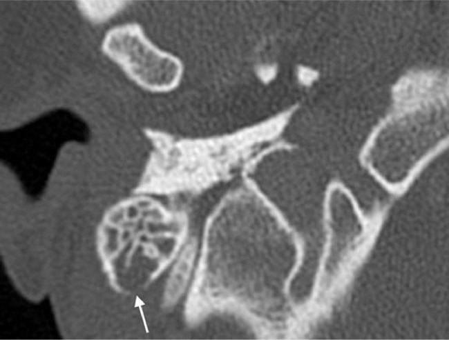
Axial section from high resolution CT of the temporal bone demonstrating opacification of the right mastoid air cells with focal bony resorption at the posterior cortex (arrow).
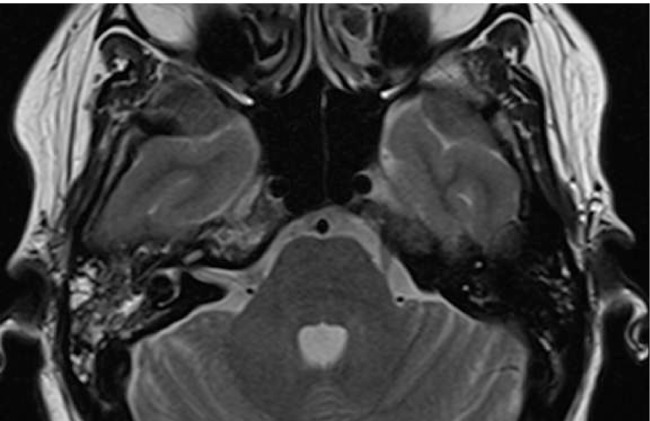
Axial T2-weighted MRI of the temporal bone reveals mixed intensity signal changes within the right mastoid air cells. Note that the right petrous apex has also increased T2-weighted signal.
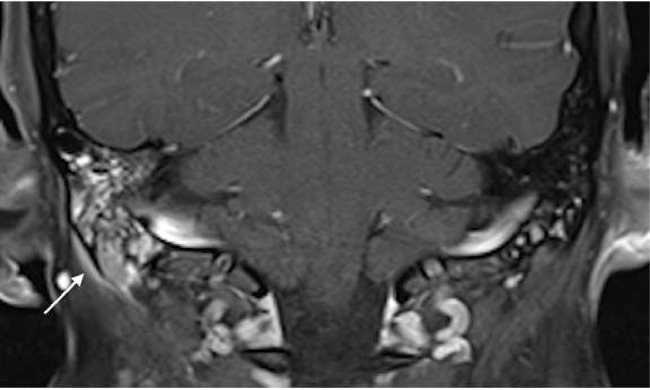
Coronal T1-weighted contrast-enhanced fat saturated image of the right temporal bone demonstrating heterogeneous enhancement within the right mastoid air cells with accompanying subperiosteal soft tissue thickening and enhancement beneath the mastoid tip (arrow).
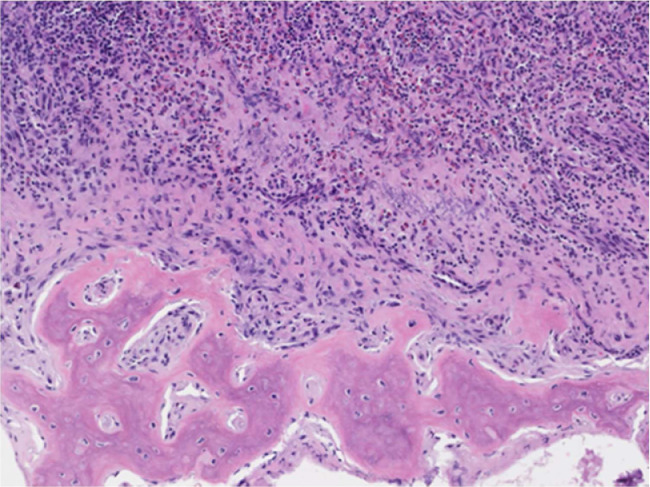
Bony lesion shows infiltration of polygonal cells with eosinophilic cytoplasm and oval nuclei (Langerhans cells); admixed with numerous eosinophils and lymphocytes; ×100 (H&E).
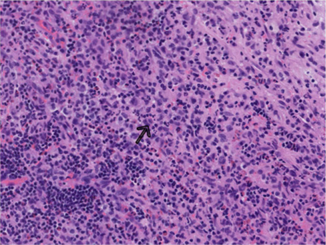
Cells with nuclei containing linear grooves (arrow), typical cytologic feature of Langerhans cells; ×200 (H&E).
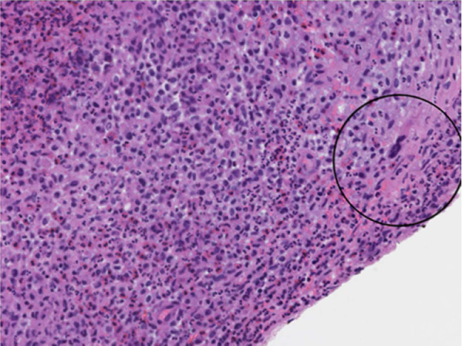
Multinucleated form (circled); ×200 (H&E).
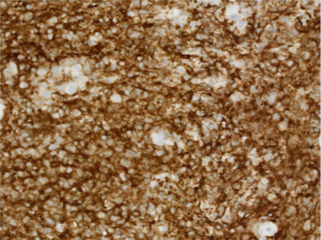
Immunohistochemistry showing presence of Langerhans cell CD1a antigen. CD1a; ×200.
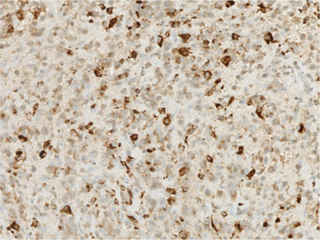
Immunohistochemistry showing presence of Langerhans cell CD68 protein; ×200.
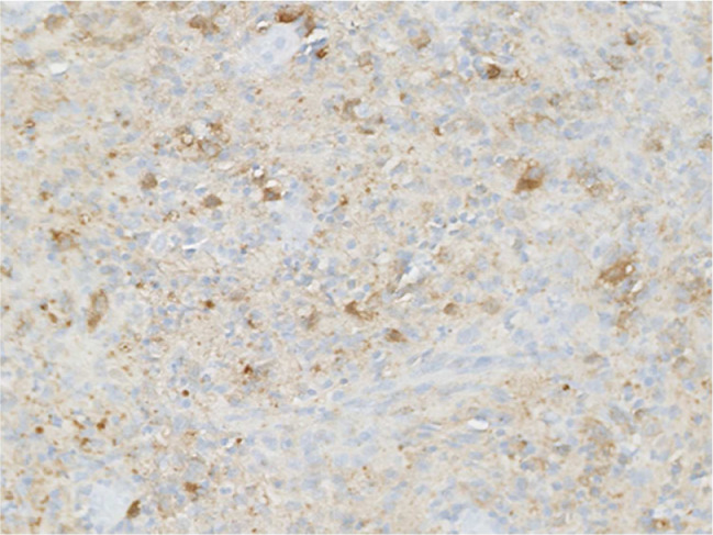
Immunohistochemistry showing presence of Langerhans cell S100 protein; ×200.
References
-
- Swerdlow S.H., Campo E., Harris N.L., Jaffe E.S., Pileri S.A., Stein H., Thiele J., Vardiman J.W. Fourth Edition. World Health Organization; 2017. WHO Classification of Tumors of Hematopoietic and Lymphoid Tissues; pp. 400–439. Revised edition.
-
- Stalemark H., Laurencikas E., Karis J. Incidence of Langerhans cell histiocytosis in children: a population-based study. Pediatr Blood Cancer. 2008;51:76–81. - PubMed
-
- Salotti J.A., Nanduri V., Pearce M.S. Incidence and clinical features of Langerhans cell histiocytosis in the UK and Ireland. Arch Dis Child. 2009;94:376–380. - PubMed
-
- Guyot-Goubin A., Donadieu J., Barkaoui M. Descriptive epidemiology of childhood Langerhans cell histiocytosis in France, 2000-2004. Pediatr Blood Cancer. 2008;51:71–75. - PubMed
Publication types
LinkOut - more resources
Full Text Sources
Research Materials
