. 2019 Apr 18:6:918-928.
doi: 10.1016/j.mex.2019.04.010. eCollection 2019.

RELi protocol: Optimization for protein extraction from white, brown and beige adipose tissues

Affiliations

RELi protocol: Optimization for protein extraction from white, brown and beige adipose tissues

R Diaz Marin et al. MethodsX. .

Abstract

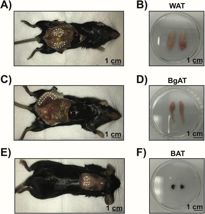
Global obesity rates have reached pandemic proportions, increasing the risk of metabolic complications for hundreds of millions of individuals worldwide. Gaining insight on adipose tissue biology and understanding how fat pads behave during obesity is critical to investigate metabolic syndromes. Elucidation of cellular signaling pathways engaged by adipose tissue both in health and disease requires standardized protocols for protein extraction that yield consistently pure samples. A recurrent problem of currently available protocols is lipid or detergent contamination in extracted protein samples, which renders protein quantification inaccurate and, as a consequence, consistency and reproducibility of protein loading become unreliable. To overcome this problem, we improved the process of adipose tissue protein extraction by improving tissue lysis and decreasing lipid contamination. Here we describe the Removal of Excess Lipids (RELi) protocol to obtain increased yields of total proteins extracted from adipose tissue. The RELi protocol allows accurate and reproducible adipose tissue sample preparation for Western blot analysis and other investigative techniques requiring adipose tissue-derived proteins.

Keywords: Adipose tissue; BAT; Protein extraction; WAT; Western blot.


Figures

None
Graphical abstract
Fig. 1
Fig. 1
Location of white, brown and beige adipose tissue in mice. A) Representative image of epididymal white adipose tissue (WAT) in male C57BL/6 mice (region delineated by dashed line). B) Epididymal adipose tissue without glands and removed from animals. C) Representative image of interscapular brown adipose tissue (BAT) under the white adipose tissue layer in male C57BL/6 mice (region delineated by dashed line). D) Interscapular adipose tissue removed from animals after extraction of white adipose tissue layer. E) Representative image of subcutaneous beige adipose tissue (BgAT) in male C57BL/6 mice (region delineated by dashed line). F) Subcutaneous BgAT adipose tissue removed from animals.
Fig. 2
Fig. 2
RELi protein extraction method compared to the old standard method. The schematic illustrates the different steps of protein extraction using the CST and RELi methods applied to distinct adipose tissues: I) Tissue collection, II) Tissue lysis, III) Centrifugation and removal of excess lipids, and IV) Protein quantification.
Fig. 3
Fig. 3
Assessment of protein extraction efficiency with the CST and RELi methods. Western blot analysis was performed on proteins extracted from 3 independent samples following the CST and RELi method. A–C) SimplyBlue SafeStaining stained gel showing protein extracts from WAT (A), BgAT (B) and BAT (C). MW, protein molecular weight standard; CST, CST method and RELi, Removal of Excess Lipids method. D) Quantification of total protein loaded for WAT, BAT and BgAT following SimplyBlue SafeStaining. E) Western blot analysis of β-actin (˜42 KDa) in WAT, BgAT and BAT extracts. F) Quantification of total protein loaded for WAT, BAT and BgAT. Data shown as mean ± S.E.M. of triplicate wells and are representative of two independent experiments; *p < 0.05, Student’s unpaired t-test.
Fig. 4
Fig. 4
Assessment of quality and uniformity of adipose tissue proteins extracted with the CST and RELi methods. A–C) Western blot analysis of adipose tissue cellular compartments: Cytosol (β-actin ˜42 KDa), Stromal vascular fraction (eNOS ˜140 KDa), Mitochondria (BCL2 ˜26 KDa), Lipid associated proteins (Perilipin A ˜68 KDa), Nuclear protein (Histone H3 ˜15 KDa) in WAT, BgAT and BAT extracts; C (Cytosol), SVF (Stromal vascular fraction), M (Mitochondria), LD (Lipid droplet associated), N (Nuclear).

References

    1. Lau D.C., Dhillon B., Yan H., Szmitko P.E., Verma S. Adipokines: molecular links between obesity and atheroslcerosis. Am. J. Physiol. Heart Circ. Physiol. 2005;288(5):H2031–41. - PubMed
    1. Smith R.E. Thermoregulatory and adaptive behavior of brown adipose tissue. Science. 1964;146(3652):1686–1689. - PubMed
    1. Wu J., Bostrom P., Sparks L.M., Ye L., Choi J.H., Giang A.H. Beige adipocytes are a distinct type of thermogenic fat cell in mouse and human. Cell. 2012;150(2):366–376. - PMC - PubMed
    1. Park A., Kim W.K., Bae K.H. Distinction of white, beige and brown adipocytes derived from mesenchymal stem cells. World J. Stem Cells. 2014;6(1):33–42. - PMC - PubMed
    1. Petrovic N., Walden T.B., Shabalina I.G., Timmons J.A., Cannon B., Nedergaard J. Chronic peroxisome proliferator-activated receptor gamma (PPARgamma) activation of epididymally derived white adipocyte cultures reveals a population of thermogenically competent, UCP1-containing adipocytes molecularly distinct from classic brown adipocytes. J. Biol. Chem. 2010;285(10):7153–7164. - PMC - PubMed

LinkOut - more resources