

Close Relationship between cIAP2 and Human ARDS Induced by Severe H7N9 Infection
- PMID: 31080811
- PMCID: PMC6475567
- DOI: 10.1155/2019/2121357
Close Relationship between cIAP2 and Human ARDS Induced by Severe H7N9 Infection
Abstract
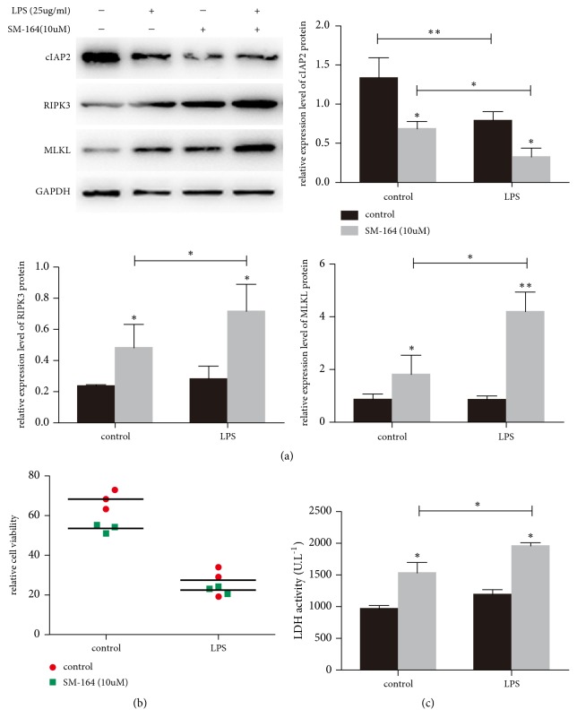
Background: cIAP2 is involved in necroptosis as a key upstream regulation factor. We aimed to investigate the role of cIAP2 in ARDS/ALI induced by H7N9 virus through regulating the RIPK1/3 necroptosis pathway.
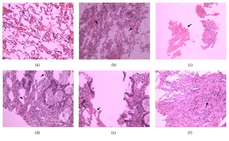
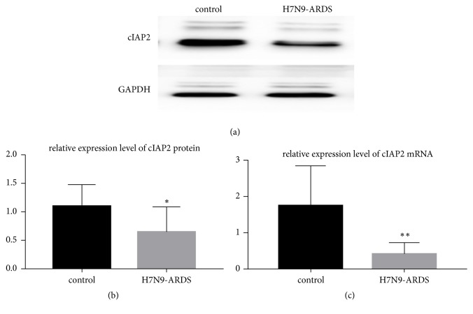
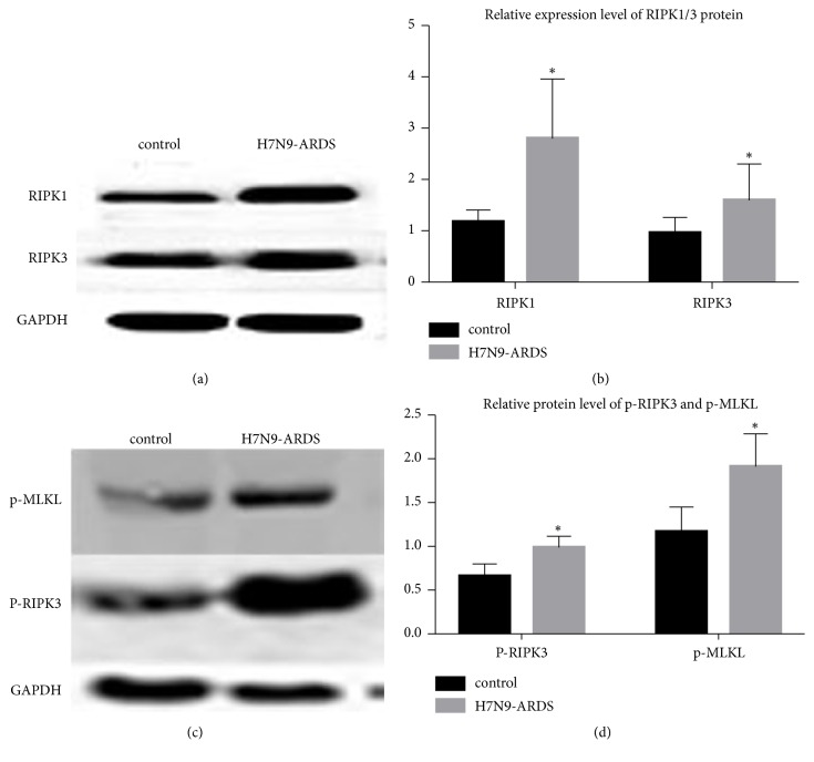
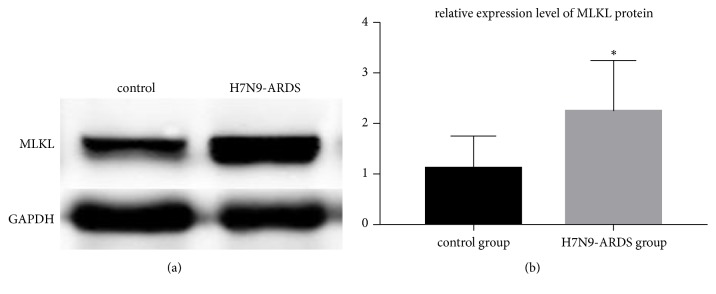
Methods: Lung tissues of 11 patients who died from ARDS-complicated H7N9 infection between 2013 and 2016 were obtained as the H7N9-ARDS group. Lung tissues near benign lung nodules were acquired as the control group. Histological changes were evaluated by H&E staining. Protein levels of cIAP2, RIPK1, RIPK3, p-RIPK3, MLKL, and p-MLKL in the lung tissues were detected by Western Blot. The mRNA levels of cIAP2, RIPK1, and RIPK3 were detected by real-time PCR.
Results: H7N9 virus infection had a high mortality, with ARDS being the leading cause of death. The protein level of cIAP2 in the experimental group was lower than that in the control group (P<0.05). However, the experimental group showed higher RIPK1, RIPK3, and p-RIPK3 protein levels than the control group (P<0.05), as well as the expression level of MLKL and p-MLKL protein, which is a key downstream protein in necroptosis (P<0.05).
Conclusion: In tissues from patients with fatal H7N9, downregulation of cIAP2 and induction of necroptosis was observed. We could speculate that necroptosis of the pulmonary epithelium is associated with severe H7N9 infection leading to ARDS. Thus, necroptosis inhibition may be a novel therapy for H7N9 influenza virus.
Figures


Similar articles
-
A cytosolic heat shock protein 90 and co-chaperone p23 complex activates RIPK3/MLKL during necroptosis of endothelial cells in acute respiratory distress syndrome.J Mol Med (Berl). 2020 Apr;98(4):569-583. doi: 10.1007/s00109-020-01886-y. Epub 2020 Feb 19. J Mol Med (Berl). 2020. PMID: 32072232
-
Cellular inhibitor of apoptosis protein cIAP2 protects against pulmonary tissue necrosis during influenza virus infection to promote host survival.Cell Host Microbe. 2014 Jan 15;15(1):23-35. doi: 10.1016/j.chom.2013.12.003. Cell Host Microbe. 2014. PMID: 24439895
-
Activation of necroptosis in a rat model of acute respiratory distress syndrome induced by oleic acid.Sheng Li Xue Bao. 2016 Oct 25;68(5):661-668. Sheng Li Xue Bao. 2016. PMID: 27778032
-
H7N9 influenza A virus activation of necroptosis in human monocytes links innate and adaptive immune responses.Cell Death Dis. 2019 Jun 5;10(6):442. doi: 10.1038/s41419-019-1684-0. Cell Death Dis. 2019. PMID: 31165725 Free PMC article.
-
The role of necroptosis in common respiratory diseases in children.Front Pediatr. 2022 Jul 25;10:945175. doi: 10.3389/fped.2022.945175. eCollection 2022. Front Pediatr. 2022. PMID: 35967568 Free PMC article. Review.
Cited by
-
Die Another Way: Interplay between Influenza A Virus, Inflammation and Cell Death.Viruses. 2020 Apr 4;12(4):401. doi: 10.3390/v12040401. Viruses. 2020. PMID: 32260457 Free PMC article. Review.
-
Targeting alveolar epithelial cells with lipid micelle-encapsulated necroptosis inhibitors to alleviate acute lung injury.Commun Biol. 2025 Apr 6;8(1):573. doi: 10.1038/s42003-025-08010-1. Commun Biol. 2025. PMID: 40188179 Free PMC article.
-
RIPK1 in the inflammatory response and sepsis: Recent advances, drug discovery and beyond.Front Immunol. 2023 Apr 5;14:1114103. doi: 10.3389/fimmu.2023.1114103. eCollection 2023. Front Immunol. 2023. PMID: 37090690 Free PMC article. Review.
-
Necroptosis in Pulmonary Diseases: A New Therapeutic Target.Front Pharmacol. 2021 Sep 14;12:737129. doi: 10.3389/fphar.2021.737129. eCollection 2021. Front Pharmacol. 2021. PMID: 34594225 Free PMC article. Review.
-
Moderate Fever Cycles as a Potential Mechanism to Protect the Respiratory System in COVID-19 Patients.Front Med (Lausanne). 2020 Sep 11;7:564170. doi: 10.3389/fmed.2020.564170. eCollection 2020. Front Med (Lausanne). 2020. PMID: 33043037 Free PMC article. Review.
References
MeSH terms
Substances
LinkOut - more resources
Full Text Sources
Medical
Miscellaneous
