Review
. 2019 May 8;5(5):e01588.
doi: 10.1016/j.heliyon.2019.e01588. eCollection 2019 May.

Syntheses of hydroxyapatite from natural sources

Affiliations
Review

Syntheses of hydroxyapatite from natural sources

N A S Mohd Pu'ad et al. Heliyon. .

Abstract

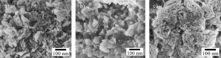
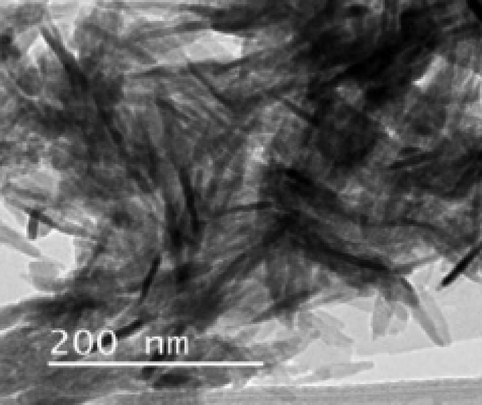
Waste materials from natural sources are important resources for extraction and recovery of valuable compounds. Transformation of these waste materials into valuable materials requires specific techniques and approaches. Hydroxyapatite (HAp) is a biomaterial that can be extracted from natural wastes. HAp has been widely used in biomedical applications owing to its excellent bioactivity, high biocompatibility, and excellent osteoconduction characteristics. Thus, HAp is gaining prominence for applications as orthopaedic implants and dental materials. This review summarizes some of the recent methods for extraction of HAp from natural sources including mammalian, aquatic or marine sources, shell sources, plants and algae, and from mineral sources. The extraction methods used to obtain hydroxyapatite are also described. The effect of extraction process and natural waste source on the critical properties of the HAp such as Ca/P ratio, crystallinity and phase assemblage, particle sizes, and morphology are discussed herein.

Keywords: Biological sciences; Biomedical engineering; Biotechnology; Health sciences; Natural product chemistry.


Figures

Fig. 1
Fig. 1
Summary of processes for synthesising natural HAp.
Fig. 2
Fig. 2
Micrographs of HAp extracted from mammalian sources using calcination. a) SEM micrograph of bovine bone calcined at 800 °C. (Adapted from , with permission from Elsevier). b) SEM micrograph of camel bone calcined at 1000 °C. (Adapted by permission from , Copyright, 2017). c). TEM micrograph of HAp synthesized from pig bone at 1000 °C. (Adapted from , with permission from Elsevier).
Fig. 3
Fig. 3
Micrographs of HAp extracted from mammalian sources using alkaline heat treatment method. a) FESEM micrograph of bovine bone. (Adapted from , with permission from Elsevier). b) SEM micrograph of bovine bone. (Adapted from , with permission from Elsevier).
Fig. 4
Fig. 4
Micrographs of HAp extracted from aquatic or marine sources. a) SEM micrograph of fish bone calcined at 900 °C. (Adapted from , with permission from Elsevier). b) TEM micrograph of fish scale extracted using alkaline heat treatment. (Adapted from , with permission from Elsevier). c) SEM micrograph of fish bones calcined at 600 °C. (Adapted from , with permission from Elsevier). d) TEM micrograph of fish scale extracted using alkaline heat treatment. (Adapted from , with permission from Elsevier). e) FESEM micrograph of fish scale calcined at 1000 °C. (Adapted from , with permission from Elsevier). f) SEM micrograph of fish scale calcined at 1200 °C. (Adapted from , with permission from Elsevier).
Fig. 5
Fig. 5
SEM micrographs of the HAp extracted from different sources using a combination of methods. a) SEM micrograph of HAp extracted from egg shells extracted using combination of calcination and wet chemical precipitation method. (Adapted from , with permission from Elsevier). b) TEM micrograph of HAp extracted from sea shells extracted using a combination of calcination and wet chemical precipitation method. (Adapted from , with permission from Elsevier). c) SEM micrograph of HAp extracted from mussel shells extracted using a combination of calcination and microwave irradiation treatment method. (Adapted from , with permission from Elsevier).
Fig. 6
Fig. 6
SEM micrographs of HAp extracted from plant sources using different methods. a) SEM micrograph of HAp extracted from a mixture of egg shell and pomelo peel using hydrothermal method. (Adapted from , with permission from Elsevier). b) SEM micrograph of HAp extracted from a mixture of egg shell and grape peel using hydrothermal method. (Adapted from , with permission from Elsevier). c) SEM micrograph of HAp extracted from a mixture of egg shell and sweet potato peel using hydrothermal method. (Adapted from , with permission from Elsevier).
Fig. 7
Fig. 7
SEM micrograph of HAp extracted from limestone extracted using a combination of calcination and ultrasonic irradiation. (Adapted from , with permission from Elsevier).

References

    1. Szcze A., Ho L., Chibowski E. Synthesis of hydroxyapatite for biomedical applications. Adv. Colloid Interface Sci. 2017:1–10. - PubMed
    1. Billiet T., Vandenhaute M., Schelfhout J., Van Vlierberghe S., Dubruel P. A review of trends and limitations in hydrogel-rapid prototyping for tissue engineering. Biomaterials. 2012;33:6020–6041. - PubMed
    1. Raghavendra G.M., Varaprasad K., Jayaramudu T. Chapter 2 - biomaterials: design, development and biomedical applications. In: Thomas S., Grohens Y., Ninan N., editors. Nanotechnology Applications for Tissue Engineering. William Andrew Publishing; Oxford: 2015. pp. 21–44.
    1. Temenoff J.S., Mikos A.G. Pearson/Prentice Hall; 2008. Biomaterials: the Intersection of Biology and Materials Science.
    1. Lertcumfu N., Jaita P., Manotham S., Jarupoom P., Eitssayeam S., Pengpat K., Rujijanagul G. Properties of calcium phosphates ceramic composites derived from natural materials. Ceram. Int. 2016;42:10638–10644.