. 2019 Jun 1;142(6):1547-1560.
doi: 10.1093/brain/awz107.

The clinical spectrum of the congenital myasthenic syndrome resulting from COL13A1 mutations

Affiliations

The clinical spectrum of the congenital myasthenic syndrome resulting from COL13A1 mutations

Pedro M Rodríguez Cruz et al. Brain. .

Abstract

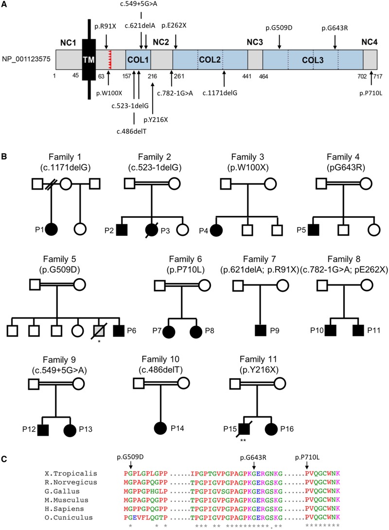
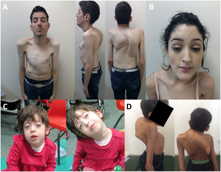
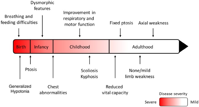
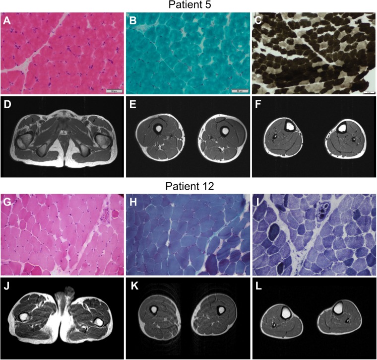
Next generation sequencing techniques were recently used to show mutations in COL13A1 cause synaptic basal lamina-associated congenital myasthenic syndrome type 19. Animal studies showed COL13A1, a synaptic extracellular-matrix protein, is involved in the formation and maintenance of the neuromuscular synapse that appears independent of the Agrin-LRP4-MuSK-DOK7 acetylcholine receptor clustering pathway. Here, we report the phenotypic spectrum of 16 patients from 11 kinships harbouring homozygous or heteroallelic mutations in COL13A1. Clinical presentation was mostly at birth with hypotonia and breathing and feeding difficulties often requiring ventilation and artificial feeding. Respiratory crisis related to recurrent apnoeas, sometimes triggered by chest infections, were common early in life but resolved over time. The predominant pattern of muscle weakness included bilateral ptosis (non-fatigable in adulthood), myopathic facies and marked axial weakness, especially of neck flexion, while limb muscles were less involved. Other features included facial dysmorphism, skeletal abnormalities and mild learning difficulties. All patients tested had results consistent with abnormal neuromuscular transmission. Muscle biopsies were within normal limits or showed non-specific changes. Muscle MRI and serum creatine kinase levels were normal. In keeping with COL13A1 mutations affecting both synaptic structure and presynaptic function, treatment with 3,4-diaminopyridine and salbutamol resulted in motor and respiratory function improvement. In non-treated cases, disease severity and muscle strength improved gradually over time and several adults recovered normal muscle strength in the limbs. In summary, patients with COL13A1 mutations present mostly with severe early-onset myasthenic syndrome with feeding and breathing difficulties. Axial weakness is greater than limb weakness. Disease course improves gradually over time, which could be consistent with the less prominent role of COL13A1 once the neuromuscular junction is mature. This report emphasizes the role of collagens at the human muscle endplate and should facilitate the recognition of this disorder, which can benefit from pharmacological treatment.

Keywords: 3,4-diaminopyridine; COL13A1; congenital myasthenic syndromes; salbutamol; synaptic basal lamina.


Figures

Figure 1
Figure 1
Schematic representation of full-length COL13A1, location of genetic variants and pedigrees of families reported. (A) COL13A1_001 (NP_001123575) consist of a short intracellular domain (NC1), a single transmembrane domain (TM), and the extracellular region with three collagenous domains (COL1–3) separated by short non-collagenous domains (NC2–3). The proprotease recognition site is labelled in red. Numbers indicate the amino acid residues composing each domain and the location of the pathogenic variants identified. Because COL13A1 undergoes complex alternative splicing, primary structures can vary. (B) Pedigrees of families included in this report. *An elder sibling from Patient 5 with similar symptoms died at an early age but genetic confirmation was not available. **Patient 15 was initially diagnosed with congenital insensitivity to pain with anhidrosis at the age of 1 year due to a homozygous NTRK1 mutation (Mardy et al., 1999). Patient 16, heterozygous for the NTRK1 variant, was diagnosed with a myasthenic syndrome. This prompted the re-evaluation of the proband in whom a myasthenic pattern of muscle activation was found on neurophysiological studies (Raspall-Chaure et al., 2005). Circles = female; squares = male; filled symbols = affected; open symbols = unaffected; strikethrough = deceased. (C) Protein alignments were performed using the Clustal Omega multiple sequence alignment program (https://www.ebi.ac.uk/Tools/msa/clustalo/).
Figure 2
Figure 2
Clinical features of patients with COL13A1 mutations. (A and B) Presence of bilateral ptosis and dysmorphic features including elongated face, micrognathia and low-set ears (Patients 12 and 13). (C) Marked weakness in neck flexors and truncal muscles with poor head control (Patient 1). (D) Severe scoliosis and associated thoracic kyphosis with restricted lung capacity (Patient 5).
Figure 3
Figure 3
Course of disease in patients with COL13A1-CMS. The proposed course of disease in COL13A1-CMS based on the observations made from the cases reported in this study. Axial weakness remained severe throughout the disease course while limb weakness and bulbar weakness improved over time. The respiratory function also improved with time but some adult patients had respiratory crisis or needed non-invasive ventilation most likely due to morphological abnormalities of the chest and the spine causing reduced vital capacity.
Figure 4
Figure 4
Clinical investigations of patients with COL13A1 mutations. Muscle biopsy from the quadriceps muscle in Patient 5 at the age of 5 years showed mild changes on haematoxylin and eosin stain (A) and modified Gomori trichrome (B), and type 1 fibre predominance on the ATPase 4.3 enzyme histochemical stain (C). Muscle MRI of his pelvis and lower limbs at the age of 13 years was normal (DF). Muscle biopsy from the biceps brachialis in Patient 12 at the age of 17 years showed mild variability of fibre size on haematoxylin and eosin stain (G). Modified Gomori trichrome staining showed a mild increase in perimysial connective tissue (H) and nicotinamide adenine dinucleotide tetrazolium reductase (NADH-TR) stain showed mild disruption of the myofibrillar architecture (I). Muscle MRI of his pelvis and lower limbs showed no abnormalities (JL).
Figure 5
Figure 5
Muscle endplate studies. (A) Muscle biopsy from quadriceps femoris in Patient 1 at age 6 months were labelled with Alexa Fluor® 488-fasciculin and Alexa Fluor® 594-α-bungarotoxin and analysed using confocal microscopy and ImageJ software. The contour of the motor endplates corresponding to the fasciculine staining (green) was selected (yellow line) and then applied to the red channel. As shown by the arrows, the α-bungarotoxin staining (red) did not completely fill the selection area. (B) Staining for the presynaptic marker S100β showed terminal Schwann cells reaching the muscle endplates. It appeared that the presynaptic marker S100β did not fully cover some of the muscle endplates, although an age-matched control muscle biopsy to compare with was not available.

References

    1. Adzhubei IA, Schmidt S, Peshkin L, Ramensky VE, Gerasimova A, Bork P et al. . A method and server for predicting damaging missense mutations. Nat Methods 2010; 7: 248. - PMC - PubMed
    1. Belaya K, Finlayson S, Slater CR, Cossins J, Liu WW, Maxwell S et al. . Mutations in DPAGT1 cause a limb-girdle congenital myasthenic syndrome with tubular aggregates. Am J Hum Genet 2012; 91: 193–201. - PMC - PubMed
    1. Clausen L, Cossins J, Beeson D. Beta-2 adrenergic receptor agonists enhance AChR clustering in C2C12 myotubes: implications for therapy of myasthenic disorders. J Neuromuscul Dis 2018; 5: 231–40. - PMC - PubMed
    1. Cossins J, Belaya K, Hicks D, Salih MA, Finlayson S, Carboni N et al. . Congenital myasthenic syndromes due to mutations in ALG2 and ALG14. Brain 2013; 136: 944–56. - PMC - PubMed
    1. D’Archy CE, Bjorksten A, Yiu EM, Bankier A, Gillies R, McLean CA et al. . King-Denborough syndrome caused by a novel mutation in the ryanodine receptor gene. Neurology 2008; 71: 776–8. - PubMed

Publication types