. 2019 Sep;73(9):e22116.
doi: 10.1002/syn.22116. Epub 2019 May 17.

Nicotine excites VIP interneurons to disinhibit pyramidal neurons in auditory cortex

Affiliations

Nicotine excites VIP interneurons to disinhibit pyramidal neurons in auditory cortex

Caitlin E Askew et al. Synapse. 2019 Sep.

Abstract

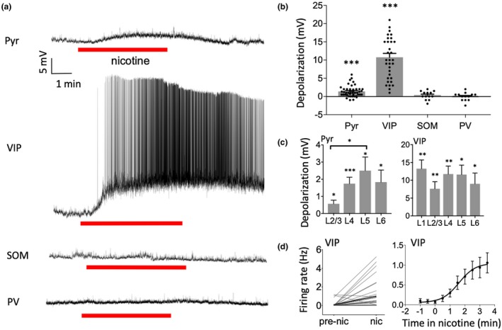
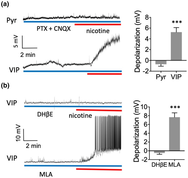
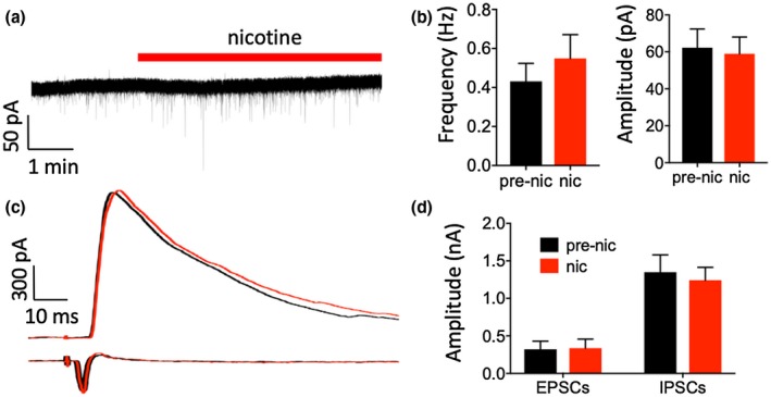
Nicotine activates nicotinic acetylcholine receptors and improves cognitive and sensory function, in part by its actions in cortical regions. Physiological studies show that nicotine amplifies stimulus-evoked responses in sensory cortex, potentially contributing to enhancement of sensory processing. However, the role of specific cell types and circuits in the nicotinic modulation of sensory cortex remains unclear. Here, we performed whole-cell recordings from pyramidal (Pyr) neurons and inhibitory interneurons expressing parvalbumin (PV), somatostatin (SOM), and vasoactive intestinal peptide (VIP) in mouse auditory cortex, in vitro. Bath application of nicotine strongly depolarized and excited VIP neurons, weakly depolarized Pyr neurons, and had no effect on the membrane potential of SOM or PV neurons. The use of receptor antagonists showed that nicotine's effects on VIP and Pyr neurons were direct and indirect, respectively. Nicotine also enhanced the frequency of spontaneous inhibitory postsynaptic currents (sIPSCs) in Pyr, VIP, and SOM, but not PV, cells. Using Designer Receptors Exclusively Activated by Designer Drugs (DREADDs), we show that chemogenetic inhibition of VIP neurons prevents nicotine's effects on Pyr neurons. Since VIP cells preferentially contact other inhibitory interneurons, we suggest that nicotine drives VIP cell firing to disinhibit Pyr cell somata, potentially making Pyr cells more responsive to auditory stimuli. In parallel, activation of VIP cells also directly inhibits Pyr neurons, likely altering integration of other synaptic inputs. These cellular and synaptic mechanisms likely contribute to nicotine's beneficial effects on cognitive and sensory function.

Keywords: VIP; interneuron; nicotine; nicotinic acetylcholine receptor; parvalbumin; pyramidal neuron; somatostatin.


Conflict of interest statement

The authors declare no competing financial interests. The data that support the findings of this study are available from the corresponding author upon reasonable request.

Figures

Figure 1
Figure 1
Nicotine selectively depolarized Pyr and VIP neurons. (a) Example recordings showing effect of nicotine (1 µM in bath) on membrane potential for each cell type. (b) Group data demonstrating nicotine‐induced depolarization in Pyr and VIP cells and no effect on SOM or PV cells. Data points represent individual cells. (c) Nicotinic depolarization by layer for Pyr (left) and VIP (right) neurons. Depolarization was stronger in deeper layers for Pyr neurons. (d) Nicotine increased action potential firing rate in VIP cells (left) and firing appears to peak after several minutes (right, firing rate examined in 30 s bins). For statistical comparisons in this and subsequent figures, asterisks indicate: *p < 0.05, **p < 0.01, ***p < 0.001
Figure 2
Figure 2
Nicotine directly depolarized VIP neurons via β2‐containing nAChRs and indirectly depolarized Pyr neurons. (a) Example recordings demonstrating nicotine's effect on membrane potential in the presence of 50 µM PTX and 10 µM CNQX (left). Group data (right) showing that PTX and CNQX prevented nicotinic depolarization in Pyr cells but not VIP cells. (b) Example recordings in VIP cells in the presence of 1 µM DHβE (top left) or 10 nM MLA (bottom left). DHβE prevented the nicotinic depolarization of VIP cells while MLA did not
Figure 3
Figure 3
Nicotine increased the frequency of sIPSCs in Pyr, SOM, and VIP neurons. (a) Example recordings showing nicotine's effects on sIPSCs for each cell type (0 mV holding potential). (b) Group data showing that nicotine increased the frequency of sIPSCs in Pyr, VIP, and SOM cells with no effect on PV cells (left). Nicotine did not alter the mean amplitude of sIPSCs in any cell type (right). (c) Same data as in B but for individual cells, to show the consistency of nicotine's effects. (d) Nicotine enhanced the frequency of sIPSCs in Pyr cells in all layers
Figure 4
Figure 4
Nicotine had no effect on EPSCs or evoked IPSCs in Pyr neurons. (a) Example recording of sEPSCs in a Pyr neuron (holding potential, −52 mV, see Results). (b) Group data showing that nicotine had no effect on the frequency (left) or amplitude (right) of sEPSCs. (c) Example recordings of evoked IPSCs (top, holding potential 0 mV) and evoked EPSCs (bottom, holding potential −52 mV) in two separate Pyr neurons. (d) Group data showing that nicotine had no effect on the peak amplitude of evoked EPSCs or evoked IPSCs
Figure 5
Figure 5
Nicotine depolarized and enhanced sIPSC frequency in Pyr neurons via VIP neurons. (a) Coronal section with immunohistochemistry against DAPI (blue), mCherry from the HM4D construct (red), and VIP (green) in HM4D‐transduced auditory cortex. Inset shows co‐labeling of mCherry and VIP in an example cell. (b) Example recordings from Pyr neurons in HM4D‐expressing mice demonstrating that CNO application prevents nicotine's effects on membrane potential (top) and sIPSCs (bottom). (c) Group data from Pyr cells in HM4D‐expressing mice; nicotine depolarized Pyr cells and CNO prevented the nicotinic depolarization of Pyr cells. (d) Group data from Pyr cells in HM4D‐expressing mice; nicotine enhanced the frequency of sIPSCs and CNO prevented the nicotinic enhancement of sIPSC frequency

References

    1. Alexander, G. M. , Rogan, S. C. , Abbas, A. I. , Armbruster, B. N. , Pei, Y. , Allen, J. A. , … Roth, B. L. (2009). Remote control of neuronal activity in transgenic mice expressing evolved G protein‐coupled receptors. Neuron, 63(1), 27–39. 10.1016/j.neuron.2009.06.014 - DOI - PMC - PubMed
    1. Amat, S. B. , Rowan, M. J. M. , Gaffield, M. A. , Bonnan, A. , Kikuchi, C. , Taniguchi, H. , & Christie, J. M. (2017). Using c‐kit to genetically target cerebellar molecular layer interneurons in adult mice. PLoS One, 12(6), e0179347 10.1371/journal.pone.0179347 - DOI - PMC - PubMed
    1. Arroyo, S. , Bennett, C. , & Hestrin, S. (2014). Nicotinic modulation of cortical circuits. Frontiers in Neural Circuits, 8, 30 10.3389/fncir.2014.00030 - DOI - PMC - PubMed
    1. Askew, C. , Intskirveli, I. , & Metherate, R. (2017). Systemic nicotine increases gain and narrows receptive fields in A1 via integrated cortical and subcortical actions. eNeuro, 4(3). 10.1523/ENEURO.0192-17.2017 - DOI - PMC - PubMed
    1. Bell, L. A. , Bell, K. A. , & McQuiston, A. R. (2015). Acetylcholine release in mouse hippocampal CA1 preferentially activates inhibitory‐selective interneurons via alpha4beta2* nicotinic receptor activation. Frontiers in Cellular Neuroscience, 9, 115 10.3389/fncel.2015.00115 - DOI - PMC - PubMed

Publication types