Review
. 2019 Aug;157(2):349-364.e1.
doi: 10.1053/j.gastro.2019.03.072. Epub 2019 May 10.

Pathogenesis and Cells of Origin of Barrett's Esophagus

Affiliations
Review

Pathogenesis and Cells of Origin of Barrett's Esophagus

Jianwen Que et al. Gastroenterology. 2019 Aug.

Abstract

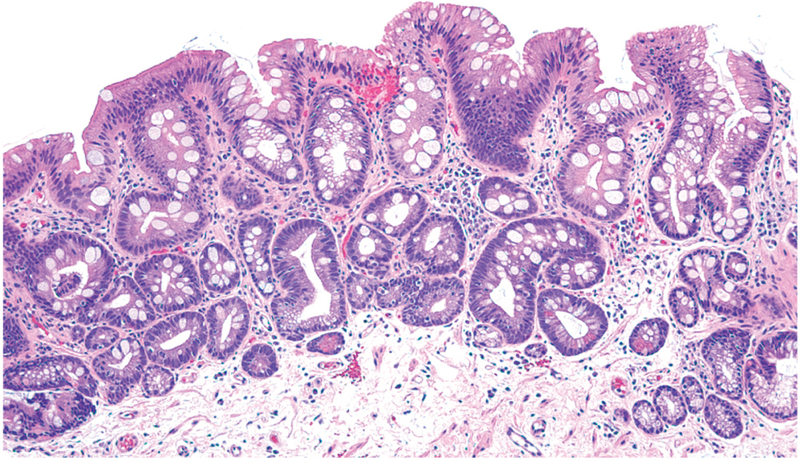
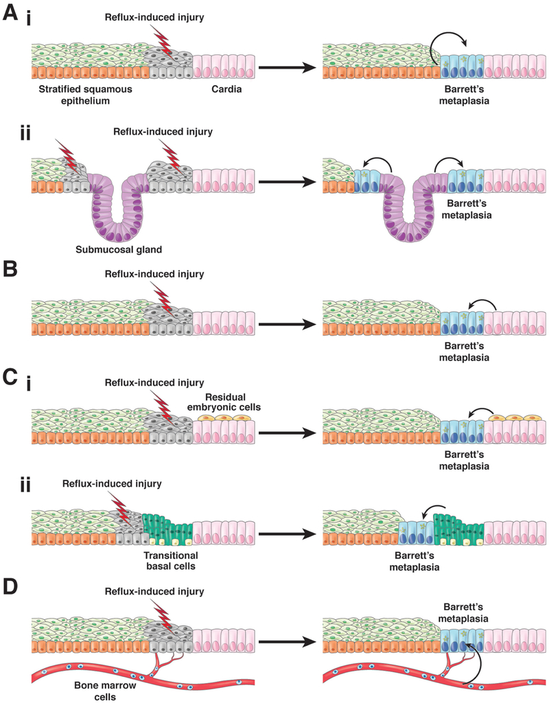
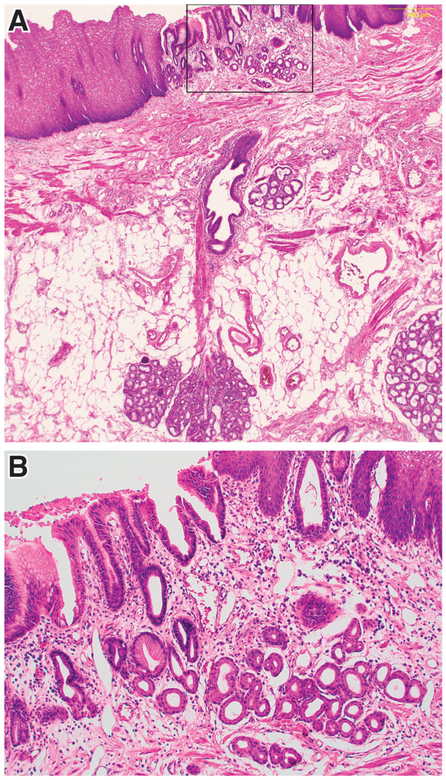
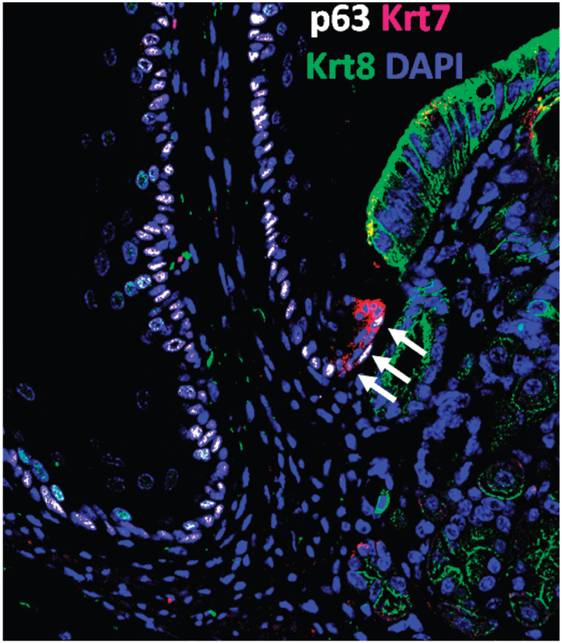
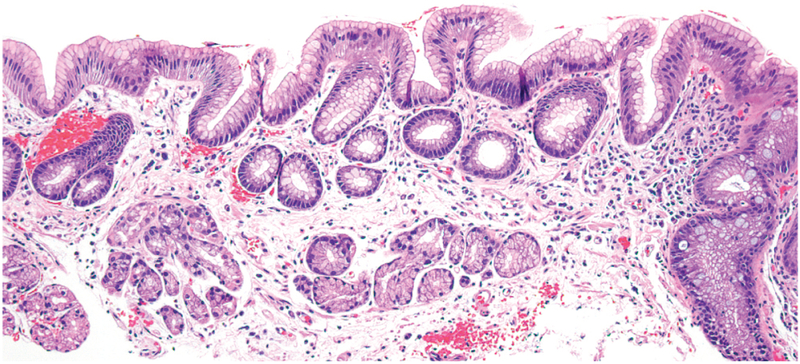
In patients with Barrett's esophagus (BE), metaplastic columnar mucosa containing epithelial cells with gastric and intestinal features replaces esophageal squamous mucosa damaged by gastroesophageal reflux disease. This condition is estimated to affect 5.6% of adults in the United States, and is a major risk factor for esophageal adenocarcinoma. Despite the prevalence and importance of BE, its pathogenesis is incompletely understood and there are disagreements over the cells of origin. We review mechanisms of BE pathogenesis, including transdifferentiation and transcommitment, and discuss potential cells of origin, including basal cells of the squamous epithelium, cells of esophageal submucosal glands and their ducts, cells of the proximal stomach, and specialized populations of cells at the esophagogastric junction (residual embryonic cells and transitional basal cells). We discuss the concept of metaplasia as a wound-healing response, and how cardiac mucosa might be the precursor of the intestinal metaplasia of BE. Finally, we discuss shortcomings in current diagnostic criteria for BE that have important clinical implications.

Keywords: Esophageal Adenocarcinoma; Esophagogastric Junction; Gastroesophageal Reflux Disease; Metaplasia; Progenitor Cells.


Figures

Figure 1.
Figure 1.
Barrett’s intestinal metaplasia with mucin-secreting gastric foveolar-type cells and prominent intestinal-type goblet cells. (Photomicrograph provided by Robert Genta, H&E, 20X)
Figure 2.
Figure 2.
Proposed cells of origin for BE. 1) Cells native to the esophagus including (1a) squamous epithelial cells that undergo reflux-induced transdifferentiation or transcommitment to produce the columnar cells of Barrett’s metaplasia and (1b) Progenitor cells in esophageal submucosal glands and/or their ducts. 2) Progenitor cells in the gastric cardia. 3) Specialized populations of cells at the esophago-gastric junction migrate into the reflux-damaged esophagus including (3a) residual embryonic cells (RECs) or (3b) transitional basal cells (TBCs). 4) Circulating bone marrow cells. For all of these proposed progenitor cells, reflux-induced injury to the esophageal squamous mucosa is assumed to initiate the metaplastic process, perhaps by stimulating progenitor cell migration into the damaged esophagus via a wound-healing process. In addition, reflux is assumed to induce the transcommitment of the progenitor cells to produce the multiple columnar cell types of Barrett’s metaplasia. (Figure modified from Jiang et al.)
Figure 3.
Figure 3.
In a patient with BE who had an island of columnar epithelium in squamous mucosa, an ESMG and duct is present directly beneath the columnar island. A) Low-power image showing the ESMG, duct and overlying columnar island (H&E, 10X). B) Higher-power image of the columnar island enclosed by the rectangle in Figure 3A (H&E, 20X). (Photomicrographs provided by Dr. Shannon J. McCall)
Figure 4.
Figure 4.
Transitional basal cells at the mouse squamo-columnar junction. The arrows point to transitional basal cells (p63+ Krt7+ Krt8-negative) located at the squamo-columnar junction. Note the neighboring squamous cells (p63+, Krt7-negative, Krt8-negative) on the left and the columnar gastric cells (p63-negative, Krt7-negative, Krt8+) on the right (see details in Jiang et al).
Figure 5.
Figure 5.
Cardiac mucosa comprised exclusively of mucus-secreting cells and glands. (Photomicrograph provided by Robert Genta, H&E, 20X)

References

    1. Spechler SJ, Souza RF. Barrett’s esophagus. N Engl J Med 2014; 371:836–45. - PubMed
    1. Shaheen NJ, Falk GW, Iyer PG, Gerson LB; American College of Gastroenterology. ACG Clinical Guideline: Diagnosis and Management of Barrett's Esophagus. Am J Gastroenterol 2016; 111:30–50. - PMC - PubMed
    1. Hayeck TJ, Kong CY, Spechler SJ, Gazelle GS, Hur C. The prevalence of Barrett’s esophagus in the US: estimates from a simulation model confirmed by SEER data. Dis Esoph 2010; 23:451–7. - PMC - PubMed
    1. Thrift AP. Barrett’s esophagus and esophageal adenocarcinoma: How common are they really? Dig Dis Sci 2018; 63:1988–96. - PubMed
    1. Virchow R Ueber metaplasie: Vortrag, gehalten auf dem internationalen medicinischen Congress in Kopenhagen. Virchows Archiv 1884; 97:410–30.

Publication types

MeSH terms

Supplementary concepts