. 2019 Jul 1:92:71-81.
doi: 10.1016/j.actbio.2019.05.019. Epub 2019 May 10.

Zwitterionic poly-carboxybetaine coating reduces artificial lung thrombosis in sheep and rabbits

Affiliations

Zwitterionic poly-carboxybetaine coating reduces artificial lung thrombosis in sheep and rabbits

Rei Ukita et al. Acta Biomater. .

Abstract

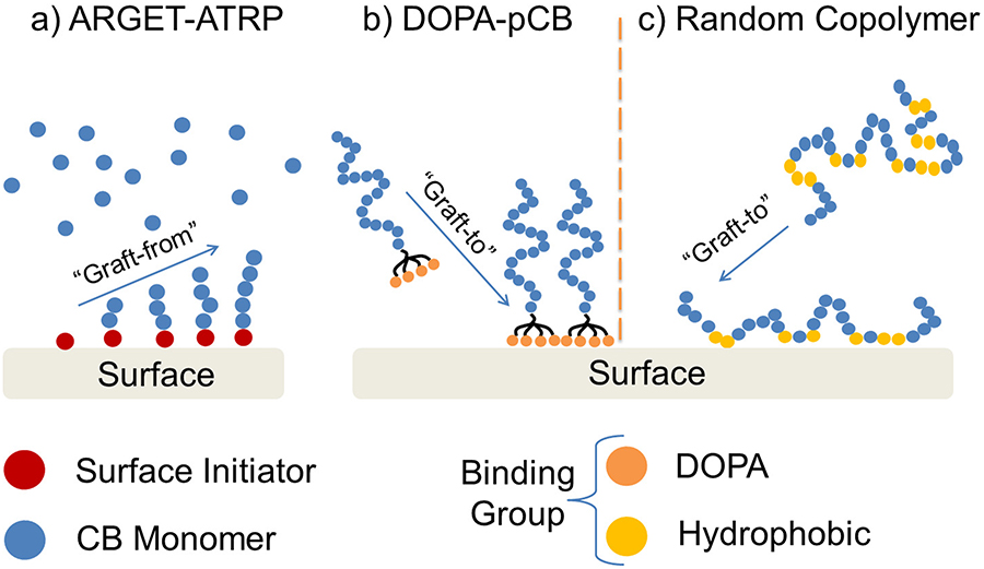
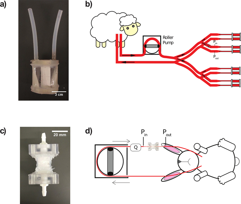
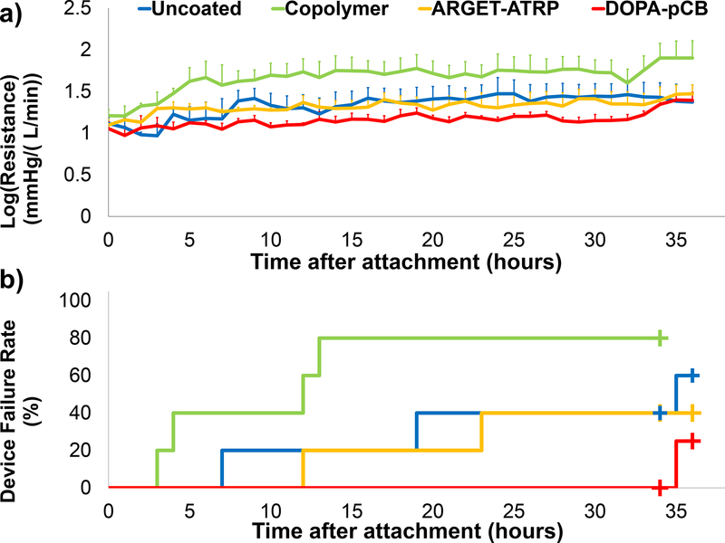
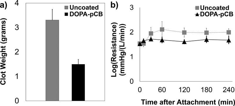
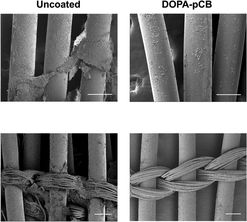
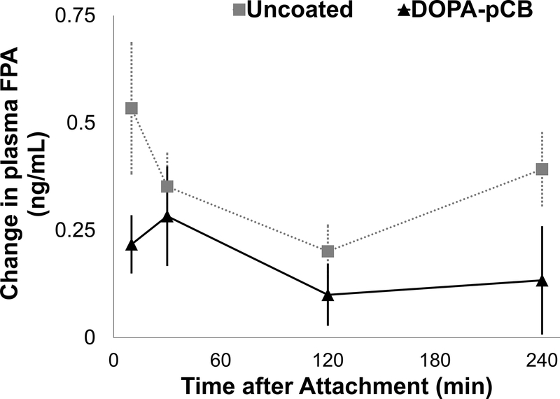
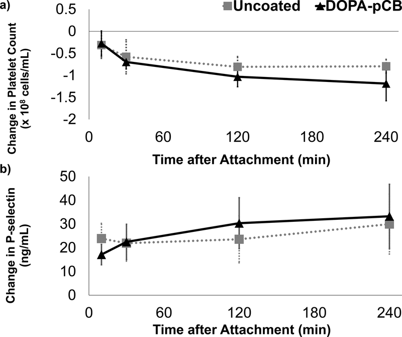
Current artificial lungs fail in 1-4 weeks due to surface-induced thrombosis. Biomaterial coatings may be applied to anticoagulate artificial surfaces, but none have shown marked long-term effectiveness. Poly-carboxybetaine (pCB) coatings have shown promising results in reducing protein and platelet-fouling in vitro. However, in vivo hemocompatibility remains to be investigated. Thus, three different pCB-grafting approaches to artificial lung surfaces were first investigated: 1) graft-to approach using 3,4-dihydroxyphenylalanine (DOPA) conjugated with pCB (DOPA-pCB); 2) graft-from approach using the Activators ReGenerated by Electron Transfer method of atom transfer radical polymerization (ARGET-ATRP); and 3) graft-to approach using pCB randomly copolymerized with hydrophobic moieties. One device coated with each of these methods and one uncoated device were attached in parallel within a veno-venous sheep extracorporeal circuit with no continuous anticoagulation (N = 5 circuits). The DOPA-pCB approach showed the least increase in blood flow resistance and the lowest incidence of device failure over 36-hours. Next, we further investigated the impact of tip-to-tip DOPA-pCB coating in a 4-hour rabbit study with veno-venous micro-artificial lung circuit at a higher activated clotting time of 220-300 s (N ≥ 5). Here, DOPA-pCB reduced fibrin formation (p = 0.06) and gross thrombus formation by 59% (p < 0.05). Therefore, DOPA-pCB is a promising material for improving the anticoagulation of artificial lungs. STATEMENT OF SIGNIFICANCE: Chronic lung diseases lead to 168,000 deaths each year in America, but only 2300 lung transplantations happen each year. Hollow fiber membrane oxygenators are clinically used as artificial lungs to provide respiratory support for patients, but their long-term viability is hindered by surface-induced clot formation that leads to premature device failure. Among different coatings investigated for blood-contacting applications, poly-carboxybetaine (pCB) coatings have shown remarkable reduction in protein adsorption in vitro. However, their efficacy in vivo remains unclear. This is the first work that investigates various pCB-coating methods on artificial lung surfaces and their biocompatibility in sheep and rabbit studies. This work highlights the promise of applying pCB coatings on artificial lungs to extend its durability and enable long-term respiratory support for lung disease patients.

Keywords: 3,4-Dihydroxyphenylalanine (DOPA); Artificial lung; Carboxybetaine; Thrombosis; Zwitterion.


Figures

Figure 1:
Figure 1:
Schematics illustrating different grafting techniques for pCB, such as graft-from approach using a) ARGET-ATRP, and graft-to approaches using b) DOPA molecules and c) random copolymerization of CB and hydrophobic monomers. Chemical structures are shown in Figure S1 in Supplementary Information.
Figure 2:
Figure 2:
Fabricated artificial lungs and animal extracorporeal circuit diagrams: a) miniature artificial lung (fiber surface area = 0.1 m2); b) sheep veno-venous parallel extracorporeal circuit featuring miniature artificial lungs coated with one of four methods (uncoated control, graft-to DOPA-pCB, graft-from ARGET-ATRP, graft-to copolymer). Each device’s thromboresistance is tracked by measuring flow rate (Q) with flow probe and pressure drop (Pin – Pout) with pressure transducers to calculate resistance; c) micro artificial lung used for rabbit study (fiber surface area = 400 cm2); d) rabbit veno-venous extracorporeal circuit, with or without tip-to-tip pCB coating
Figure 3:
Figure 3:
X-ray photoemission spectroscopy analysis of coated fiber surfaces in miniature artificial lung: a) survey scan of uncoated and pCB-coated surfaces; the dotted box shows that nitrogen was present on the three coated surfaces but not on the uncoated surface; b) high-resolution scan of DOPA-pCB surface shows the types of carbon bonds (top) and nitrogen (bottom) that correspond to CB moieties
Figure 4:
Figure 4:
a) Device log(resistance) for each coating type over the 36-hour period (N = 5 for uncoated, ARGET-ATRP, Copolymer; N = 4 for DOPA-pCB; error bars = S.E.M. b) Device failure curve plotted over the 36-hour period for the same set of devices; plus signs (+) indicate censored events.
Figure 5:
Figure 5:
Quantitative measurements of thrombus formation: a) total clot formation inside the micro artificial lung at end of the 4-hour rabbit study, p < 0.05, N ≥ 4; b) log(Resistance) for micro-artificial lung in the 4-hour rabbit study, N ≥ 5, p = 0.18. Error bars = S.E.M. for both figures
Figure 6:
Figure 6:
Scanning electron microscope images of uncoated (left column) and DOPA-pCB (right column). Hollow fibers (top row) and finer weaving fibers (bottom row) are both shown. Scale bar = 200 μm for all four panels
Figure 7:
Figure 7:
The change in plasma fibrinopeptide A(FPA) level from baseline measurements, p = 0.06, N ≥ 5, error bar = S.E.M.
Figure 8:
Figure 8:
Platelet activity and consumption during rabbit study: a) change in platelet count from baseline measurement, N ≥ 4; b) change in plasma p-selectin level from baseline measurement, N ≥ 4. Error bar = S.E.M. for both figures

References

    1. Murphy SL, Xu J, Kochanek KD, Curtin SC, Arias E, Deaths: Final data for 2015, Natl. Vital Stat. Reports 66 (2017). doi:10.1136/vr.h753. - DOI - PubMed
    1. Cystic Fibrosis Foundation Patient Registry 2016 Annual Data Report, Bethesda, Maryland, 2017.
    1. Olson AL, Swigris JJ, Lezotte DC, Norris JM, Wilson CG, Brown KK, Mortality from Pulmonary Fibrosis Increased in the United States from 1992 to 2003, Am. J. Respir. Crit. Care Med. 176 (2007) 277–284. doi:10.1164/rccm.200701-044OC. - DOI - PubMed
    1. Hutchinson JP, McKeever TM, Fogarty AW, Navaratnam V, Hubbard RB, Increasing global mortality from idiopathic pulmonary fibrosis in the twenty-first century., Ann. Am. Thorac. Soc 11 (2014) 1176–1185. doi:10.1513/AnnalsATS.201404-145OC. - DOI - PubMed
    1. Valapour M, Lehr CJ, Skeans MA, Smith JM, Carrico R, Uccellini K, Lehman R, Robinson A, Israni AK, Snyder JJ, Kasiske BL, OPTN/SRTR 2016 Annual Data Report: Lung, Am. J. Transplant 18 (2018) 363–433. doi:10.1111/ajt.14562. - DOI - PubMed

Publication types