Review
. 2019 May 11;8(5):445.
doi: 10.3390/cells8050445.

Strategies to Interfere with Tumor Metabolism through the Interplay of Innate and Adaptive Immunity

Affiliations
Review

Strategies to Interfere with Tumor Metabolism through the Interplay of Innate and Adaptive Immunity

Javier Mora et al. Cells. .

Abstract

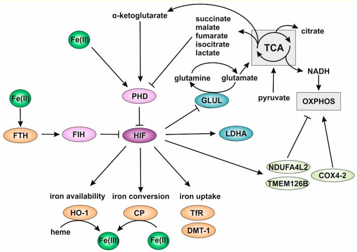
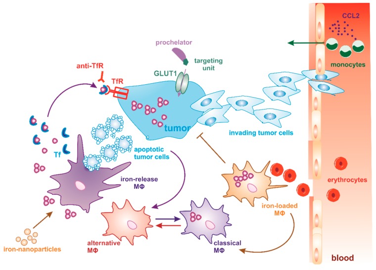
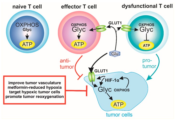
The inflammatory tumor microenvironment is an important regulator of carcinogenesis. Tumor-infiltrating immune cells promote each step of tumor development, exerting crucial functions from initiation, early neovascularization, to metastasis. During tumor outgrowth, tumor-associated immune cells, including myeloid cells and lymphocytes, acquire a tumor-supportive, anti-inflammatory phenotype due to their interaction with tumor cells. Microenvironmental cues such as inflammation and hypoxia are mainly responsible for creating a tumor-supportive niche. Moreover, it is becoming apparent that the availability of iron within the tumor not only affects tumor growth and survival, but also the polarization of infiltrating immune cells. The interaction of tumor cells and infiltrating immune cells is multifaceted and complex, finally leading to different activation phenotypes of infiltrating immune cells regarding their functional heterogeneity and plasticity. In recent years, it was discovered that these phenotypes are mainly implicated in defining tumor outcome. Here, we discuss the role of the metabolic activation of both tumor cells and infiltrating immune cells in order to adapt their metabolism during tumor growth. Additionally, we address the role of iron availability and the hypoxic conditioning of the tumor with regard to tumor growth and we describe the relevance of therapeutic strategies to target such metabolic characteristics.

Keywords: T cells; cancer cell metabolism; hypoxia; iron chelator; iron metabolism; tumor-associated macrophages.


Conflict of interest statement

The authors declare no conflict of interest.

Figures

Figure 1
Figure 1
Hypoxia inducible factor (HIF) as a central mediator in iron homeostasis. Degradation of hypoxia inducible factors (HIFs) is mediated by prolyl hydroxylases (PHDs), which are regulated by various tricarboxylic acid (TCA) cycle metabolites. TCA is fueled by glutamine and pyruvate from glycolysis and provides electrons via NADH for oxidative phosphorylation (OXPHOS). OXPHOS is regulated by NADH dehydrogenase [ubiquinone] 1 alpha subcomplex subunit 4-like 2 (NDUFA4L2), the complex I assembly factor TMEM126B, and cytochrome c oxidase subunit (COX) 4-2. Inhibition of glutamine synthase (GLUL) under hypoxia could facilitate fuel for TCA by glutamate. While ferrous iron (Fe(II)) and α-ketoglutarate are essential cofactors for PHDs, succinate, malate, fumarate, isocitrate, and lactate act as inhibitors. Lactate is produced by the HIF-target lactate dehydrogenase (LDH). Another HIF limiting protein is factor inhibiting HIF (FIH), which is regulated by binding of the iron storage protein ferritin heavy chain (FTH). HIF in turn modulates iron metabolism by enhancing the transcription of heme oxygenase 1 (HO-1), ceruloplasmin (CP), the iron transporter transferrin (TfR) and divalent metal transporter 1 (DMT-1). HO-1 removes ferric iron (Fe(III)) from heme and CP converts extracellular Fe(II) to Fe(III).
Figure 2
Figure 2
Dynamics and targeting of MΦ phenotypes in the tumor microenvironment. Macrophages (MΦs) can be targeted to reprogram the immunosuppressive tumor microenvironment and consequently enhance anti-tumor response. One strategy is to prevent the systemic mobilization and recruitment of monocytes to the tumor site by specifically blocking chemokine gradients such as CCL2. Instead of completely depleting MΦs, therapies aim at reprogramming alternatively polarized MΦs towards the anti-tumor, classical MΦ phenotype. With regard to their iron-regulated gene profile, two different MΦ phenotypes are distinguished. The iron-release phenotype is determined by the phagocytosis of apoptotic tumor cells, whereby iron is recycled from engulfed cells and secreted to the microenvironment, where it is rapidly bound to transferrin (Tf). Tf-bound iron is then taken up by tumor cells through the Tf receptor (TfR) and promotes tumor proliferation and growth. The engulfment of apoptotic tumor cells also promotes the anti-inflammatory MΦ phenotype. In order to control iron availability in tumor cells, targeted iron-chelation therapy (prochelator strategy) is used. This is accomplished by conjugating the prochelator with a glucose targeting unit, which is recognized by glucose transporter (GLUT1) on tumor cells. Another possibility is the use of TfR antibodies, which has shown strong anti-neoplastic effects. In contrast, iron-loaded MΦs reside in close vicinity to tumor vessels, where they are exposed to high levels of erythrocytes leaking into the tumor. Iron-loaded MΦs also adopt a pro-inflammatory phenotype. Iron nanoparticles are currently under investigation to reduce pro-tumor MΦ functions and, thus, tumor growth.
Figure 3
Figure 3
T cell Tumor cell metabolism dictates T cell effector function. Naïve T cells produce ATP predominantly by oxidative phosphorylation. After activation, a metabolic switch towards glycolysis is associated with T cell effector functions. Increased expression of the glucose transporter GLUT1 is essential for ATP production in effector T cells. Due to HIF-1α stabilization in tumor cells, GLUT1 expression is enhanced, which, in turn, leads to accumulation of glucose in tumor cells used for glycolysis, whereby OXPHOS is reduced. As a consequence, sufficient glucose is not available to maintain T cell effector functions and T cells adopt a dysfunctional phenotype due to reduced ATP production. Thus, targeting tumor hypoxia, e.g. by metformin, is a novel strategy to improve T cell function and response to immune-check point blockade.

References

    1. Hui L., Chen Y. Tumor microenvironment: Sanctuary of the devil. Cancer Lett. 2015;368:7–13. doi: 10.1016/j.canlet.2015.07.039. - DOI - PubMed
    1. Xu L., Vaupel P., Bai S., Menze B., Shi K. Computational Simulation of Tumor Hypoxia Based on In Vivo Microvasculature Assessed in a Dorsal Skin Window Chamber. Adv. Exp. Med. Biol. 2017;977:109–117. doi: 10.1007/978-3-319-55231-6_15. - DOI - PubMed
    1. Kim J., Bae J.-S. Tumor-Associated Macrophages and Neutrophils in Tumor Microenvironment. Mediat. Inflamm. 2016;2016:6058147. doi: 10.1155/2016/6058147. - DOI - PMC - PubMed
    1. Brüne B., Weigert A., Dehne N. Macrophage Polarization In The Tumor Microenvironment. Redox Biol. 2015;5:419. doi: 10.1016/j.redox.2015.09.028. - DOI - PubMed
    1. Dehne N., Mora J., Namgaladze D., Weigert A., Brüne B. Cancer cell and macrophage cross-talk in the tumor microenvironment. Curr. Opin. Pharmacol. 2017;35:12–19. doi: 10.1016/j.coph.2017.04.007. - DOI - PubMed

Publication types

LinkOut - more resources