Review
. 2019 May 11;20(9):2336.
doi: 10.3390/ijms20092336.

Flavivirus Replication Organelle Biogenesis in the Endoplasmic Reticulum: Comparison with Other Single-Stranded Positive-Sense RNA Viruses

Affiliations
Review

Flavivirus Replication Organelle Biogenesis in the Endoplasmic Reticulum: Comparison with Other Single-Stranded Positive-Sense RNA Viruses

Masashi Arakawa et al. Int J Mol Sci. .

Abstract

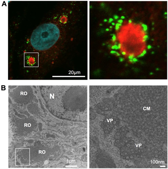
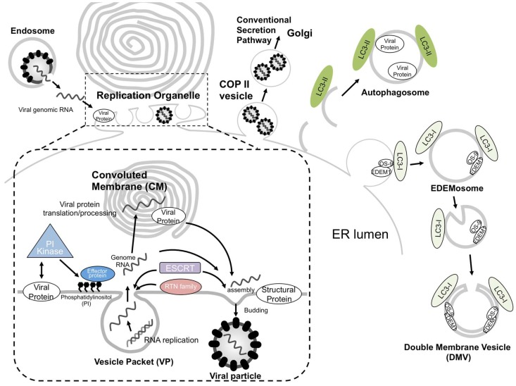
Some single-stranded positive-sense RNA [ssRNA(+)] viruses, including Flavivirus, generate specific organelle-like structures in the host endoplasmic reticulum (ER). These structures are called virus replication organelles and consist of two distinct subdomains, the vesicle packets (VPs) and the convoluted membranes (CMs). The VPs are clusters of small vesicle compartments and are considered to be the site of viral genome replication. The CMs are electron-dense amorphous structures observed in proximity to the VPs, but the exact roles of CMs are mostly unknown. Several recent studies have revealed that flaviviruses recruit several host factors that are usually used for the biogenesis of other conventional organelles and usurp their function to generate virus replication organelles. In the current review, we summarize recent studies focusing on the role of host factors in the formation of virus replication organelles and discuss how these intricate membrane structures are organized.

Keywords: ESCRT; autophagy; endoplasmic reticulum; flavivirus; phosphatidylinositol; reticulons.


Conflict of interest statement

The authors declare that the research was conducted in the absence of any commercial or financial relationships that could be construed as a potential conflict of interest.

Figures

Figure 1
Figure 1
Viral replication organelle in Japanese encephalitis virus (JEV)-infected cells. (A). fluorescent imaging of JEV replication organelle. At 48 h post-infection, JEV infected Vero cells were fixed, permeabilized, and stained with anti-nonstructural protein 4B (NS4B; red) and anti-double stranded RNA (dsRNA; Green) antibodies. Nuclear DNA was stained with DAPI (blue). Scale bar: 20 microns. (B). Viral replication organelle structure visualized using transmission electron microscopy (TEM). At 48 h post-infection, JEV infected Vero cells were fixed, ultra-thin section, and observed using TEM. N, nucleus; RO, viral replication organelle; CM, convoluted membrane; VP, vesicle packet. Scale bar = 1 micron (left) or 100 nm (right).
Figure 2
Figure 2
Schematic model of single-stranded positive-sense RNA viruses (ssRNA(+)) virus replication organelle biogenesis in the endoplasmic reticulum. After entry of the virus via the endocytosis pathway, its positive-stranded RNA genome is released into the cytoplasm by membrane fusion. Viral non-structural proteins are directly or indirectly interacted with lipid modifiers, which deform the endoplasmic reticulum (ER) membranes to create vesicle packets (VPs) for the replication of their genomic RNA. Newly synthesized ssRNA(+) is transported outside the VP to be used as a template for further viral protein synthesis or, alternatively, to the site of progeny virus particle assembly. The site of virus particle assembly is generally close to a VP pore. Reticulon (RTN) family proteins are involved in VP formation, and endosomal sorting complex required for transport (ESCRT) proteins can function in VP or virus particle formation. Progeny virus particles are sorted into coat protein complex II (COP II) vesicles and released via the conventional secretion pathway. Apart from this pathway, autophagosomes or EDEMosomes have been proposed to be involved in the formation of the ssRNA(+) virus replication compartment.

References

    1. Santos J.C., Enninga J. At the crossroads: Communication of bacteria-containing vacuoles with host organelles. Cell. Microbiol. 2016;18:330–339. doi: 10.1111/cmi.12567. - DOI - PubMed
    1. Aktepe T.E., Mackenzie J.M. Shaping the flavivirus replication complex: It is curvaceous! Cell. Microbiol. 2018;20:e12884. doi: 10.1111/cmi.12884. - DOI - PMC - PubMed
    1. Klema V.J., Padmanabhan R., Choi K.H. Flaviviral Replication Complex: Coordination between RNA Synthesis and 5′-RNA Capping. Viruses. 2015;7:4640–4656. doi: 10.3390/v7082837. - DOI - PMC - PubMed
    1. Romero-Brey I., Bartenschlager R. Endoplasmic Reticulum: The Favorite Intracellular Niche for Viral Replication and Assembly. Viruses. 2016;8:160. doi: 10.3390/v8060160. - DOI - PMC - PubMed
    1. Romero-Brey I., Bartenschlager R. Membranous replication factories induced by plus-strand RNA viruses. Viruses. 2014;6:2826–2857. doi: 10.3390/v6072826. - DOI - PMC - PubMed

MeSH terms

LinkOut - more resources