

Incomplete Healing as a Cause of Aging: The Role of Mitochondria and the Cell Danger Response
- PMID: 31083530
- PMCID: PMC6627909
- DOI: 10.3390/biology8020027
Incomplete Healing as a Cause of Aging: The Role of Mitochondria and the Cell Danger Response
Abstract
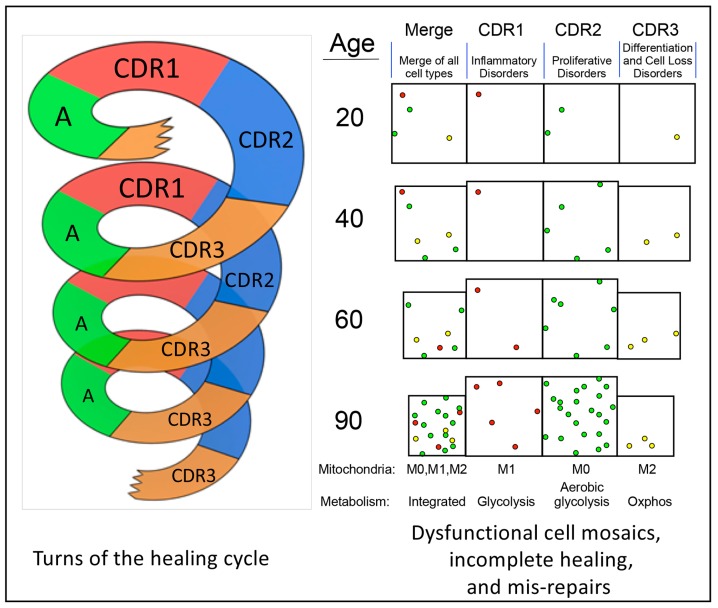
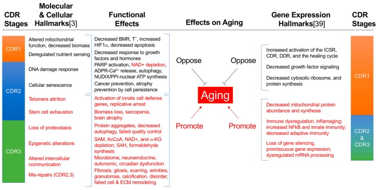
The rate of biological aging varies cyclically and episodically in response to changing environmental conditions and the developmentally-controlled biological systems that sense and respond to those changes. Mitochondria and metabolism are fundamental regulators, and the cell is the fundamental unit of aging. However, aging occurs at all anatomical levels. At levels above the cell, aging in different tissues is qualitatively, quantitatively, and chronologically distinct. For example, the heart can age faster and differently than the kidney and vice versa. Two multicellular features of aging that are universal are: (1) a decrease in physiologic reserve capacity, and (2) a decline in the functional communication between cells and organ systems, leading to death. Decreases in reserve capacity and communication impose kinetic limits on the rate of healing after new injuries, resulting in dyssynchronous and incomplete healing. Exercise mitigates against these losses, but recovery times continue to increase with age. Reinjury before complete healing results in the stacking of incomplete cycles of healing. Developmentally delayed and arrested cells accumulate in the three stages of the cell danger response (CDR1, 2, and 3) that make up the healing cycle. Cells stuck in the CDR create physical and metabolic separation-buffer zones of reduced communication-between previously adjoining, synergistic, and metabolically interdependent cells. Mis-repairs and senescent cells accumulate, and repeated iterations of incomplete cycles of healing lead to progressively dysfunctional cellular mosaics in aging tissues. Metabolic cross-talk between mitochondria and the nucleus, and between neighboring and distant cells via signaling molecules called metabokines regulates the completeness of healing. Purinergic signaling and sphingolipids play key roles in this process. When viewed against the backdrop of the molecular features of the healing cycle, the incomplete healing model provides a new framework for understanding the hallmarks of aging and generates a number of testable hypotheses for new treatments.
Keywords: cell danger response; crabtree effect; de-emergence; healing cycle; integrated cell stress response; metabokines; mitochondria; pasteur effect; purinergic signaling; sphingolipids.
Conflict of interest statement
RKN is an unpaid scientific advisory board member for the Autism Research Institute (ARI) and the Open Medicine Foundation (OMF). Financial supporters for this study had no role in data analysis, interpretation, writing, or publication of this work.
Figures

References
-
- McCay C.M., Crowell M.F. Prolonging the life span. Sci. Mon. 1934;39:405–414.
Publication types
LinkOut - more resources
Full Text Sources
Molecular Biology Databases
