. 2019 May 13;14(5):e0215752.
doi: 10.1371/journal.pone.0215752. eCollection 2019.

Influence of hepatic fibrosis and inflammation: Correlation between histopathological changes and Gd-EOB-DTPA-enhanced MR imaging

Affiliations

Influence of hepatic fibrosis and inflammation: Correlation between histopathological changes and Gd-EOB-DTPA-enhanced MR imaging

N Verloh et al. PLoS One. .

Abstract

Objective: To evaluate the influence of an active inflammatory process in the liver on Gd-EOB-DTPA-enhanced MR imaging in patients with different degrees of fibrosis/cirrhosis.

Material and methods: Overall, a number of 91 patients (61 men and 30 women; mean age 58 years) were included in this retrospective study. The inclusion criteria for this study were Gd-EOB-DTPA-enhanced MRI of the liver and histopathological evaluation of fibrotic and inflammatory changes. T1-weighted VIBE sequences of the liver with fat suppression were evaluated to determine the relative signal change (RE) between native and hepatobiliary phase (20min). In simple and multiple linear regression analyses, the influence of liver fibrosis/cirrhosis (Ishak score) and the histopathological degree of hepatitis (Modified Hepatic Activity Index, mHAI) on RE were evaluated.

Results: RE decreased significantly with increasing liver fibrosis/cirrhosis (p < 0.001) and inflammation (mHAI, p = 0.004). In particular, a correlation between RE and periportal or periseptal boundary zone hepatitis (moth feeding necrosis, mHAI A, p = 0.001) and portal inflammation (mHAI D, p < 0.001) was observed. In multiple linear regression analysis, both the degree of inflammation and the degree of fibrosis were significant predictors for RE (p < 0.01).

Conclusion: The results of this study suggest that the MR-based hepatic enhancement index RE is not only influenced by the degree of fibrosis, but also by the degree of inflammation.


Conflict of interest statement

The authors have declared that no competing interests exist.

Figures
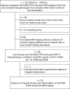
Fig 1
Fig 1. Flowchart of patient inclusion.
The criteria for inclusion in this study were Gd-EOB-DTPA-enhanced MRI of the liver as well as evaluation of histopathological liver samples concerning fibrotic and inflammation findings (within three months of the examination) from a patient cohort analyzed in 2015.
Fig 2
Fig 2. The level of inflammation plotted against the level of fibrosis/cirrhosis.
The figure shows the level of inflammation, as characterized by the histologic activity index (mHAI) in comparison to the Ishak-Score. A dash indicates the mean of each group in the data cloud.
Fig 3
Fig 3
Representative images of a case with (A) high hepatic enhancement (RE: 1.29), absent inflammation (mHAI 0) and no fibrotic marker (ISHAK 0) is shown in comparison to a case with (B) low hepatic enhancement (RE: 0.30) paired with active inflammation (mHAI 9) and fibrosis (ISHAK 4). The first column represents the non-contrasted MR image while the second column represents the hepatobiliary phase. The corresponding histopathological imaging of the evaluated hepatic sections is shown in the third column. All displayed MR images have the same window and center level. The scale bar on histopathological images represents 500 μm.
Fig 4
Fig 4
Relative enhancement (RE) values plotted with corresponding mHAI (A) and Ishak-Score (B) separately and combined (C). A dash indicates the mean of each group in the data cloud. Each dot represents the median of the combined model in graph C. The corresponding correlations with RE as the dependent variable are as followed: mHAI, r2 = 0.142, p = < 0.001; Ishak-Score, r2 = 0.792, p < 0.001. The combined model accounts for an R2 of 0.815.
Fig 5
Fig 5
Relative enhancement (RE) values plotted against the combined model for patients with unknown/non-parenchymal disease (A), known inflammatory- (B), and non-inflammatory-parenchymal diseases (C). Each dot represents the median of the combined model. The corresponding correlations with RE as the dependent variable are as followed: A, R2 = 0.503; B, R2 = 0.849: C, R2 = 0.812.
Fig 6
Fig 6
Relative enhancement (RE) values plotted against the combined model for patients with non-tumorous (A), benign tumorous (B), and malignant tumorous diseases (C). Each dot represents the median of the combined model. The corresponding correlations with RE as the dependent variable are as followed: A, R2 = 0.852; B, no significant correlation: C, R2 = 0.751. mHAI, histologic activity index.

References

    1. Friedman SL. Mechanisms of hepatic fibrogenesis. Gastroenterology. 2008;134(6):1655–69. Epub 2008/05/13. 10.1053/j.gastro.2008.03.003 - DOI - PMC - PubMed
    1. Lee UE, Friedman SL. Mechanisms of Hepatic Fibrogenesis. Best practice & research Clinical gastroenterology. 2011;25(2):195–206. 10.1016/j.bpg.2011.02.005 - DOI - PMC - PubMed
    1. Czaja AJ. Hepatic inflammation and progressive liver fibrosis in chronic liver disease. World Journal of Gastroenterology: WJG. 2014;20(10):2515–32. 10.3748/wjg.v20.i10.2515 - DOI - PMC - PubMed
    1. Hintermann E, Ehser J, Bayer M, Pfeilschifter JM, Christen U. Mechanism of autoimmune hepatic fibrogenesis induced by an adenovirus encoding the human liver autoantigen cytochrome P450 2D6. Journal of autoimmunity. 2013;44:49–60. Epub 2013/07/03. 10.1016/j.jaut.2013.05.001 . - DOI - PubMed
    1. Regev A, Berho M, Jeffers LJ, Milikowski C, Molina EG, Pyrsopoulos NT, et al. Sampling error and intraobserver variation in liver biopsy in patients with chronic HCV infection. The American journal of gastroenterology. 2002;97(10):2614–8. Epub 2002/10/19. 10.1111/j.1572-0241.2002.06038.x . - DOI - PubMed