Review
. 2019 Jul;292(1):1-14.
doi: 10.1148/radiol.2019180760. Epub 2019 May 14.

Digital Breast Tomosynthesis: Concepts and Clinical Practice

Affiliations
Review

Digital Breast Tomosynthesis: Concepts and Clinical Practice

Alice Chong et al. Radiology. 2019 Jul.

Abstract

Digital breast tomosynthesis (DBT) is emerging as the standard of care for breast imaging based on improvements in both screening and diagnostic imaging outcomes. The additional information obtained from the tomosynthesis acquisition decreases the confounding effect of overlapping tissue, allowing for improved lesion detection, characterization, and localization. In addition, the quasi three-dimensional information obtained from the reconstructed DBT data set allows a more efficient imaging work-up than imaging with two-dimensional full-field digital mammography alone. Herein, the authors review the benefits of DBT imaging in screening and diagnostic breast imaging.


Figures

Figure 1a:
Figure 1a:
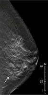
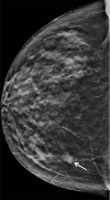
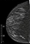
Images in 57-year-old woman with cancer detected only with digital breast tomosynthesis (DBT). (a–c) Craniocaudal DBT image (a) demonstrates a spiculated mass (arrow) not visible on synthetic craniocaudal (b) or mediolateral oblique (c) mammograms. (d) On the basis of the location in the DBT craniocaudal stack, the mass (arrow) can be localized on DBT image obtained in mediolateral oblique projection. (e) The mass (arrow) is better demonstrated on DBT images in mediolateral projection. (f) US examination shows that finding corresponds to an irregular, hypoechoic mass. Pathologic examination of biopsy specimen revealed invasive ductal carcinoma that was estrogen receptor positive, progesterone receptor positive, and human epidermal growth factor negative. F = foot, H = head, L = lateral, M = medial.
Figure 1b:
Figure 1b:
Images in 57-year-old woman with cancer detected only with digital breast tomosynthesis (DBT). (a–c) Craniocaudal DBT image (a) demonstrates a spiculated mass (arrow) not visible on synthetic craniocaudal (b) or mediolateral oblique (c) mammograms. (d) On the basis of the location in the DBT craniocaudal stack, the mass (arrow) can be localized on DBT image obtained in mediolateral oblique projection. (e) The mass (arrow) is better demonstrated on DBT images in mediolateral projection. (f) US examination shows that finding corresponds to an irregular, hypoechoic mass. Pathologic examination of biopsy specimen revealed invasive ductal carcinoma that was estrogen receptor positive, progesterone receptor positive, and human epidermal growth factor negative. F = foot, H = head, L = lateral, M = medial.
Figure 1c:
Figure 1c:
Images in 57-year-old woman with cancer detected only with digital breast tomosynthesis (DBT). (a–c) Craniocaudal DBT image (a) demonstrates a spiculated mass (arrow) not visible on synthetic craniocaudal (b) or mediolateral oblique (c) mammograms. (d) On the basis of the location in the DBT craniocaudal stack, the mass (arrow) can be localized on DBT image obtained in mediolateral oblique projection. (e) The mass (arrow) is better demonstrated on DBT images in mediolateral projection. (f) US examination shows that finding corresponds to an irregular, hypoechoic mass. Pathologic examination of biopsy specimen revealed invasive ductal carcinoma that was estrogen receptor positive, progesterone receptor positive, and human epidermal growth factor negative. F = foot, H = head, L = lateral, M = medial.
Figure 1d:
Figure 1d:
Images in 57-year-old woman with cancer detected only with digital breast tomosynthesis (DBT). (a–c) Craniocaudal DBT image (a) demonstrates a spiculated mass (arrow) not visible on synthetic craniocaudal (b) or mediolateral oblique (c) mammograms. (d) On the basis of the location in the DBT craniocaudal stack, the mass (arrow) can be localized on DBT image obtained in mediolateral oblique projection. (e) The mass (arrow) is better demonstrated on DBT images in mediolateral projection. (f) US examination shows that finding corresponds to an irregular, hypoechoic mass. Pathologic examination of biopsy specimen revealed invasive ductal carcinoma that was estrogen receptor positive, progesterone receptor positive, and human epidermal growth factor negative. F = foot, H = head, L = lateral, M = medial.
Figure 1e:
Figure 1e:
Images in 57-year-old woman with cancer detected only with digital breast tomosynthesis (DBT). (a–c) Craniocaudal DBT image (a) demonstrates a spiculated mass (arrow) not visible on synthetic craniocaudal (b) or mediolateral oblique (c) mammograms. (d) On the basis of the location in the DBT craniocaudal stack, the mass (arrow) can be localized on DBT image obtained in mediolateral oblique projection. (e) The mass (arrow) is better demonstrated on DBT images in mediolateral projection. (f) US examination shows that finding corresponds to an irregular, hypoechoic mass. Pathologic examination of biopsy specimen revealed invasive ductal carcinoma that was estrogen receptor positive, progesterone receptor positive, and human epidermal growth factor negative. F = foot, H = head, L = lateral, M = medial.
Figure 1f:
Figure 1f:
Images in 57-year-old woman with cancer detected only with digital breast tomosynthesis (DBT). (a–c) Craniocaudal DBT image (a) demonstrates a spiculated mass (arrow) not visible on synthetic craniocaudal (b) or mediolateral oblique (c) mammograms. (d) On the basis of the location in the DBT craniocaudal stack, the mass (arrow) can be localized on DBT image obtained in mediolateral oblique projection. (e) The mass (arrow) is better demonstrated on DBT images in mediolateral projection. (f) US examination shows that finding corresponds to an irregular, hypoechoic mass. Pathologic examination of biopsy specimen revealed invasive ductal carcinoma that was estrogen receptor positive, progesterone receptor positive, and human epidermal growth factor negative. F = foot, H = head, L = lateral, M = medial.
Figure 2a:
Figure 2a:
Images in 78-year-old woman with mass in right breast. (a, b) Images from synthetic mammography (SM) show lesion in inner breast on craniocaudal view (arrow in a) that is not well seen on mediolateral view (b). (c) The position of the lesion (arrow) on the DBT craniocaudal stack helped localize the mass (arrow) to the lower inner part of the breast. (d) With this information, the mass (arrow) is now detected on DBT image in mediolateral oblique view. (e) The patient proceeded directly to US examination, which showed a suspicious irregular hypoechoic mass. Pathologic examination of biopsy specimen revealed invasive ductal carcinoma that was estrogen receptor positive, progesterone receptor positive, and human epidermal growth factor 2 positive. F = foot, H = head, L = lateral, M = medial.
Figure 2b:
Figure 2b:
Images in 78-year-old woman with mass in right breast. (a, b) Images from synthetic mammography (SM) show lesion in inner breast on craniocaudal view (arrow in a) that is not well seen on mediolateral view (b). (c) The position of the lesion (arrow) on the DBT craniocaudal stack helped localize the mass (arrow) to the lower inner part of the breast. (d) With this information, the mass (arrow) is now detected on DBT image in mediolateral oblique view. (e) The patient proceeded directly to US examination, which showed a suspicious irregular hypoechoic mass. Pathologic examination of biopsy specimen revealed invasive ductal carcinoma that was estrogen receptor positive, progesterone receptor positive, and human epidermal growth factor 2 positive. F = foot, H = head, L = lateral, M = medial.
Figure 2c:
Figure 2c:
Images in 78-year-old woman with mass in right breast. (a, b) Images from synthetic mammography (SM) show lesion in inner breast on craniocaudal view (arrow in a) that is not well seen on mediolateral view (b). (c) The position of the lesion (arrow) on the DBT craniocaudal stack helped localize the mass (arrow) to the lower inner part of the breast. (d) With this information, the mass (arrow) is now detected on DBT image in mediolateral oblique view. (e) The patient proceeded directly to US examination, which showed a suspicious irregular hypoechoic mass. Pathologic examination of biopsy specimen revealed invasive ductal carcinoma that was estrogen receptor positive, progesterone receptor positive, and human epidermal growth factor 2 positive. F = foot, H = head, L = lateral, M = medial.
Figure 2d:
Figure 2d:
Images in 78-year-old woman with mass in right breast. (a, b) Images from synthetic mammography (SM) show lesion in inner breast on craniocaudal view (arrow in a) that is not well seen on mediolateral view (b). (c) The position of the lesion (arrow) on the DBT craniocaudal stack helped localize the mass (arrow) to the lower inner part of the breast. (d) With this information, the mass (arrow) is now detected on DBT image in mediolateral oblique view. (e) The patient proceeded directly to US examination, which showed a suspicious irregular hypoechoic mass. Pathologic examination of biopsy specimen revealed invasive ductal carcinoma that was estrogen receptor positive, progesterone receptor positive, and human epidermal growth factor 2 positive. F = foot, H = head, L = lateral, M = medial.
Figure 2e:
Figure 2e:
Images in 78-year-old woman with mass in right breast. (a, b) Images from synthetic mammography (SM) show lesion in inner breast on craniocaudal view (arrow in a) that is not well seen on mediolateral view (b). (c) The position of the lesion (arrow) on the DBT craniocaudal stack helped localize the mass (arrow) to the lower inner part of the breast. (d) With this information, the mass (arrow) is now detected on DBT image in mediolateral oblique view. (e) The patient proceeded directly to US examination, which showed a suspicious irregular hypoechoic mass. Pathologic examination of biopsy specimen revealed invasive ductal carcinoma that was estrogen receptor positive, progesterone receptor positive, and human epidermal growth factor 2 positive. F = foot, H = head, L = lateral, M = medial.
Figure 3a:
Figure 3a:
Images in 62-year-old woman with an enlarged axillary lymph node at screening mammography. (a) Full-field digital mammogram in mediolateral oblique view demonstrates an enlarged low left axillary lymph node (arrow). (b, c) DBT images show subtle architectural distortion (arrow) in superior part of breast on mediolateral oblique view (b) and in central part of breast on craniocaudal view (c). (d) US examination demonstrates a suspicious hypoechoic mass at the 12 o’clock location. Pathologic examination of biopsy specimen revealed invasive lobular carcinoma.
Figure 3b:
Figure 3b:
Images in 62-year-old woman with an enlarged axillary lymph node at screening mammography. (a) Full-field digital mammogram in mediolateral oblique view demonstrates an enlarged low left axillary lymph node (arrow). (b, c) DBT images show subtle architectural distortion (arrow) in superior part of breast on mediolateral oblique view (b) and in central part of breast on craniocaudal view (c). (d) US examination demonstrates a suspicious hypoechoic mass at the 12 o’clock location. Pathologic examination of biopsy specimen revealed invasive lobular carcinoma.
Figure 3c:
Figure 3c:
Images in 62-year-old woman with an enlarged axillary lymph node at screening mammography. (a) Full-field digital mammogram in mediolateral oblique view demonstrates an enlarged low left axillary lymph node (arrow). (b, c) DBT images show subtle architectural distortion (arrow) in superior part of breast on mediolateral oblique view (b) and in central part of breast on craniocaudal view (c). (d) US examination demonstrates a suspicious hypoechoic mass at the 12 o’clock location. Pathologic examination of biopsy specimen revealed invasive lobular carcinoma.
Figure 3d:
Figure 3d:
Images in 62-year-old woman with an enlarged axillary lymph node at screening mammography. (a) Full-field digital mammogram in mediolateral oblique view demonstrates an enlarged low left axillary lymph node (arrow). (b, c) DBT images show subtle architectural distortion (arrow) in superior part of breast on mediolateral oblique view (b) and in central part of breast on craniocaudal view (c). (d) US examination demonstrates a suspicious hypoechoic mass at the 12 o’clock location. Pathologic examination of biopsy specimen revealed invasive lobular carcinoma.
Figure 4a:
Figure 4a:
Images in 40-year-old woman who presented for screening with an asymmetry seen at full-field digital mammography. (a) Synthetic mammogram (SM) in mediolateral oblique projection demonstrates asymmetry (arrow) in upper part of the breast. (b–d) On sequential DBT reconstructions, each component of the asymmetry is discernible as separate, individual normal structures that are superimposed as a summation artifact on the SM image. (e) SM in the craniocaudal view demonstrates no abnormality. L = lateral, M = medial.
Figure 4b:
Figure 4b:
Images in 40-year-old woman who presented for screening with an asymmetry seen at full-field digital mammography. (a) Synthetic mammogram (SM) in mediolateral oblique projection demonstrates asymmetry (arrow) in upper part of the breast. (b–d) On sequential DBT reconstructions, each component of the asymmetry is discernible as separate, individual normal structures that are superimposed as a summation artifact on the SM image. (e) SM in the craniocaudal view demonstrates no abnormality. L = lateral, M = medial.
Figure 4c:
Figure 4c:
Images in 40-year-old woman who presented for screening with an asymmetry seen at full-field digital mammography. (a) Synthetic mammogram (SM) in mediolateral oblique projection demonstrates asymmetry (arrow) in upper part of the breast. (b–d) On sequential DBT reconstructions, each component of the asymmetry is discernible as separate, individual normal structures that are superimposed as a summation artifact on the SM image. (e) SM in the craniocaudal view demonstrates no abnormality. L = lateral, M = medial.
Figure 4d:
Figure 4d:
Images in 40-year-old woman who presented for screening with an asymmetry seen at full-field digital mammography. (a) Synthetic mammogram (SM) in mediolateral oblique projection demonstrates asymmetry (arrow) in upper part of the breast. (b–d) On sequential DBT reconstructions, each component of the asymmetry is discernible as separate, individual normal structures that are superimposed as a summation artifact on the SM image. (e) SM in the craniocaudal view demonstrates no abnormality. L = lateral, M = medial.
Figure 4e:
Figure 4e:
Images in 40-year-old woman who presented for screening with an asymmetry seen at full-field digital mammography. (a) Synthetic mammogram (SM) in mediolateral oblique projection demonstrates asymmetry (arrow) in upper part of the breast. (b–d) On sequential DBT reconstructions, each component of the asymmetry is discernible as separate, individual normal structures that are superimposed as a summation artifact on the SM image. (e) SM in the craniocaudal view demonstrates no abnormality. L = lateral, M = medial.
Figure 5a:
Figure 5a:
Images in 48-year-old man who presented with palpable lump in right breast. (a, b) Image from digital breast tomosynthesis (DBT) in mediolateral view (a) demonstrates area of fat necrosis (arrow) not well seen on full-field digital mammogram in mediolateral view (b). Patient reported being in a motor vehicle collision several months earlier, with direct trauma to the breast. No further workup was needed due to the typical appearance of fat necrosis on DBT image.
Figure 5b:
Figure 5b:
Images in 48-year-old man who presented with palpable lump in right breast. (a, b) Image from digital breast tomosynthesis (DBT) in mediolateral view (a) demonstrates area of fat necrosis (arrow) not well seen on full-field digital mammogram in mediolateral view (b). Patient reported being in a motor vehicle collision several months earlier, with direct trauma to the breast. No further workup was needed due to the typical appearance of fat necrosis on DBT image.

References

    1. Sechopoulos I. A review of breast tomosynthesis. Part I. The image acquisition process. Med Phys 2013;40(1):014301. - PMC - PubMed
    1. Sechopoulos I, Ghetti C. Optimization of the acquisition geometry in digital tomosynthesis of the breast. Med Phys 2009;36(4):1199–1207. - PubMed
    1. Vedantham S, Karellas A, Vijayaraghavan GR, Kopans DB. Digital breast tomosynthesis: state of the art. Radiology 2015;277(3):663–684. - PMC - PubMed
    1. Bissonnette M, Hansroul M, Masson E, et al. . Digital breast tomosynthesis using an amorphous selenium flat panel detector. In: Flynn MJ, ed. Proceedings of SPIE: medical imaging 2005—physics of medical imaging. Vol 5745. Bellingham, Wash: International Society for Optics and Photonics, 2005; 529–540.
    1. Durand MA, Wang S, Hooley RJ, Raghu M, Philpotts LE. Tomosynthesis-detected architectural distortion: management algorithm with radiologic-pathologic correlation. RadioGraphics 2016;36(2):311–321. - PubMed

Publication types