. 2019 Aug:102:150-158.
doi: 10.1016/j.jaut.2019.05.002. Epub 2019 May 10.

Transitional B cells in quiescent SLE: An early checkpoint imprinted by IFN

Affiliations

Transitional B cells in quiescent SLE: An early checkpoint imprinted by IFN

Yannick Dieudonné et al. J Autoimmun. 2019 Aug.

Abstract

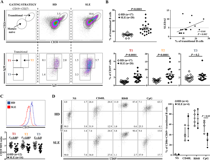
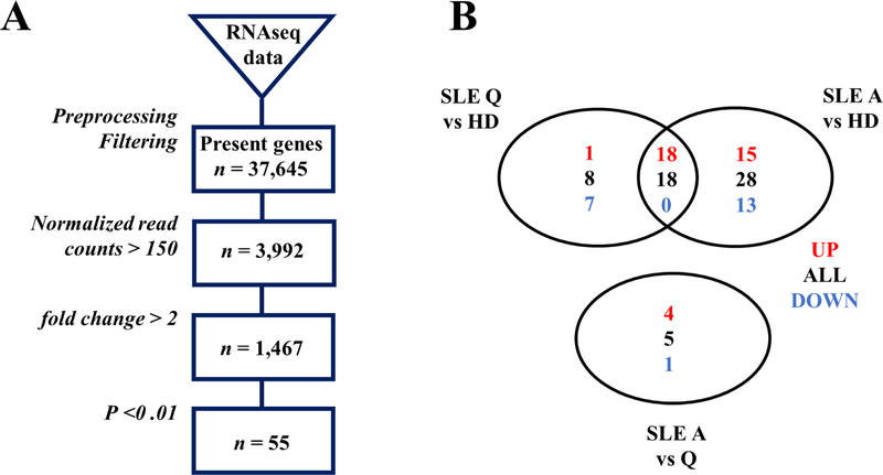
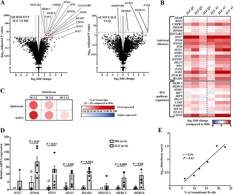
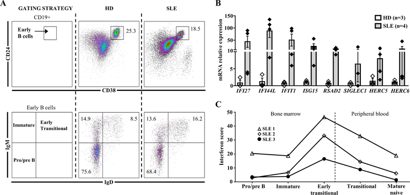
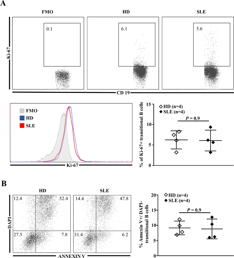
Systemic lupus (SLE) is characterized by a break of B cell tolerance that plays a central role in disease pathophysiology. An early checkpoint defect occurs at the transitional stage leading to the survival of autoreactive B cells and consequently the production of pathogenic autoantibodies. The main purpose of our work was to determine whether transitional B cells, as the most immature naïve B cell subset upstream of pathogenic B cells, display specific features compared to healthy non SLE subjects. Through extensive analysis of transitional B cells from untreated or low treated, mostly Caucasian, SLE patients, we demonstrated that transitional (T1 and T2) B cell frequencies were increased in SLE and positively correlated with disease activity. SLE transitional B cells displayed defects in two closely inter-related molecules (i.e. TLR9 defective responses and CD19 downregulation). RNA sequencing of sorted transitional B cells from untreated patients revealed a predominant overexpression of interferon stimulated genes (ISGs) even out of flares. In addition, early transitional B cells from the bone marrow displayed the highest interferon score, reflecting a B cell interferon burden of central origin. Hence, the IFN signature in transitional B cells is not confined to African American SLE patients and exists in quiescent disease since the medullary stage. These results suggest that in SLE these 3 factors (i.e. IFN imprintment, CD19 downregulation and TLR9 responses impairment) could take part at the early transitional B cell stage in B cell tolerance by-pass, ultimately leading in periphery to the expansion of autoantibodies-secreting cells.

Keywords: CD19; Interferon; Systemic lupus erythematosus; TLR9; Transitional B cells.


Conflict of interest statement

CONFLICT-OF-INTEREST DISCLOSURE

The authors declare no competing financial interests.

Figures

Fig. 1
Fig. 1
(A) Representative dot plots of transitional B cells and their subsets in one HD and one SLE patient. (B) Frequency of transitional B cells (top left panel) and their subsets (bottom panel) (% of CD19+) in HDs compared to SLE patients. Correlation between transitional B cells (% of CD19+) and disease activity assessed by the SLEDAI-2K score (top right panel). (C) MFI of CD19 in transitional B cells subsets from HDs and SLE patients. (D) Frequency of CD86+CD69+ transitional B cells from HDs or SLE patients after no stimulation (NS) or in vitro stimulation with CD40L (CD40 ligand), R848 (TLR7 agonist) or CpG (TLR9 agonist) for 2 days. HDs: healthy donors; MFI: mean of fluorescence intensity; SLE: Systemic lupus erythematosus.
Fig. 2
Fig. 2
(A) Flow chart of RNA Sequencing analysis. (B) Venn Diagram representing genes significantly up (in red) or down-regulated (in blue) between quiescent SLE patients, active SLE patients and HDs. A: active; HDs: healthy donors; Q: quiescent; SLE: systemic lupus erythematosus.
Fig. 3
Fig. 3
(A) Volcano plot representation of RNA-Seq data comparing transitional B cells from HDs (n=6) versus untreated quiescent SLE patients (n=3) (left panel) and transitional B cells from untreated quiescent versus active patients (n=3) (right panel). (B) Hierarchical representation of interferon stimulated genes (ISGs) expression in transitional B cells from each patient compared to HDs. (C) Modular analysis of RNA-Seq data representing the % of transcripts over/under-expressed (P<0.01) in untreated SLE patients (quiescent, n=3; active, n=3) compared to HDs (n=6). (D) qRT-PCR of ISGs in sorted transitional B cells of quiescent SLE patients compared to HDs. (E) Correlation between transitional B cells (% of CD19+ cells) and the interferon score. HDs: healthy donors; ND: not detected; SLE: Systemic lupus erythematosus.
Fig. 4
Fig. 4
(A) Gating strategy for BM analysis of pre/proB (CD19+CD24++CD38++IgMIgD), immature (CD19+CD24++CD38++IgM+IgD) and early transitional B cells (CD19+CD24++CD38++IgM+IgD+). (B) qRT-PCR of eight interferon stimulated genes in sorted early transitional B cells from BM of SLE patients compared to control. (C) Representation of the interferon score in BM and peripheral B cell subpopulations in three SLE patients. BM: Bone marrow; ND: not detected; SLE: Systemic lupus erythematosus.
Fig. 5
Fig. 5
(A) Representative dot plots of Ki-67+ cells (top panel) and Ki-67 MFI (bottom left panel) in gated transitional B cells from one HD and one SLE patient; frequency of Ki-67+ transitional B cells from HDs and SLE patients (bottom right panel). (B) Frequency of early apoptotic (DAPI AnnexinV+) transitional B cells in HDs and SLE patients. FMO: Fluorescence Minus One; HDs: healthy donors; MFI: mean of fluorescence intensity; SLE: systemic lupus erythematosus.

References

    1. Arbuckle MR, McClain MT, Rubertone MV, Scofield RH, Dennis GJ, James JA, Harley JB, Development of autoantibodies before the clinical onset of systemic lupus erythematosus, N. Engl. J. Med 349 (2003) 1526–1533. doi:10.1056/NEJMoa021933. - DOI - PubMed
    1. Tsokos GC, Lo MS, Reis P. Costa, Sullivan KE, New insights into the immunopathogenesis of systemic lupus erythematosus, Nat. Rev. Rheumatol 12 (2016) 716–730. doi:10.1038/nrrheum.2016.186. - DOI - PubMed
    1. Tsokos GC, A new checkpoint in lupus, J. Allergy Clin. Immunol 143 (2019) 1351–1352. doi:10.1016/j.jaci.2018.12.996. - DOI - PubMed
    1. Yurasov S, Wardemann H, Hammersen J, Tsuiji M, Meffre E, Pascual V, Nussenzweig MC, Defective B cell tolerance checkpoints in systemic lupus erythematosus, J. Exp. Med 201 (2005) 703–711. doi:10.1084/jem.20042251. - DOI - PMC - PubMed
    1. Suurmond J, Atisha-Fregoso Y, Marasco E, Barlev AN, Ahmed N, Calderon SA, Wong MY, Mackay MC, Aranow C, Diamond B, Loss of an IgG plasma cell checkpoint in patients with lupus, J. Allergy Clin. Immunol 143 (2019) 1586–1597. doi:10.1016/j.jaci.2018.10.041. - DOI - PMC - PubMed

Publication types