. 2019 Jul 1:92:265-276.
doi: 10.1016/j.actbio.2019.05.023. Epub 2019 May 11.

Nonlinear elasticity of the lung extracellular microenvironment is regulated by macroscale tissue strain

Affiliations

Nonlinear elasticity of the lung extracellular microenvironment is regulated by macroscale tissue strain

Ignasi Jorba et al. Acta Biomater. .

Abstract

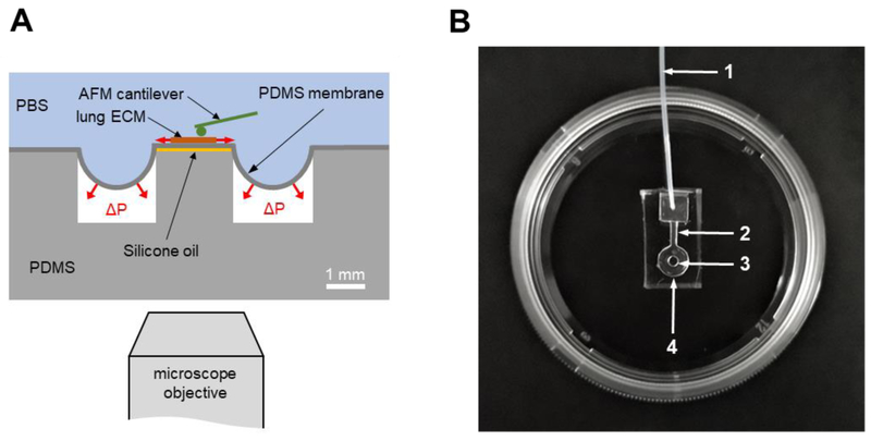
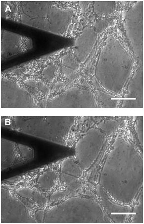
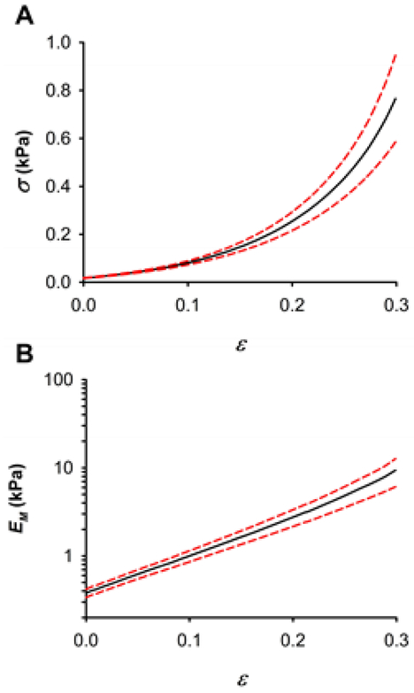
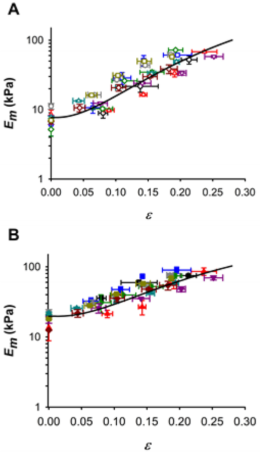
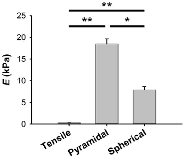
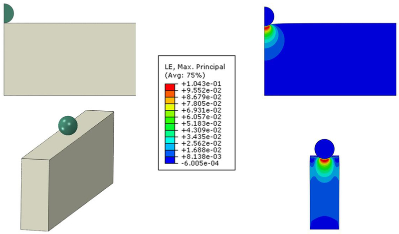
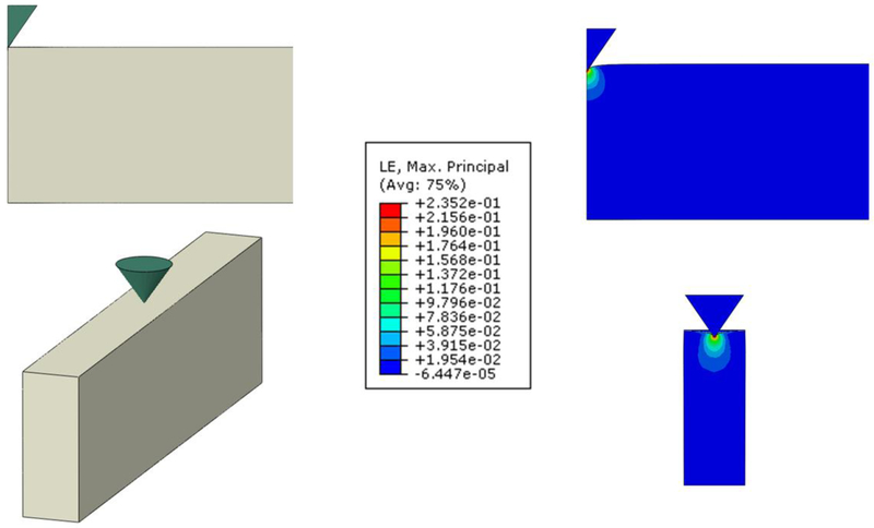
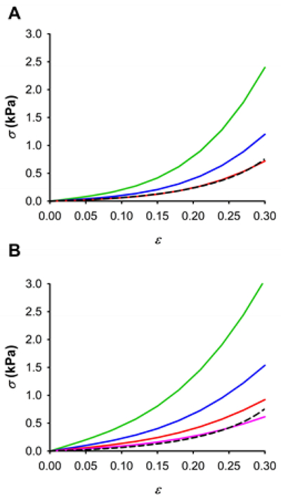
The extracellular matrix (ECM) of the lung provides physical support and key mechanical signals to pulmonary cells. Although lung ECM is continuously subjected to different stretch levels, detailed mechanics of the ECM at the scale of the cell is poorly understood. Here, we developed a new polydimethylsiloxane (PDMS) chip to probe nonlinear mechanics of tissue samples with atomic force microscopy (AFM). Using this chip, we performed AFM measurements in decellularized rat lung slices at controlled stretch levels. The AFM revealed highly nonlinear ECM elasticity with the microscale stiffness increasing with tissue strain. To correlate micro- and macroscale ECM mechanics, we also assessed macromechanics of decellularized rat lung strips under uniaxial tensile testing. The lung strips exhibited exponential macromechanical behavior but with stiffness values one order of magnitude lower than at the microscale. To interpret the relationship between micro- and macromechanical properties, we carried out a finite element (FE) analysis which revealed that the stiffness of the alveolar cell microenvironment is regulated by the global strain of the lung scaffold. The FE modeling also indicates that the scale dependence of stiffness is mainly due to the porous architecture of the lung parenchyma. We conclude that changes in tissue strain during breathing result in marked changes in the ECM stiffness sensed by alveolar cells providing tissue-specific mechanical signals to the cells. STATEMENT OF SIGNIFICANCE: The micromechanical properties of the extracellular matrix (ECM) are a major determinant of cell behavior. The ECM is exposed to mechanical stretching in the lung and other organs during physiological function. Therefore, a thorough knowledge of the nonlinear micromechanical properties of the ECM at the length scale that cells probe is required to advance our understanding of cell-matrix interplay. We designed a novel PDMS chip to perform atomic force microscopy measurements of ECM micromechanics on decellularized rat lung slices at different macroscopic strain levels. For the first time, our results reveal that the microscale stiffness of lung ECM markedly increases with macroscopic tissue strain. Therefore, changes in tissue strain during breathing result in variations in ECM stiffness providing tissue-specific mechanical signals to lung cells.

Keywords: AFM; ECM micromechanics; Multiscale lung mechanics; Tensile testing.


Conflict of interest statement

Disclosures

The authors declare no conflict of interest.

Figures

Fig. 1.
Fig. 1.
PDMS chip designed to measure ECM micromechanics with AFM under controlled stretch levels. (A) Schematic drawing of the chip consisting of a PDMS pillar at the center of a PDMS well. The well and the pillar are covered with an elastic PDMS membrane (~20 μm thick). A lung slice is adhered on top of the membrane covering the pillar. Vacuum (ΔP) applied underneath the membrane generates in-plane biaxial stretch at the central region of the membrane located above the pillar. Silicone oil placed between the pillar and the membrane avoids adhesion. The chip is covered by PBS. (B) Picture of a PDMS chip adhered to the bottom of a 35 mm culture dish. 1: vacuum inlet, 2: lateral channel, 3: pillar, 4: well.
Fig. 2.
Fig. 2.
Phase contrast image of a lung ECM slice (~20 μm thick) adhered to the membrane of a chip. (A) Unstretched state. (B) 19% strain. A V-shaped AFM cantilever is located over the ECM slice. Scale bar = 50 μm.
Fig. 3.
Fig. 3.
Macromechanics of decellularized lung parenchymal strips probed by uniaxial tensile testing. (A) Stress-strain (σ-ε) relationship of lung ECM strips. (B) Dependence of the macroscopic Young’s modulus (EM) on strip strain. Data (n = 9) are shown as mean (black solid lines) ± SE (dashed red lines).
Fig. 4.
Fig. 4.
Nonlinear micromechanics of lung ECM measured by AFM. (A) Micromechanical Young’s modulus (Em) of decellularized lung parenchymal slices (n = 9) measured with a spherical tip (radius = 2.25 μm) at different strain (ε) levels. Symbols and colors identify different ECM slices (n = 9). (B) Em measurements performed with a pyramidal tip (semi-included angle = 35°, apex radius = 100 nm). Solid lines are a fit of the Yeoh model (Eq. 12). Data are mean ± SE (n = 9).
Fig. 5.
Fig. 5.
Multiscale stiffness of lung ECM. Young’s modulus of lung ECM at the unstretched state measured with macroscale tensile testing and with microscale AFM measurements using spherical and pyramidal tips (indentation depth of 0.7 μm). Data are mean ± SE (n = 9). * and ** are p < 0.05 and p < 0.01, respectively.
Fig. 6.
Fig. 6.
FE simulations of a spherical tip (radius = 2.25 μm) indenting a Yeoh hyperelastic material of different dimensions. Strain distribution at indentation δ = 0.7 μm in the X-Y plane of a vertical cylinder 16.8 μm in height and 200 μm in radius (top, half-space model) and a cuboid with width = 6.6 μm, length = 400 μm and height = 16.8 μm (bottom, acellular alveolar wall model). Color scale is the common logarithmic maximal principal strain distribution (averaged at nodes). Maximum strain is 0.1 in both cases.
Fig. 7.
Fig. 7.
FE simulations of a blunted conical tip (semi-included angle = 35°, apex radius = 100 nm) indenting a Yeoh hyperelastic material of different dimensions. Strain distribution at indentation δ = 0.7 μm in the XY plane of a vertical cylinder 16.8 μm in height and 200 μm in radius (top, half-space model) and a cuboid with width = 6.6 μm, length = 400 μm and height = 16.8 μm (bottom, acellular alveolar wall model). Color scale is the common logarithmic maximal principal strain distribution (averaged at nodes). Maximum strain is 0.32 and 0.24 in the half-space and the acellular alveolar wall models, respectively.
Fig. 8.
Fig. 8.
Porosity model of lung ECM. Fit of the Yeoh porous model to the macroscopic experimental stress-strain relationships (σ-ε) measured by tensile testing (dashed black line) for different material adjustments using spherical (A) and pyramidal (B) tip measurements at different levels of tissue fraction ratio: 0.2 (green), 0.1 (blue) and 0.06 (red), 0.04 (pink).

References

    1. Badylak SF, Freytes DO, Gilbert TW, Extracellular matrix as a biological scaffold material: Structure and function, Acta Biomater. 5(1) (2009) 1–13. - PubMed
    1. Suki B, Ito S, Stamenovic D, Lutchen KR, Ingenito EP, Biomechanics of the lung parenchyma: critical roles of collagen and mechanical forces, J. Appl. Physiol 98 (2005) 1892–1899. - PubMed
    1. Suki B, Stamenovic D, Hubmayr RD, Lung Parenchymal Mechanics, Compr Physiol 1 (2011) 1317–1351. - PMC - PubMed
    1. Discher DE, Smith L, Cho S, Colasurdo M, Garcia AJ, Safran S, Matrix Mechanosensing: From Scaling Concepts in 'Omics Data to Mechanisms in the Nucleus, Regeneration, and Cancer, Annual review of biophysics 46 (2017) 295–315. - PMC - PubMed
    1. Engler AJ, Carag-Krieger C, Johnson CP, Raab M, Tang HY, Speicher DW, Sanger JW, Sanger JM, Discher DE, Embryonic cardiomyocytes beat best on a matrix with heart-like elasticity: scar-like rigidity inhibits beating, J. Cell Sci 121 (2008) 3794–3802. - PMC - PubMed

Publication types