Radiation-induced brain cavernomas in elderly: review of the literature and a rare case report
- PMID: 31085976
- PMCID: PMC6625569
- DOI: 10.23750/abm.v90i5-S.8328
Radiation-induced brain cavernomas in elderly: review of the literature and a rare case report
Abstract
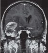
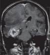
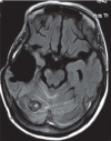
Radiation-induced brain cavernomas have been mainly reported in children who underwent radiotherapy for medulloblastoma, leukemia, or low-grade glioma. Otherwise, the "de novo" appearance of a cavernoma in an elderly long-survivor patient after resection and radiotherapy of a glioblastoma is a rare event. We report the case of a 62-year-old female patient who underwent surgical resection of a right temporal glioblastoma, followed by radiation therapy of the operative field and surrounding brain and concomitant adjuvant temozolomide. Four years after the operation, a follow-up Magnetic Resonance revealed a good tumor control and a small round lesion at the superior surface of the right cerebellar hemisphere, close to the margins of the previous irradiation field. The radiological items were consistent with a cavernous angioma. Because of the small size of the malformation and the absence of related symptoms, no treatment was performed. The patient died for tumor progression 86 months after the initial operation, with unchanged cerebellar cavernoma. The occurrence of a cavernous angioma in an elderly patient after radiotherapy for brain glioblastoma is an exceptional event; the distribution of radiotherapy-induced cavernous malformations reported in current literature is presented and the mechanism of their formation is discussed.
Figures


References
-
- Keezer MR, Del Maestro R. Radiation-induced cavernous hemangiomas: case report and literature review. Can J Neurol Sci. 2009;36:303–10. - PubMed
-
- Nimjee SM, Powers CJ, Bulsara KR. Review of the literature on de novo formation of cavernous malformations of the central nervous system after radiation therapy. Neurosurg Focus. 2006;21:e4. - PubMed
-
- Caruso R, Colonnese C, Elefante A, Innocenzi G, Raguso M, Gagliardi FM. Use of spiral computerized tomography angiography in patients with cerebral aneurysm. Our experience. J Neurosurg Sci. 2002;46:4–9. - PubMed
-
- Elefante A, Peca C, Del Basso De Caro ML, et al. Symptomatic spinal cord metastasis from cerebral oligodendroglioma. Neurol Sci. 2012;33:609–13. - PubMed
Publication types
MeSH terms
LinkOut - more resources
Full Text Sources
Medical
Molecular Biology Databases

