Review
. 2019 May 13;20(9):2363.
doi: 10.3390/ijms20092363.

A Symphony of Signals: Intercellular and Intracellular Signaling Mechanisms Underlying Circadian Timekeeping in Mice and Flies

Affiliations
Review

A Symphony of Signals: Intercellular and Intracellular Signaling Mechanisms Underlying Circadian Timekeeping in Mice and Flies

Sara Hegazi et al. Int J Mol Sci. .

Abstract

The central pacemakers of circadian timekeeping systems are highly robust yet adaptable, providing the temporal coordination of rhythms in behavior and physiological processes in accordance with the demands imposed by environmental cycles. These features of the central pacemaker are achieved by a multi-oscillator network in which individual cellular oscillators are tightly coupled to the environmental day-night cycle, and to one another via intercellular coupling. In this review, we will summarize the roles of various neurotransmitters and neuropeptides in the regulation of circadian entrainment and synchrony within the mammalian and Drosophila central pacemakers. We will also describe the diverse functions of protein kinases in the relay of input signals to the core oscillator or the direct regulation of the molecular clock machinery.

Keywords: Drosophila; central pacemaker; circadian rhythms; entrainment; intercellular and intracellular signaling; neuropeptides; neurotransmitters; protein kinases; suprachiasmatic nucleus; synchrony.


Conflict of interest statement

The authors declare no conflict of interest.

Figures
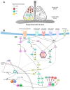
Figure 1
Figure 1
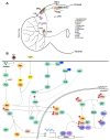
The mammalian suprachiasmatic nucleus: neurochemical composition and canonical signaling pathways that regulate the core oscillator and photic entrainment. (A) Neurochemical composition of the SCN. The SCN is divided into ventral (core) and dorsal (shell) regions. Most, if not all, of the ~20,000 SCN cells are GABAergic but differ in their neuropeptide content. VIP and AVP delineate the core and shell regions, respectively. GRP-expressing neurons are primarily localized in the medial core. Prokineticin 2 is another SCN neuropeptide that is highly expressed by core and shell SCN neurons. (B) Canonical signaling pathways within the SCN. The figure depicts the major signaling pathways and protein kinases that have so far been shown to function within the SCN. Various neuropeptides and neurotransmitters impinging on SCN neurons can activate receptors and ion channels on the plasma membrane to trigger intracellular signaling events. Activation of G-protein coupled receptors (GPCRs) can signal via Gs, Gi, and Gq proteins to activate adenylyl cyclase (AC), inhibit AC, or activate phospholipase Cβ (PLCβ), respectively. AC stimulates the production of cAMP, which in turn activates protein kinase A (PKA) and exchange protein activated by cAMP (Epac). CREB-mediated transcription can be induced by PKA and Epac. PLCβ catalyzes the hydrolysis of phosphatidylinositol 4,5-bisphosphate into diacylglycerol (DAG) and inositol 1,4,5-trisphosphate (IP3). DAG activates protein kinase C at the plasma membrane, whereas IP3 diffuses into the cytosol to induce the release of intracellular Ca2+ stores from the endoplasmic reticulum (ER). The rise in cytosolic Ca2+ levels can also be induced by ionotropic receptors, voltage-gated ion channels, G-protein gated ion channels, and receptor tyrosine kinases. Downregulation of GPCR signaling is achieved by phosphorylation of the receptor by G protein-coupled receptor kinase 2 (GRK2). Receptor-mediated Ras activation at the plasma membrane stimulates the mitogen-activated protein kinase pathway (RAF, MEK1/2, ERK1/2). PKC facilitates Ras/MAPK signaling, either by activating Ras or derepressing RKIP-mediated inhibition of Raf via phosphorylation of RKIP. MAPK/ERK activates p90 ribosomal S6 kinase 1 (RSK1) and mitogen- and stress-activated protein kinase 1 (MSK1), which in turn stimulate CREB-mediated transcription. MAPK/ERK is also an upstream activator of mammalian target of rapamycin (mTOR), which promotes translation by activating p70 S6 kinase (p70S6K) and inhibiting eukaryotic translation initiation factor 4E (eIF4E)-binding protein 1 (4E-BP1)-mediated repression of eIF4E. In terms of Ca2+ signaling, Ca2+-induced activation of Ca2+/calmodulin-dependent protein kinase II (CaMKII) can couple to cyclic guanosine monophosphate (cGMP) production through the nitric oxide synthase (NOS)/guanylyl cyclase (GC) pathway. cGMP activates protein kinase G (PKG), which promotes CREB-mediated transcription. The NOS pathway also activates the small G protein, Dexras1, which inhibits GPCR-mediated Gi activation and indirectly inhibits AC through ligand-independent activation of Gi. Many upstream signaling events converge on the MAPK/ERK pathway, a pivotal player in photic entrainment via its effects on CREB. Salt-inducible kinase 1 (SIK1) is a CRE-inducible gene that acts as a feedback inhibitor of CREB signaling through suppression of CREB-dependent transcription coactivator 1 (CRTC1). Finally, a number of protein kinases have been shown to phosphorylate clock proteins, PERIOD and CRY: these include casein kinase 1 and 2 (CK1 and CK2, respectively), glycogen synthase kinase 3 (GSK3), SIK3, and GRK2. Dashed lines denote indirect interactions.
Figure 2
Figure 2
The central clock network in Drosophila: neuronal clusters, neurochemical composition, and clock regulation by protein kinases. (A) Classification and neurochemical composition within the adult Drosophila central pacemaker. The clock network is composed of ~150 neurons that include the small and large ventral lateral neurons (s-LNvs and l-LNvs, respectively), dorsal lateral neurons (LNds), dorsal neurons (DNs), and lateral posterior neurons (LPNs). Different neurochemicals colocalize in the same neuron within a cluster. The PDF-expressing s-LNvs project into the dorsal protocerebrum, and the l-LNvs project contralaterally and into the optic lobe. Glutamate, DH31 and Allostatin-C are expressed in overlapping and non-overlapping subsets of DN1p neurons. While Allostatin-C and glutamate have been shown to co-localize within the same neurons, it is not clear whether DH31 co-localizes with either Allostatin-C or glutamate in the same DN1p neurons. CCHa1, CCHamide1; DH31, Diuretic Hormone 31; ITP, Ion Transport Peptide; NPF, Neuropeptide F; sNPF, Short Neuropeptide F; PDF, Pigment Dispersing Factor. (B) Regulation of the clockwork via protein kinases in the Drosophila pacemaker. The primary clock feedback loop where CLK/CYC dimers initiate the transcription of per and tim genes. The phosphorylated PER/TIM complex then translocates to the nucleus to repress CLK/CYC activity. Several protein kinases are involved in mediating the nuclear translocation and degradation of PER and TIM within the nuclear and cytoplasmic compartments. In the nucleus, kinases act to repress the CLK/CYC transcriptional complex via the phosphorylation and degradation of CLK. PER and TIM must also undergo degradation in the nucleus to reset the loop. The asterisk (*) refers to the action of NEMO in priming PER for DBT-mediated phosphorylation. Also shown is the role of PDF-PDFR signaling in stabilizing PER and TIM proteins. See text for detailed description of the depicted pathways. AC3, Adenylyl Cyclase 3; AKT, Protein Kinase B; cAMP, cyclic adenosine monophosphate; CK2, Casein Kinase 2; CLK, Clock; CRY, Cryptochrome; CYC, Cycle; DBT, Doubletime; Gsα60A, stimulatory G protein α subunit 60A; GTP, guanosine triphosphate; MAPK, Mitogen-Activated Protein Kinase; MEK, MAPK/ERK Kinase; NEMO, NEMO kinase; p38, p38 MAPK; PER, Period; PKA, Protein Kinase A; Ras, Ras-GTPase; Rheb, Rheb GTPase; SGG, Shaggy; S6KII, Ribosomal S6 Kinase II; TIM, Timeless; TOR, Target of Rapamycin. Phosphate groups are depicted in red circles (P); dashed lines show indirect effects through other signaling molecules; dissociated proteins indicate degradation; upward pointing arrows placed beside molecules show stabilization and/or accumulation.

References

    1. Partch C.L., Green C.B., Takahashi J.S. Molecular architecture of the mammalian circadian clock. Trends Cell Biol. 2014;24:90–99. doi: 10.1016/j.tcb.2013.07.002. - DOI - PMC - PubMed
    1. Ralph M.R., Foster R.G., Davis F.C., Menaker M. Transplanted suprachiasmatic nucleus determines circadian period. Science. 1990;247:975–978. doi: 10.1126/science.2305266. - DOI - PubMed
    1. Albrecht U. Timing to Perfection: The Biology of Central and Peripheral Circadian Clocks. Neuron. 2012;74:246–260. doi: 10.1016/j.neuron.2012.04.006. - DOI - PubMed
    1. Wagner S., Castel M., Gainer H., Yarom Y. GABA in the mammalian suprachiasmatic nucleus and its role in diurnal rhythmicity. Nature. 1997;387:598–603. doi: 10.1038/42468. - DOI - PubMed
    1. Abrahamson E.E., Moore R.Y. Suprachiasmatic nucleus in the mouse: Retinal innervation, intrinsic organization and efferent projections. Brain Res. 2001;916:172–191. doi: 10.1016/S0006-8993(01)02890-6. - DOI - PubMed

LinkOut - more resources