

Melanoma-Derived Extracellular Vesicles: Focus on Their Proteome
- PMID: 31086060
- PMCID: PMC6630787
- DOI: 10.3390/proteomes7020021
Melanoma-Derived Extracellular Vesicles: Focus on Their Proteome
Abstract
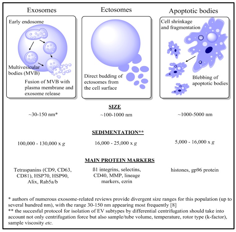
Malignant melanoma is one of the most aggressive types of cancer, and its incidence is increasing rapidly each year. Despite the extensive research into improved diagnostic and treatment methods, early detection and disease constraint still present significant challenges. As successful isolation protocols have been developed, extracellular vesicles (EVs) have become the subject of extensive investigation in terms of their role in cancer progression and as a possible source of disease biomarkers. Besides functional studies, quantitative and qualitative proteomics have recently emerged as promising tools for the advancement of melanoma biomarkers. Nevertheless, the amount of data concerning the proteome of melanoma-derived EVs is still very limited. In this review we cover the current knowledge on protein content of melanoma-derived EVs, with a focus on their potential role in the development and progression of melanomas.
Keywords: cancer; ectosomes; exosomes; extracellular vesicles; melanoma; proteomics.
Conflict of interest statement
The authors declare no conflict of interest.
Figures
References
Publication types
Grants and funding
LinkOut - more resources
Full Text Sources
Research Materials
