. 2019 May 13;8(5):34.
doi: 10.1038/s41389-019-0144-0.

Mechanism for oral tumor cell lysyl oxidase like-2 in cancer development: synergy with PDGF-AB

Affiliations

Mechanism for oral tumor cell lysyl oxidase like-2 in cancer development: synergy with PDGF-AB

Faranak Mahjour et al. Oncogenesis. .

Abstract

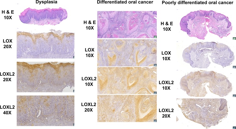
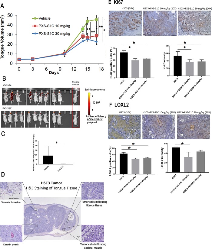
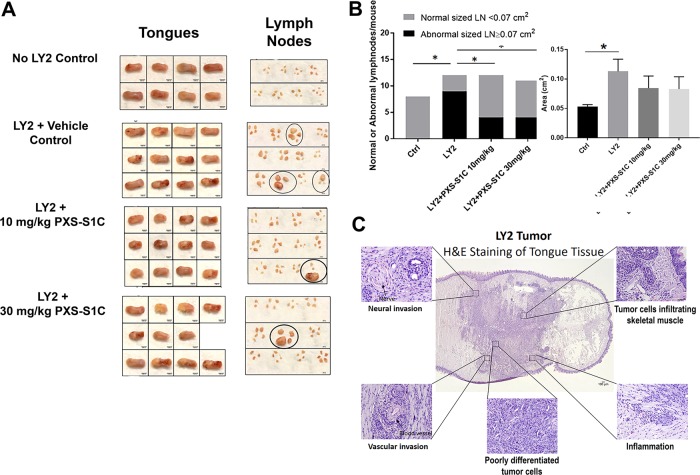
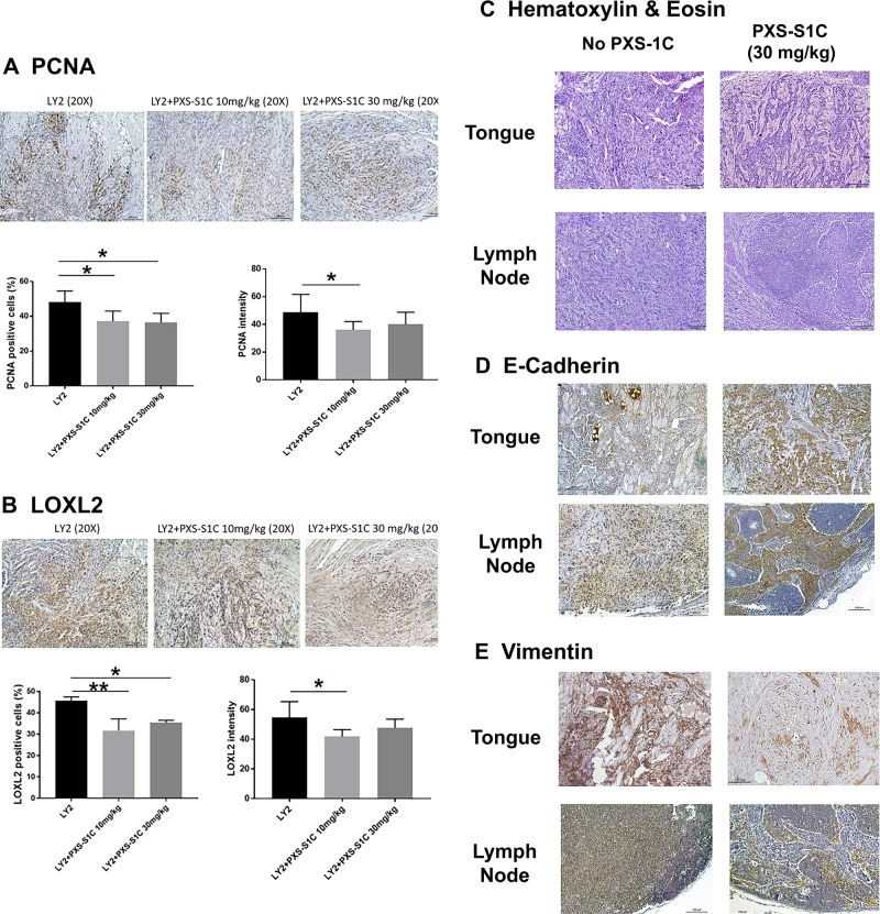
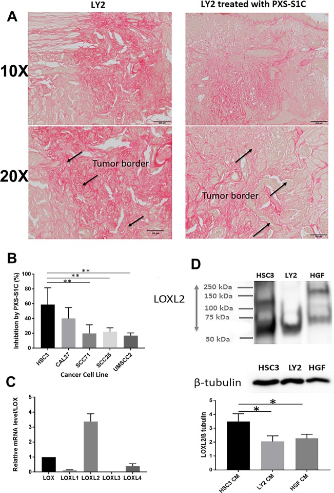
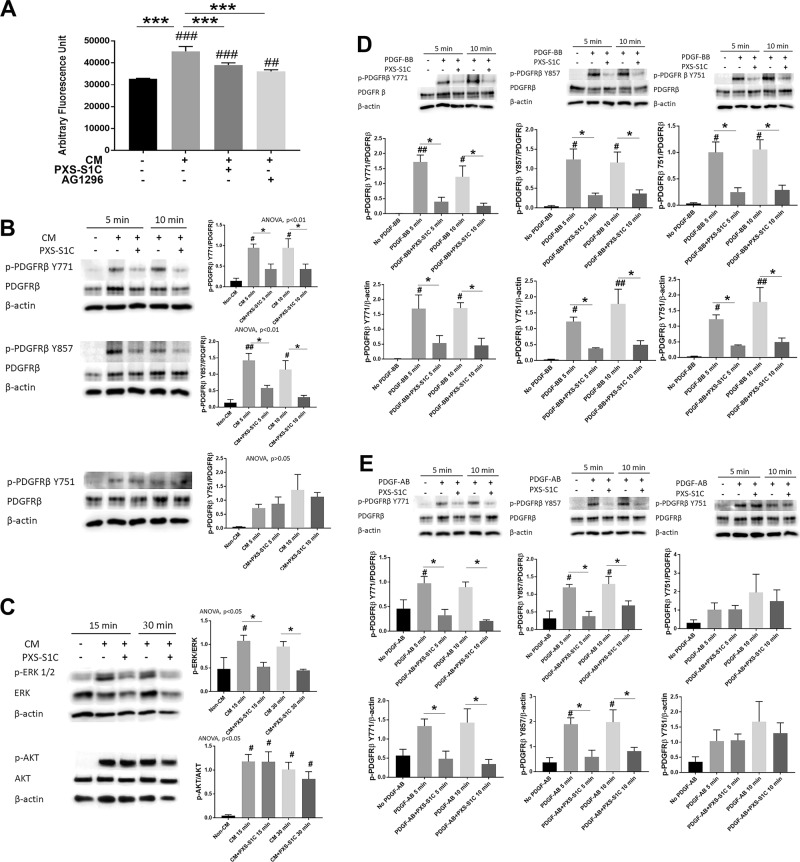
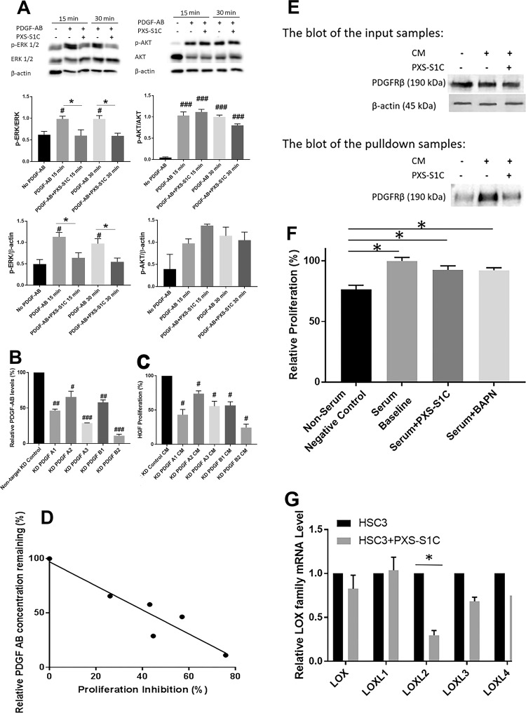
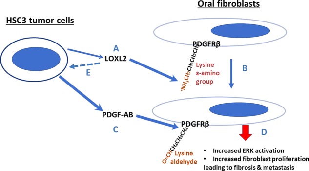
Extracellular lysyl oxidases (LOX and LOXL1-LOXL4) are critical for collagen biosynthesis. LOXL2 is a marker of poor survival in oral squamous cell cancer. We investigated mechanisms by which tumor cell secreted LOXL2 targets proximal mesenchymal cells to enhance tumor growth and metastasis. This study identified the first molecular mechanism for LOXL2 in the promotion of cancer via its enzymatic modification of a non-collagenous substrate in the context of paracrine signaling between tumor cells and resident fibroblasts. The role and mechanism of active LOXL2 in promoting oral cancer was evaluated and employed a novel LOXL2 small molecule inhibitor, PSX-S1C, administered to immunodeficient, and syngeneic immunocompetent orthotopic oral cancer mouse models. Tumor growth, histopathology, and metastases were monitored. In vitro mechanistic studies with conditioned tumor cell medium treatment of normal human oral fibroblasts were carried out in the presence and absence of the LOXL2 inhibitor to identify signaling mechanisms promoted by LOXL2 activity. Inhibition of LOXL2 attenuated cancer growth and lymph node metastases in the orthotopic tongue mouse models. Immunohistochemistry data indicated that LOXL2 expression in and around tumors was decreased in mice treated with the inhibitor. Inhibition of LOXL2 activity by administration of PXS-S1C to mice reduced tumor cell proliferation, accompanied by changes in morphology and in the expression of epithelial to mesenchymal transition markers. In vitro studies identified PDGFRβ as a direct substrate for LOXL2, and indicated that LOXL2 and PDGF-AB together secreted by tumor cells optimally activated PDGFRβ in fibroblasts to promote proliferation and the tendency toward fibrosis via ERK activation, but not AKT. Optimal fibroblast proliferation in vitro required LOXL2 activity, while tumor cell proliferation did not. Thus, tumor cell-derived LOXL2 in the microenvironment directly targets neighboring resident cells to promote a permissive local niche, in addition to its known role in collagen maturation.


Conflict of interest statement

P.C.T. has served as a consultant for Pharmaxis Corporation, and Pharmaxis partially funded this work. The remaining authors declare that they have no conflict of interest.

Figures

Fig. 1
Fig. 1. Histology and immunohistochemistry of human dysplasia and oral cancer biopsies for LOX and LOXL2.
Biopsy samples were selected by the pathology service at Boston University Henry M. Goldman School of Dental Medicine and tissue sections were prepared and stained. Slides made from one selected subject from 3 to 5 subjects sampled in each category of dysplasia, differentiated oral cancer, and poorly differentiated oral cancer, respectively, are shown. Stained slides were imaged using an automated slide imager, and images were processed using Case Viewer software version 2.2 (Budapest, Hungary). Data indicate that LOXL2 was highly expressed in a variety of cancer cells and associated mesenchymal cells in human oral cancer, while LOX expression was more restricted
Fig. 2
Fig. 2. LOXL2 promotes human tongue orthotopic cancer growth and metastasis in mice.
a PXS-S1C attenuates human tongue tumor growth in mice, and b and c PSX-S1C significantly decreases tumor cell spreading. Tumor volume of mouse tongues were measured every 3 days. Data are means ± SD. ANOVA, p: 0.0001, Tukey’s multiple comparisons test, **p < 0.001, ***p < 0.0001 indicate difference among the groups (n = 8 per group). b IVIS imaging for red fluorescent protein-labeled HSC3 cells 21 days after commencing injections of vehicle or PXS-S1C (30 mg/kg). The fluorescence signals were optimized for DsRed protein at excitation 570 nm and emission 620 nm. c Quantification of fluorescence signal area shows a significant difference between PXS-S1C-treated and non-treated groups. Data are mean ± SD. Student’s t-test, *p < 0.05 indicates difference between the groups. d Histology of HSC3 cell orthotopic tongue tumors. Hematoxylin and eosin staining a mouse tongue is shown 18 days after implantation. The photo is representative of histological features of HSC3 orthotopic tumors. The images were taken at ×4 and ×20 magnifications. Scale bar = 100 µm. PXS-S1C attenuates expression of (e) Ki67 and (f) LOXL2 in LY2 orthotopic tumors in immunodeficient mice. e Immunohistochemistry staining of tongue sections with anti-Ki-67 antibody shows that PXS-S1C reduced Ki-67 staining in orthotopic HSC3 tumors in mice. Scale bar = 100 µm. Data are mean ± SD. ANOVA, p < 0.01, Tukey’s multiple comparisons test, *p < 0.05 indicate difference among the groups (n = 8 per group). f Staining of tongue tissues with anti-LOXL2 antibody shows that PXS-S1C reduced LOXL2 staining in orthotopic HSC3 tumors in mice. Scale bar = 100 µm. Data are mean ± SD. ANOVA, p < 0.01, Tukey’s multiple comparisons test, *p < 0.05 indicate difference among the groups (n = 8 per group)
Fig. 3
Fig. 3. LOXL2 promotes syngeneic orthotopic tongue tumor growth and metastasis to cervical lymph nodes in immunocompetent mice.
a Gross features of tongues and lymph nodes of the mice in all groups. Circles mark grossly oversized lymph nodes. b Number of the mice with abnormal size of lymph nodes (left panel) was reduced by the treatment with PXS-S1C. Number of the mice with normal versus abnormal size of lymph nodes. Normal sized LN in control mice = 0.053 ± 0.01 cm2, *p < 0.05. Chi-Square test (4 × 2 analysis) p = 0.008, Chi-Square test, p = 0.008, indicates difference in number of normal and abnormal sized LN among the groups. Average size of the lymph nodes in each group (right panel). ANOVA, p < 0.05, Tukey’s multiple comparison test *p < 0.05 indicates difference between the groups. Fisher’s exact test (2 × 2 analysis). Control vs. LY2, p = 0.001, mice injected with LY2 have larger LNs than controls. Fisher’s exact test (2 × 2 analysis) LY2 vs. both LY2 + PXS-S1C 10 and 30 mg/kg group ogether, p = 0.03, LOX inhibitor reduces the frequency of mice having enlarged LNs. Fisher’s exact test (2 × 2 analysis), LY2 + PXS-S1C 10 mg/kg vs. LOXL2 + 30 mg/kg, p > 0.05 There is no statistical difference between the two different doses of LOX inhibitor. c Histology of LY2 orthotopic tongue tumors. Hematoxylin and eosin staining of LY2 orthotopic tongue tumor. The images are representative of histological features of LY2 tumor. The images were taken at ×4 and ×20 objectives. Scale bar = 100 µm
Fig. 4
Fig. 4. LOXL2 promotes proliferation and EMT of primary tongue and cervical lymph node metastases.
PXS-S1C attenuates expression of (a) PCNA and (b) LOXL2 in LY2 orthotopic tongue tumors in immunocompetent mice after 6 weeks of treatment, scale bar = 100 µm. Data are mean ± SD. ANOVA, p < 0.05, Tukey’s multiple comparisons test, *p < 0.05 indicate difference among the groups (n = 12 per group). PSX-S1C treatment of LY2 tongue orthotopic tumors in mice alters tongue and cervical lymph node (c) tumor cell morphology, (d) E-cadherin expression and (e) vimentin expression. The images were taken at ×10 and ×20 magnifications. Scale bar = 100 µm. Images are from mice at the 6-week time point
Fig. 5
Fig. 5. LOXL2 promotes collagen accumulation in syngeneic tongue oral cancer in mice, while human oral tumor cells-derived LOXL2 stimulates oral fibroblast proliferation in vitro.
a Collagen accumulation in orthotopic tongue LY2 tumors by Sirius red staining of LY2 tumors in the tongue. A representative image is shown from one of 12 mice at the 6-week time point. Scale bar = 100 µm. Treatment with PXS-S1C appeared to reduce the amount of collagen, particularly at the apparent interfaces of tumor with surrounding non-tumor tissue. The images were taken at ×10 and ×20 magnifications. b Stimulation of human gingival fibroblast proliferation induced by CM of different oral cancer cell lines was inhibited by PXS-S1C treatment. Gingival fibroblasts were serum depleted for 24 h and then treated with cancer cell CM with and without PXS-S1C (1 µM) in serum-free conditions for 24 h, and DNA accumulation measured by CyQUANT assays: [(HSC3 CM)–(HSC3 + PXS-S1C)]/HSC3 CM × 100]. Data are means ± SD. Experiments were done with six replicate samples for each cell line. ANOVA, p < 0.0001, Tukey’s multiple comparison test. **p < 0.001 indicate differences among different groups. c LOXL2 is the most abundantly expressed paralogue by HSC3 cells. RNAs isolated from serum-depleted HSC3 cells was subjected to qPCR for all five lysyl oxidase paralogues using Taqman probes. Data are means ± SEM. This experiment was performed three times independently with triplicate samples. The RNA levels were normalized to 18S rRNA. ANOVA One way, P < 0.01 among all LOX family members. d LOXL2 protein is secreted at high levels by HSC3 cells. CM from human HSC3 cells, mouse LY2 cells and normal human gingival fibroblasts were collected under serum-free conditions, concentrated by 25-fold, and subjected to western blotting and visualized with anti-LOXL2 antibody. β-tubulin was used as a loading control. Data are means ± SEM. This experiment was done three times independently. ANOVA, p < 0.002, Tukey’s multiple comparison test *p < 0.05 indicate difference among the groups
Fig. 6
Fig. 6. HSC3 oral tumor cell-derived LOXL2 stimulates oral fibroblast proliferation and cell signaling in collaboration with PDGF-AB.
LOXL2 inhibitor PXS-S1C attenuates HSC3 CM-stimulated human oral fibroblast (a) proliferation, (b) phosphorylation of PDGFRβ at the Y771 and Y857 but not Y751 residues, and (c) ERK activation, but not AKT. a Human gingival fibroblast proliferation was reduced after 24-h treatment with PXS-S1C (1 µM) or AG 1296 (5 µM) in the HSC3 CM as determined by the CyQUANT assay. Data are means SEM. This experiment was done three times independently with six replicate samples. ANOVA, p < 0.0001, Dunnett’s multiple comparisons test, ***p < 0.0001 indicates significant difference between treated groups; while ##p < 0.001, ###p < 0.0001 indicate significant differences from non-CM group. b and c Gingival fibroblasts were serum depleted and then treated with non-CM, and CM with and without PXS- S1C (1 µM) and cell layer protein samples were subjected to western blot. Data are means ± SEM. The experiment was performed with three times independently with primary human gingival fibroblasts isolated from three different donors. Representative blots are shown. Data from all three experiments were subjected to quantitative analyses. Sidak’s multiple comparison test, *p < 0.05 indicates a significant difference from PXS-S1C treated group. Dunnett’s multiple comparison test, #p < 0.05, ##p < 0.001 indicate significant differences from Non-CM group. d PXS-S1C attenuates PDGF-BB stimulated phosphorylation of all three PDGFRβ phosphorylation sites Y771, Y857 and Y751, and AKT activation in oral fibroblasts. Gingival fibroblasts were serum depleted and then treated with no PDGF-BB, and PDGF-BB (10 ng/ml) with and without PXS-S1C (1 µM). The protein samples were subjected to western blot. Data are means ± SEM. The experiment was done with three times independently with primary human gingival fibroblasts isolated from three different donors. Representative blots are shown. Data from all three experiments were subjected to quantitative analyses. Sidak’s multiple comparison test, *p < 0.05, **p < 0.001, and ***p < 0.0001 indicate difference from PXS-S1C-treated group. Dunnett’s multiple comparison test, #p < 0.05, ##p < 0.001, ###p < 0.0001 indicate difference from No PDGF group. e PDGF-AB mimics the effects of HSC3 cell CM on oral fibroblasts in phosphorylation of PDGFRβ. Gingival fibroblasts were serum starved and then treated with no PDGF-AB, and PDGF-AB (10 ng/ml) with and without PXS-S1C (1 µM). The protein samples were subjected to western blot. Data are means ± SEM. The experiment was done with three times independently with primary human gingival fibroblasts isolated from three different donors. Representative blots are shown. Data from all three experiments were subjected to quantitative analyses. Sidak’s multiple comparison test, *p < 0.05 indicates difference from PXS-S1C-treated group. Dunnett’s multiple comparison test, #p < 0.05 indicates difference from No PDGF group
Fig. 7
Fig. 7. PDGF-AB, but not PDGF-BB, is secreted by HSC3 tumor cells, PDGFRβ is a substrate for LOXL2, and LOXL2 promotes its own synthesis in HSC3 cells.
a PDGF-AB mimics the effects of HSC3 cancer cell CM on oral fibroblasts in phosphorylation of ERK1/2, and not AKT. Oral fibroblasts were serum depleted and then treated with no PDGF-AB, and PDGF-AB (10 ng/ml) with and without PXS-S1C (1 µM). The protein samples were subjected to western blot. Data are means ± SEM. The experiment was done with three times independently with primary human gingival fibroblasts isolated from three different donors. Representative blots are shown. Data from all three experiments were subjected to quantitative analyses. Sidak’s multiple comparison test, *p < 0.05 indicates difference from PXS-S1C-treated group. Dunnett’s multiple comparison test, #p < 0.05 and ###p < 0.0001 indicate difference from No PDGF group. b PDGF-A and PDGF-B knockdown in HSC3 cells blocks HSC3 CM stimulation of oral fibroblast proliferation. The concentration of PDGF-AB ligand was decreased significantly in CM of knockdown HSC3 cells as compared with non-target control cell CM. PDGF-A and PDGF-B were, respectively, knocked down with independent shRNAs (A1, A2, or A3 for PDGF-A; and B1 and B2 for PDGF’B). The concentration of PDGF-AB ligand in knocked down HSC3 CM was measured using a PDGF-AB-specific ELISA kit. Data are mean ± SD. This experiment was done with triplicate samples. ANOVA, p < 0.0001, Dunnett’s multiple comparisons test #p < 0.05, ##p < 0.001, ###p < 0.0001 indicate difference from Control HSC3 group. c The proliferation of oral fibroblasts treated with knocked down HSC3 medium for 24 h was assessed by CyQUANT assay. Data are mean ± SEM. This experiment was performed three times independently with primary human gingival fibroblasts isolated from three different donors. ANOVA, p: 0.0001, Dunnett’s multiple comparisons test: #p < 0.05, indicate difference from HSC3 Control group. d The degree of PDGF-A or PDGF-B knockdown correlate linearly with decreased proliferative responses to HSC3 CM. The relationship between fibroblast proliferation inhibition and the relative level of PDGF-AB concentration was analyzed using linear regression. Correlation coefficient r: −0.93, R squared: 0.87, p-value: 0.006. Data indicate that PDGF-AB specifically is the ligand in HSC3 CM that stimulates oral fibroblast proliferation. e Carbonyl pull down assay for PDGFRβ in oral fibroblasts treated with HSC3 CM in the absence or presence of PXS-S1C. Human oral fibroblasts were treated with HSC3 CM in the absence or presence of 1 µM PXS-S1C followed by biotin hydrazide derivitization and affinity pulldown with a streptavidin affinity resin (Neutravidin). Input samples and proteins eluted by boiling in SDS–PAGE were subjected to Western blotting for PDGFRβ. Data are representative of two experiments with the same outcome from two different gingival fibroblast donors. f PXS-S1C and BAPN did not inhibit serum-stimulated proliferative response of HSC3 tumor cells. HSC3 cells were serum-depleted overnight and treated with PXS-S1C (1 µM) or BAPN (0.5 mM) in medium containing 2.5% serum for serum stimulation of a proliferative response. Data are means ± SEM. ANOVA, p < 0.0001, Tukey’s multiple comparisons *p < 0.05 indicates difference among the groups. g PXS-S1C decreased the expression of LOXL2 in HSC3 cells in vitro. Relative LOXL2 mRNA levels in HSC3 cell line with and without PXS-S1C after 24 h treatment was measured. Data are means ± SEM. This experiment was done three times independently with triplicate samples. ANOVA, p: 0.04, Sidak’s multiple comparisons test *p < 0.05 indicates difference from non-treated HSC3 group. The RNA levels were normalized to 18S rRNA
Fig. 8
Fig. 8. Summary of interactions between oral tumor cells and fibroblasts that contribute to oral cancer development.
LOXL2 secreted by tumor cells (a) oxidizes lysine residues on PDGFRβ in proximal fibroblasts (b), in addition, to its classical role in collagen maturation (not shown). PDGF-AB secreted by tumor cells is consequently able to more efficiently stimulate PDGF signaling (c), resulting in increased ERK1/2 activation and cell proliferation (d). LOXL2 production by tumor cells is required for maintaining LOXL2 synthesis is a feed-forward pathway (e), whose mechanism remains to be determined

References

    1. Cooper JS, et al. National Cancer Database report on cancer of the head and neck: 10-year update. Head Neck. 2009;31:748–758. doi: 10.1002/hed.21022. - DOI - PubMed
    1. Musharraf SG, et al. Metabolite profiling of preneoplastic and neoplastic lesions of oral cavity tissue samples revealed a biomarker pattern. Sci. Rep. 2016;6:38985. doi: 10.1038/srep38985. - DOI - PMC - PubMed
    1. da Silva SD, et al. Advances and applications of oral cancer basic research. Oral Oncol. 2011;47:783–791. doi: 10.1016/j.oraloncology.2011.07.004. - DOI - PubMed
    1. Wreesmann VB, et al. Influence of extracapsular nodal spread extent on prognosis of oral squamous cell carcinoma. Head Neck. 2016;38(Suppl. 1):E1192–E1199. doi: 10.1002/hed.24190. - DOI - PMC - PubMed
    1. Moriwaki K, et al. TRKB tyrosine kinase receptor is a potential therapeutic target for poorly differentiated oral squamous cell carcinoma. Oncotarget. 2018;9:25225–25243. doi: 10.18632/oncotarget.25396. - DOI - PMC - PubMed