. 2019 May 13;9(1):7295.
doi: 10.1038/s41598-019-43870-8.

The importance of inter-individual Kupffer cell variability in the governance of hepatic toxicity in a 3D primary human liver microtissue model

Affiliations

The importance of inter-individual Kupffer cell variability in the governance of hepatic toxicity in a 3D primary human liver microtissue model

Ali Kermanizadeh et al. Sci Rep. .

Abstract

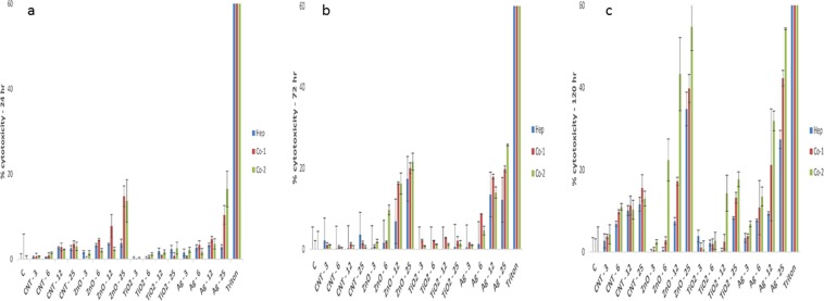
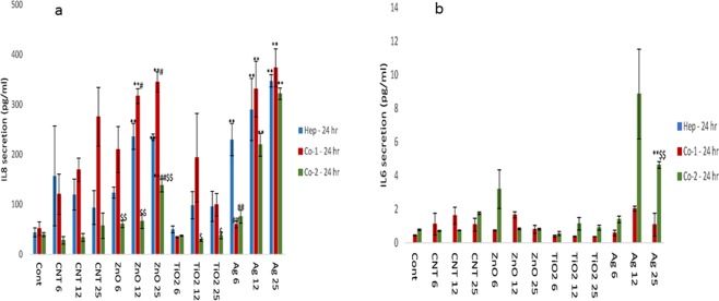
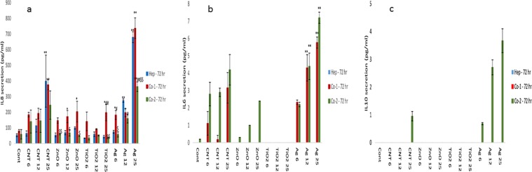
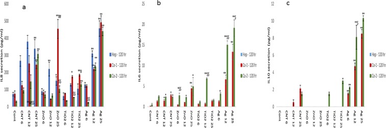
The potential for nanomaterial (NM) translocation to secondary organs is a realistic prospect, with the liver one of the most important target organs. Traditional in vitro or ex vivo hepatic toxicology models are often limiting and/or troublesome (i.e. short life-span reduced metabolic activity, lacking important cell populations, high inter-individual variability, etc.). Building on previous work, this study utilises a 3D human liver microtissue (MT) model (MT composed of mono-culture of hepatocytes or two different co-culture MT systems with non-parenchymal cell (NPC) fraction sourced from different donors) to investigate the importance of inter-donor variability of the non-parenchymal cell population in the overall governance of toxicological response following exposure to a panel of NMs. To the best of our knowledge, this is the first study of its kind to investigate inter-donor variability in hepatic NPC population. The data showed that the Kupffer cells were crucial in dictating the overall hepatic toxicity following exposure to the materials. Furthermore, a statistically significant difference was noted between the two co-culture MT models. However, the trend for particle-induced biological responses was similar between the co-cultures (cytotoxicity, cytokine production and caspase activity). Therefore, despite the recognition of some discrepancies in the absolute values between the co-culture models, the fact that the trends and patterns of biological responses were comparable between the multi-cellular models we propose the 3D liver MT to be a valuable tool in particle toxicology.


Conflict of interest statement

The authors declare no competing interests.

Figures

Figure 1
Figure 1
Cytotoxicity in human liver MT (mono-culture of hepatocytes only, co-culture of hepatocytes with NPC fraction from donor 1 and co-culture of hepatocytes with NPC from donor 2) following single or repeated exposures to a panel of engineered NMs for up 120 hr. The cytotoxicity as measured by AK release via ToxiLight™ cytotoxicity assay (a) 24 hr (b) 72 hr and (c) 120 hr (NM treatments µg/ml). Triton-X was utilised as a positive control in the cytotoxicity assay (24 hr exposure). The values represent mean ± SEM (n = 3) with significance indicated by *p < 0.05 and **p < 0.005 of NM-induced effects compared to negative control, #p < 0.05 and ##p < 0.005 signifying differences between the mono-culture and the co-culture and $p < 0.05 and $$p < 0.005 highlighting variation between co-culture 1 and co-culture 2.
Figure 2
Figure 2
IL8, IL6 secretion from NM exposed human liver MT. The tissues were exposed to cell medium (cont) or NMs for a period of 24 hr (a) IL8 (b) IL6. The values represent mean ± SEM (n = 3) with significance indicated by *p < 0.05 and **p < 0.005 of NM-induced effects compared to negative control, #p < 0.05 and ##p < 0.005 signifying statistical differences between the mono-culture and the co-culture and $p < 0.05 and $$p < 0.005 highlighting variation between co-culture 1 and co-culture 2.
Figure 3
Figure 3
IL8, IL6 and IL10 secretion from NM exposed human liver MT. The tissues were exposed to cell medium (cont) or NMs for a period of 72 hr (a) IL8 (b) IL6 and (c) IL10. The values represent mean ± SEM (n = 3) with significance indicated by *p < 0.05 and **p < 0.005 of NM-induced effects compared to negative control, #p < 0.05 and ##p < 0.005 signifying statistical differences between the mono-culture and the co-culture and $p < 0.05 and $$p < 0.005 highlighting variation between co-culture 1 and co-culture 2.
Figure 4
Figure 4
IL8, IL6 and IL10 secretion from NM exposed human liver MT. The tissues were exposed to cell medium (cont) or NMs for a period of 120 hr (a) IL8 (b) IL6 and (c) IL10. The values represent mean ± SEM (n = 3) with significance indicated by *p < 0.05 and **p < 0.005 of NM-induced effects compared to negative control, #p < 0.05 and ##p < 0.005 signifying statistical differences between the mono-culture and the co-culture and $p < 0.05 and $$p < 0.005 highlighting variation between co-culture 1 and co-culture 2.
Figure 5
Figure 5
Caspase activity in 3D human liver MT. The effect of NM exposure (or positive control Camptothecin (Campto)) on caspase 3/7 activity in the human liver MT (a) 24 hr (b) 120 hr. The values represent mean ± SEM (n = 3) with significance indicated by *p < 0.05 and **p < 0.005 of NM-induced effects compared to negative control, #p < 0.05 and ##p < 0.005 signifying statistical differences between the mono-culture and the co-culture and $p < 0.05 and $$p < 0.005 highlighting variation between co-culture 1 and co-culture 2.

References

    1. Kermanizadeh A, Powell L, Stone V, Møller P. Nano delivery systems and stabilized solid drug nanoparticles for orally administered medicine - current landscape. International Journal of Nanomedicine. 2018;13:7575–7605. doi: 10.2147/IJN.S177418. - DOI - PMC - PubMed
    1. Vance ME, et al. Nanotechnology in the real world: Redeveloping the nanomaterial consumer products inventory. Beilstein Journal of Nanotechnology. 2015;6:1769–1780. doi: 10.3762/bjnano.6.181. - DOI - PMC - PubMed
    1. Johnston H, Brown D, Kermanizadeh A, Gubbins E, Stone V. Investigating the relationship between nanomaterial hazard and physicochemical properties: Informing the exploitation of nanomaterials with therapeutic and diagnosis applications. Journal of Controlled Release. 2012;164:307–313. doi: 10.1016/j.jconrel.2012.08.018. - DOI - PubMed
    1. Kermanizadeh A, Balharry D, Wallin H, Loft S, Møller P. Nanomaterial translocation - the biokinetics, tissue accumulation, toxicity and fate of materials in secondary organs - a review. Critical Reviews of Toxicology. 2015;45:837–872. doi: 10.3109/10408444.2015.1058747. - DOI - PubMed
    1. Tetley TD. Health effects of nanomaterials. Biochemical Society Transactions. 2007;35:527–531. doi: 10.1042/BST0350527. - DOI - PubMed

Publication types