. 2019 May 13;9(1):7288.
doi: 10.1038/s41598-019-43783-6.

5-Oxo-1-[(2,3,6,7-tetramethoxy-9-phenanthrenyl)methyl]-L-proline Inhibits Hepatitis C Virus Entry

Affiliations

5-Oxo-1-[(2,3,6,7-tetramethoxy-9-phenanthrenyl)methyl]-L-proline Inhibits Hepatitis C Virus Entry

Lap P Nguyen et al. Sci Rep. .

Abstract

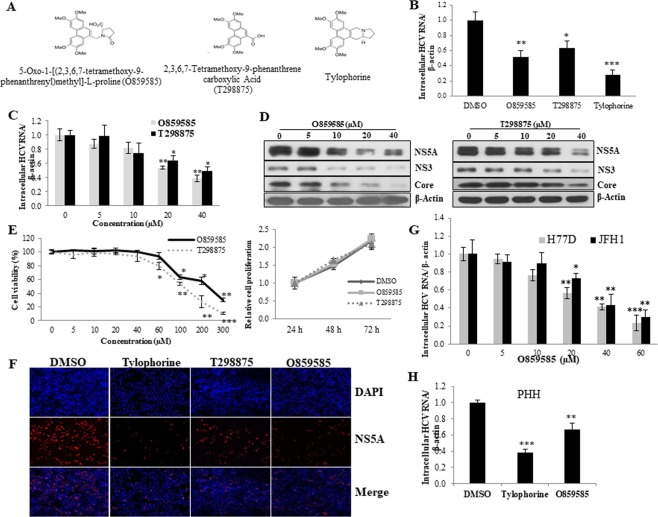
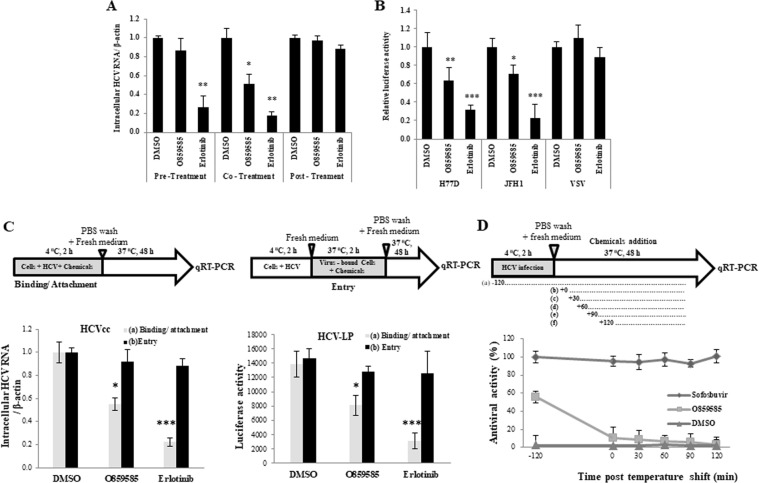
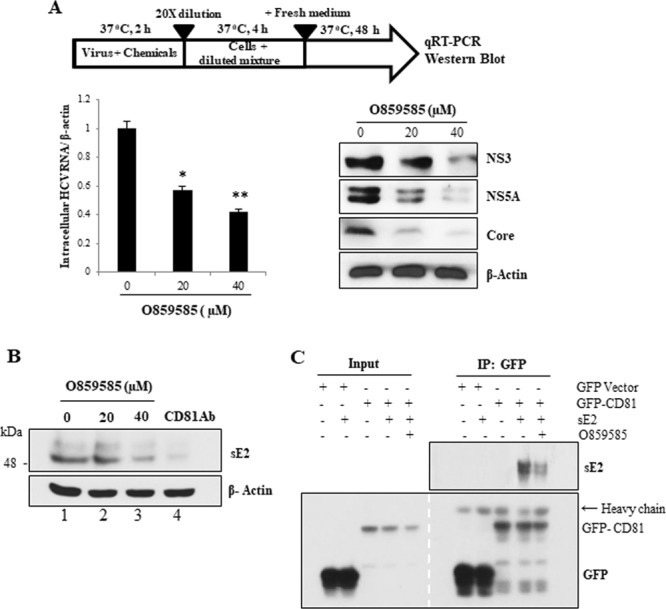
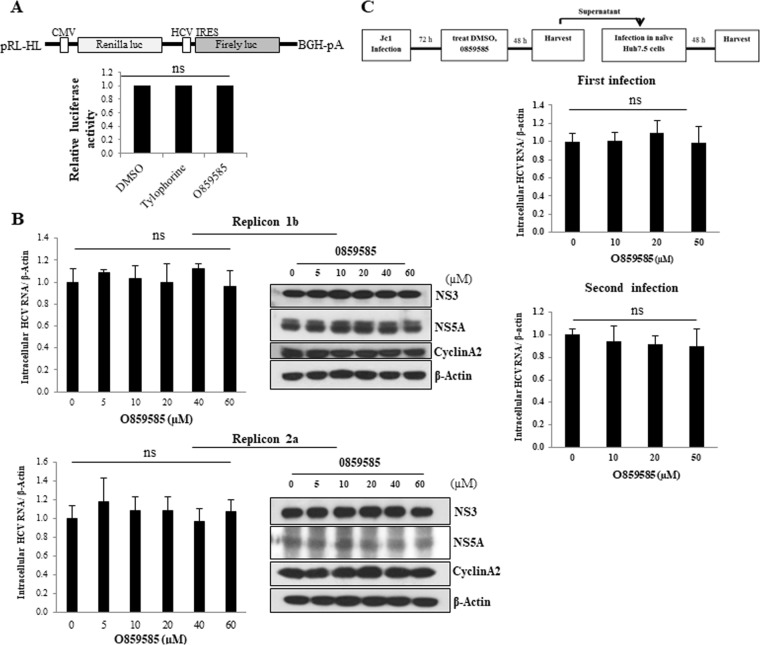
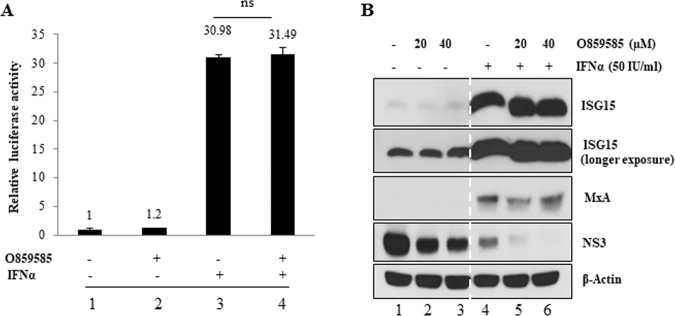
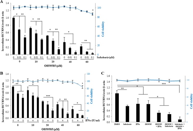
Hepatitis C virus (HCV) is the major causative agent of chronic liver diseases, including liver cirrhosis and hepatocellular carcinoma. The recent development of highly effective direct-acting antivirals (DAAs) has revolutionized the treatment of HCV patients. However, these DAAs are exorbitantly expensive for the majority of HCV patients worldwide. Moreover, these drugs still show genotypic difference in cure rate and have some resistant-associated variants. Tylophorine, a natural compound derived from Tylophora indica plants, is known to have anti-inflammatory and anti-cancerous growth activities. In the present study, we showed that two tylophorine intermediates, 5-Oxo-1-[(2,3,6,7-tetramethoxy-9-phenanthrenyl) methyl]-L-proline (O859585) and 2,3,6,7-tetramethoxy-9-phenanthrenecarboxylic acid (T298875), displayed anti-HCV activity with an EC50 of 38.25 µM for T298875 and 29.11~35.3 µM for O859585 in various HCV genotypes. We demonstrated that O859585 efficiently blocked HCV attachment by neutralizing free viral particles without affecting other stages of the HCV life cycle and interferon stimulation. O859585 interrupted binding between HCV E2 and CD81. Of note, co-treatment of O859585 with either interferon alpha (IFNα) or sofosbuvir exerted either an additive or synergistic antiviral activity in HCV-infected cells with no measurable effect on cell viability. Most importantly, O859585 in combination with IFNα and sofosbuvir exhibited synergistic effects on anti-HCV activity in primary human hepatocytes. Collectively, these data suggest that O859585 may be a novel antiviral agent for HCV therapy.


Conflict of interest statement

The authors declare no competing interests.

Figures

Figure 1
Figure 1
O859585 and T298875, tylophorine intermediates, inhibit HCV propagation. (A) Chemical structures of O859585, T298875, and tylophorine. (B) Huh7.5 cells were pretreated with either 20 µM O859585, 20 µM T298875, or 0.075 µM tylophorine for 1 h, and then infected with Jc1 for 4 h in the presence of the indicated chemicals. Cells were further cultured with fresh media containing each chemical. At 48 h postinfection, intracellular HCV RNA levels were quantified by qRT-PCR. (C,D) Huh7.5 cells were pretreated with various concentrations of either O859585 or T298875 for 1 h and then infected with Jc1 for 4 h in the presence of each chemical. Cells were further cultured with fresh media containing various concentrations of each chemical. At 48 h postinfection, intracellular HCV RNA (C) and protein (D) levels were quantified by qRT-PCR and immunoblot analysis, respectively. (E) (Left panel) Huh7.5 cells were treated with various doses of either O859585 or T298875 for 48 h and then cell toxicity was determined by using WST assay. (Right panel) Huh7.5 cells were treated with either DMSO (vehicle) or 29.65 µM O859585, or 38.25 µM T298875 for 1 h and then infected with Jc1 for 4 h in the absence or presence of each chemical. At the indicated time intervals after Jc1 infection, cell proliferation was determined by WST assay. (F) Huh7.5 cells were seeded on glass coverslips and treated with either DMSO (vehicle) or 0.075 µM tylophorine, 20 µM T298875, 20 µM O859585, respectively for 1 h and then infected with Jc1 for 4 h in the absence or presence of each chemical. At 48 h postinfection, cells were fixed in 4% paraformaldehyde and further incubated with NS5A antibody. Cells were counterstained with 4′,6-diamidino-2-phenylindole (DAPI) to label nuclei (blue signal). (G) Huh7.5 cells were pretreated with various concentrations of O859585 for 1 h and then infected with HCV derived from either genotype 1a (H77D) or genotype 2a (JFH1) for 4 h. At 48 h postinfection, intracellular HCV RNA levels were determined by qRT-PCR. (H) Primary human hepatocytes were pretreated with either DMSO, 0.075 µM tylophorine, or 20 µM O859585 for 1 h and then infected with Jc1 for 4 h in the presence of the indicated chemicals. Cells were further cultured with fresh media containing each chemical. At 48 h postinfection, intracellular HCV RNA levels were quantified by qRT-PCR. Data represent averages from three independent experiments for panel (B,C,G,H). The asterisks indicate significant differences (*p < 0.05; **p < 0.01; ***p < 0.001).
Figure 2
Figure 2
O859585 interupts HCV entry at binding/attachment step. (A) Huh7.5 cells were either pretreated with the indicated chemicals, washed twice with PBS, and then infected with HCV (pre-treatment), or infected with HCV in the presence of the indicated chemicals (co-treatment), or infected with HCV and then treated with the indicated chemicals (post-treatment) as described in Materials and methods. At 48 h postinfection, intracellular HCV RNA levels were determined by qRT-PCR. 0.1% DMSO (vehicle), 20 µM O859585, and 5 µM erlotinib were used for each experiment. (B) Huh7.5 cells were pretreated with DMSO, 20 µM O859585 or 5 µM erlotinib for 1 h. Cells were then infected with either VSVpp or HCVpp derived from either genotype 1a (H77D) or 2a (JFH1) for 6 h. At 48 h postinfection, viral entry was determined by luciferase activity. (C) For binding/attachment assay, Huh7.5 cells were incubated with either HCVcc (left panel) or HCV-LP (right panel) at 4 °C for 2 h in the presence of the indicated chemicals. The cells were washed with PBS and then bound HCV were determined either by analyzing RNA levels (left panel) or luciferase activity (right panel). For entry assay, Huh7.5 cells were incubated with either HCVcc (left panel) or HCV-LP (right panel) at 4 °C for 2 h in the presence of the indicated chemicals. The cells were washed with PBS and then temperature was shifted to 37 °C for 4 h. The cells were trypsinized and washed twice with PBS to remove free virions. Internalized HCV virions were indirectly determined either by analyzing RNA levels (left panel) or luciferase activity (right panel). (D) Huh7.5 cells were incubated with Jc1 for 2 h at 4 °C and then temperature was shifted to 37 °C. Each of 0.1% DMSO, 20 µM O859585, and 0.1 µM sofosbuvir was added at the indicated time points. At 48 h postinfection, antiviral activities in each time points were determined by analyzing HCV RNA levels. Data represent means ± SD of three independent assays. p-values are indicated by asterisks (*p < 0.05; **p < 0.01; ***p < 0.001).
Figure 3
Figure 3
O859585 neutralizes HCV virions by disrupting E2 and CD81 binding. (A) HCV Jc1 virions were mingled with various concentrations of O859585 for 2 h at 37 °C. The mixture was diluted 20-fold to make ineffective concentration of O859585 and used to infect Huh7.5 cells. At 48 h postinfection, the viral RNA (left panel) and protein (right panel) levels were analyzed by qRT-PCR and immunoblot assay, respectively. Data are means of 3 independent experiments (*p < 0.05; **p < 0.01). (B) Huh7.5 cells were incubated for 4 h with sE2 protein derived from genotype 1a in the presence of various doses of O859585 or 1 µg/ml CD81 antibody. The cells were washed with PBS to remove unbound sE2 and then total cell lysates were immunoblotted with E2 antibody. (C) HEK293T cells were transfected with GFP-CD81. At 48 h after transfection, total cell lysates were incubated with sE2 in the absence or presence of 20 µM O859585. Total mixtures were immunoprecipitated with GFP and bound protein was immunoblotted with an anti-E2 antibody.
Figure 4
Figure 4
O859585 displays no effect on IRES-mediated HCV translation, replication, and virion production. (A) A schematic diagram of the pRL-HL plasmid is shown on top. Huh7.5 cells were treated with 0.1% DMSO, 0.075 µM tylophorine or 20 µM O859585. At 1 h after treatment, Huh7.5 cells were transfected with pRL-HL dual luciferase and a pCH110 β-galactosidase plasmid. At 48 h after transfection, relative luciferase activity was determined. (B) Huh7 cells harboring HCV subgenomic replicon derived from either 1b (upper) or 2a (lower) were treated with either 0.1% DMSO or increasing doses of O859585. At 48 h after treatment, viral RNA and proteins expression levels were analyzed by qRT-PCR and immunoblot assays, respectively. (C) Schematic diagram of the experimental set-up is shown on top. (Upper) Huh7.5 cells were infected with Jc1. At 72 h postinfection, cells were treated with various concentrations with O859585. At 48 h after treatment, intracellular HCV RNA level was analyzed by qRT-PCR. (Lower) Naïve Huh7.5 cells were infected with Jc1 containing culture supernatant harvested from the first infection. At 48 h postinfection, intracellular HCV RNA level was quantified by qRT-PCR. Data are means of 3 independent experiments.
Figure 5
Figure 5
O859585 exerts no IFNα stimulation. (A) Huh7.5 cells were transfected with a reporter plasmid carrying IFN sensitive responsive elements (ISRE) upstream of the luciferase gene and then either left untreated or treated with 20 μM O859585, 50 IU/mL IFNα, individually or co-treated with O859585 and IFNα for 24 h and then luciferase activity was determined. Experiments were performed three times. ns, non-significant. (B) Huh7.5 cells infected with Jc1 were treated with the indicated combinations of O859585 and IFNα. At 48 h after treatment, total cell lysates were immunoblotted with the indicated antibodies.
Figure 6
Figure 6
O859585 in combination with IFNα and sofosbuvir exhibits an additive anti-HCV activity. (A) Huh7.5 cells were infected with Jc1 in the presence of various concentrations of O859585 for 4 h. Culture medium was replaced with fresh medium containing the indicated concentrations of sofosbuvir. At 48 h postinfection, both intracellular HCV RNA level (left) and cell viability (right) were analyzed by qRT-PCR and WST assay, respectively. (B) Huh7.5 cells were infected with Jc1 in the presence of various concentrations of O859585 for 4 h. Culture medium was replaced with fresh medium containing the indicated concentrations of IFNα. At 48 h postinfection, both intracellular HCV RNA level (left) and cell viability (right) were analyzed by qRT-PCR and WST assay, respectively. (C) Primary human hepatocytes infected with Jc1 were treated with the indicated combinations of 20 µM O859585, IFNα, and sofosbuvir. At 48 h postinfection, both intracellular HCV RNA level (left) and cell viability (right) were analyzed by qRT-PCR and WST assay, respectively. Data represent means ± SD of three independent experiments. P-values are indicated by asterisks (*p < 0.05; **p < 0.01; ***p < 0.001).

References

    1. Chung CY, et al. (4R,6S)-2-Dihydromenisdaurilide is a butenolide that efficiently inhibits hepatitis C virus entry. Sci. Rep. 2016;6:29969. doi: 10.1038/srep29969. - DOI - PMC - PubMed
    1. Mohamed AA, et al. Hepatitis C virus: A global view. World J. Hepatol. 2015;7:2676–2680. doi: 10.4254/wjh.v7.i26.2676. - DOI - PMC - PubMed
    1. Joyce MA, Tyrrell DL. The cell biology of hepatitis C virus. Microbes Infect. 2010;12:263–271. doi: 10.1016/j.micinf.2009.12.012. - DOI - PubMed
    1. Suzuki T, Ishii K, Aizaki H, Wakita T. Hepatitis C viral life cycle. Adv. Drug Deliv. Rev. 2007;59:1200–1212. doi: 10.1016/j.addr.2007.04.014. - DOI - PubMed
    1. Tsoulfas G, et al. Hepatitis C and liver transplantation. Hippokratia. 2009;13:211–215. - PMC - PubMed

Publication types

MeSH terms