. 2019 Jun;22(6):992-999.
doi: 10.1038/s41593-019-0394-3. Epub 2019 May 13.

Compulsivity and impulsivity traits linked to attenuated developmental frontostriatal myelination trajectories

Affiliations

Compulsivity and impulsivity traits linked to attenuated developmental frontostriatal myelination trajectories

Gabriel Ziegler et al. Nat Neurosci. 2019 Jun.

Abstract

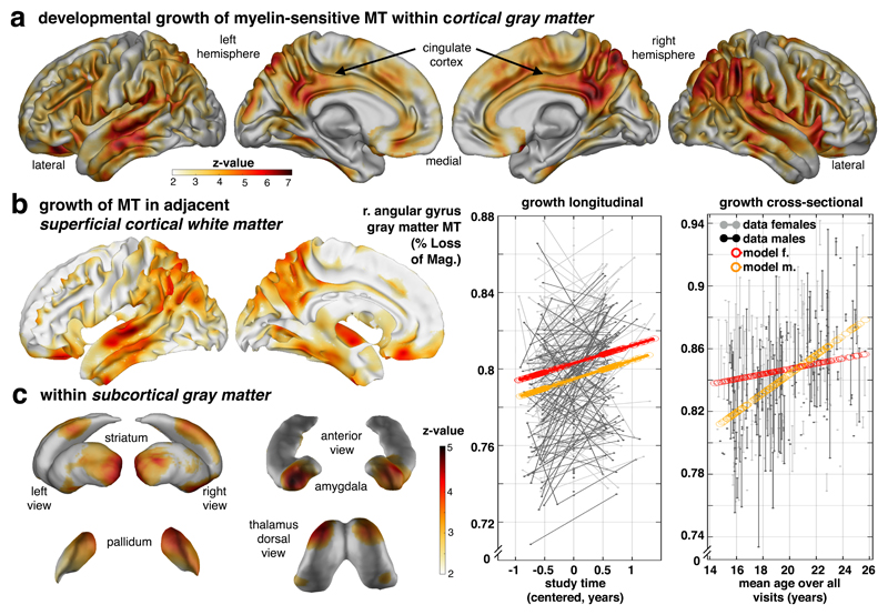
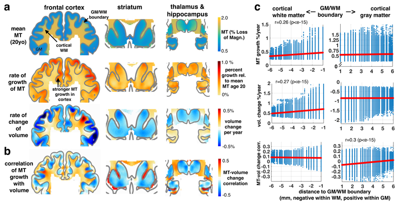
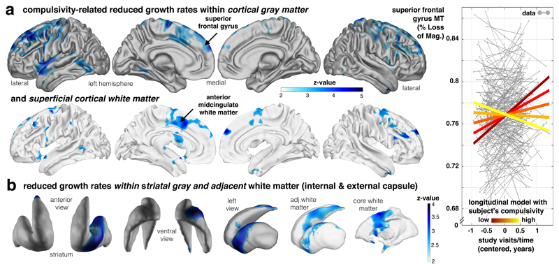
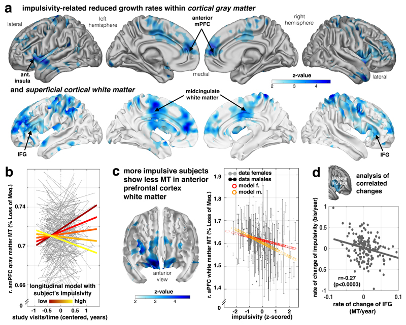
The transition from adolescence to adulthood is a period when ongoing brain development coincides with a substantially increased risk of psychiatric disorders. The developmental brain changes accounting for this emergent psychiatric symptomatology remain obscure. Capitalizing on a unique longitudinal dataset that includes in vivo myelin-sensitive magnetization transfer (MT) MRI scans, we show that this developmental period is characterized by brain-wide growth in MT trajectories within both gray matter and adjacent juxtacortical white matter. In this healthy population, the expression of common developmental traits, namely compulsivity and impulsivity, is tied to a reduced growth of these MT trajectories in frontostriatal regions. This reduction is most marked in dorsomedial and dorsolateral prefrontal regions for compulsivity and in lateral and medial prefrontal regions for impulsivity. These findings highlight that psychiatric traits of compulsivity and impulsivity are linked to regionally specific reductions in myelin-related growth in late adolescent brain development.


Conflict of interest statement

Competing Interest

E.T.B. is employed half-time by the University of Cambridge and half-time by GlaxoSmithKline and holds stock in GlaxoSmithKline. All other authors declare no competing financial interests.

Figures

Figure 1
Figure 1. Developmental growth of myelin-sensitive MT into early adulthood.
Transitioning into adulthood is characterised by marked increases in a myelin marker within cortical gray (a), white (b) and subcortical gray matter (c). Statistical maps of voxel-wise MT saturation show increase with time/visit (longitudinal) or age (cross-sectional; for specific effects of covariates, e.g. time/visit, age, sex, interactions etc., see supplementary information). (a) Gray matter MT increase (top row; statistical z-maps from one-sided Wald-test, p<.05 FDR corrected, sampling-based correction reported in Supplementary Table 1, cf Supplementary Fig. 2c, n=497/288 scans/subjects, 51.7% female, sample and test apply for panels a-c) is strongest in parietal, lateral temporal, posterior and middle cingulate, but is also present in prefrontal cortex. Longitudinal model in angular gyrus peak (mean across a 6mm sphere; coloured lines in left data plot; x-axis: relative time of scan) and adjusted data (uncoloured) shows an MT growth in both sexes, with a marked sex difference reflecting greater MT in females (see Supplementary Fig. 3c for region-specific sex differences). Corresponding cross-sectional model predictions in the same region show a similar increase with age (right data plot; y-axis: MT; x-axis: mean age over visits). (b) MT growth in adjacent cortical white matter is most pronounced in cingulate and parieto-temporal cortex with a coarse topographical correspondence to the gray matter MT effects. (c) Subcortical gray matter nuclei express MT age effects in striatum, pallidum, thalamus, amygdala and hippocampus (cf Fig. 2a-b). This growth is most pronounced in amygdala, ventral (max z-value voxel [z=4.81, p=.004 FDR], [MNI: 20 13 -11]) and posterior (z=4.47, p=0.004 FDR, [MNI: -31 -19 3]) striatum suggesting ongoing myelin-associated changes in both cortical and subcortical brain structures.
Figure 2
Figure 2. The relation between macrostructural and microstructural brain development.
(a) Coronal sections through prefrontal (left panels), striatal (middle) and thalamus/hippocampus (right; MNI: y=15, 12, -14) show more myelin-related MT in white than in gray matter with a clearly preserved white-gray matter boundary (top row, model intercept/mean, n=497/288 scans/subjects, 51.7% female, sample applies for panels a-b, all beta parameters shown for illustration of effect sizes, for statistical tests see Fig. 1 and Supplementary Fig. 4). Developmental change in MT (second row, averaged beta parameter of age and time/visit effects) illustrating the rate of change of our myelin marker in both tissue classes, with faster increase in gray matter areas. Developmental change in macrostructural brain volume (third row, averaged beta parameter of age and time/visit effects) shows a characteristic cortical shrinkage (blue colours) in gray, but an expansion in core and frontal white matter (red colours; cf Supplementary Fig. 4). Only hippocampal gray matter shows an opposite effect with continuing volume growth up to the verge of adulthood. (b) Association between microstructural myelin growth and macrostructural volume change. A positive association throughout whole-brain white matter supports the notion that myelination contributes to white matter expansion. In gray matter, a predominantly negative association in deep layers points to partial volume effects at the tissue boundary and positive associations in superficial layers (correlation was obtained from posterior covariance of beta parameters in sandwich estimator model simultaneously including longitudinal observations of both imaging modalities, unthresholded). (c) Association as a function of Euclidean distance to GM/WM boundary. Both tissue classes show consistent increase of MT (top row, Pearson’s correlation with distance and two-sided p-values from corresponding t-distribution based on n=336164/118502 voxels in cortical gray/white matter), but opposite macrostructural volume change (middle row). Association between micro- and macrostructural growth is positive in white matter, rather independent of distance to GM/WM. In gray matter, the mean association changes from negative in deep layers (i.e. myelin MT change associated with reduced gray matter volume) to more positive associations in superficial layers (i.e. MT associated with a tendency to more gray matter volume).
Figure 3
Figure 3. Compulsivity is related to altered fronto-striatal MT growth.
Longitudinal developmental change of our myelin marker is reduced in high compulsive subjects. (a) Aggregate compulsivity score is related to decreased MT increase in dorsolateral frontal gray matter (upper panel; statistical z-maps, p<.05, FDR and bootstrapping corrected; Supplementary Table 3, one-sided Wald-test, n=452/246 scans/subjects and test apply for panels a-b with available compulsivity, 50.4% female) and adjacent white matter, as well as cingulate cortex (lower panel; blue colours depicting negative time by compulsivity interactions). Subjects with higher compulsivity scores (light yellow) compared to low scoring subjects (dark red) express significantly less MT increase over visits (coloured lines in right panel indicate the interaction effect; y-axis: MT; x-axis: time of scan in years relative to each subject’s mean age over visits). (b) The above slowing in cortical myelin-related growth is mirrored by a decreased developmental growth in subcortical ventral striatum (left panel) and the adjacent white matter (right panel). These findings indicate young people with high compulsive traits express slower maturational myelin-related change in a fronto-striatal network comprising cingulate cortex and ventral striatum.
Figure 4
Figure 4. Decreased frontal growth in myelin-sensitive MT in impulsivity.
Myelin marker (MT) in frontal lobe is linked to impulsivity traits. (a) Impulsivity is associated with reduced growth of MT in lateral (inferior and middle frontal gyrus), medial prefrontal areas, motor/premotor and parietal areas in both gray (top panel) and adjacent white matter (bottom panel) depicting negative time/visit by impulsivity interactions (z maps, statistical z-maps, p<.05 FDR and bootstrapping corrected, Supplementary Table 4, one-sided Wald-test, n=497/288 subjects/scans, apply for panels a-c, 51.7% female). (b) Plot shows subjects with higher impulsivity (light yellow) compared to low scoring subjects (dark red) express significantly less MT growth over visits (coloured lines in right panel indicate the interaction effect; y-axis: MT; x-axis: time of scan in years relative to each subject’s mean age over visits). (c) More impulsive subjects show a local decrement in baseline myelin marker (peak middle frontal gyrus, p<0.05, FDR and bootstrapping corrected, Supplementary Table 5) in lateral and orbitofrontal areas (fixed for other covariates, e.g. time/visits, mean age of subject, sex). Right panel shows the plot of MT in this peak voxel over impulsivity (x-axis, z-scored) and with adjusted data (gray/black) and model predictions (red/orange, effects of interest: intercept, impulsivity, sex by impulsivity). (d) Bilateral IFG not only shows a reduced myelination process for higher impulsivity (as shown in a, b), but this reduced growth rate is more strongly expressed in subjects who manifest an accentuated impulsivity growth over study visits, such that subjects who manifest an even more restricted growth in myelin become more impulsive (Pearson’s correlation and p-value from corresponding t-distribution based on n=188 independent subjects with available follow-up scans).

References

    1. Gogtay N, et al. Dynamic mapping of human cortical development during childhood through early adulthood. Proc Natl Acad Sci U S A. 2004;101:8174–8179. - PMC - PubMed
    1. Sowell ER, Thompson PM, Holmes CJ, Jernigan TL, Toga AW. In vivo evidence for post-adolescent brain maturation in frontal and striatal regions. Nat Neurosci. 1999;2:859–861. - PubMed
    1. Paus T. Growth of white matter in the adolescent brain: myelin or axon? Brain Cogn. 2010;72:26–35. - PubMed
    1. Miller DJ, et al. Prolonged myelination in human neocortical evolution. Proc Natl Acad Sci U S A. 2012;109:16480–16485. - PMC - PubMed
    1. Perrin JS, et al. Growth of white matter in the adolescent brain: role of testosterone and androgen receptor. J Neurosci Off J Soc Neurosci. 2008;28:9519–9524. - PMC - PubMed

Publication types