. 2019 Sep;21(9):e13042.
doi: 10.1111/cmi.13042. Epub 2019 Jun 6.

The endoplasmic reticulum chaperone PfGRP170 is essential for asexual development and is linked to stress response in malaria parasites

Affiliations

The endoplasmic reticulum chaperone PfGRP170 is essential for asexual development and is linked to stress response in malaria parasites

Heather M Kudyba et al. Cell Microbiol. 2019 Sep.

Abstract

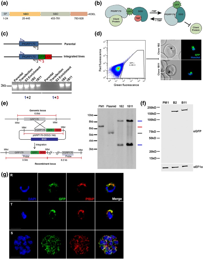
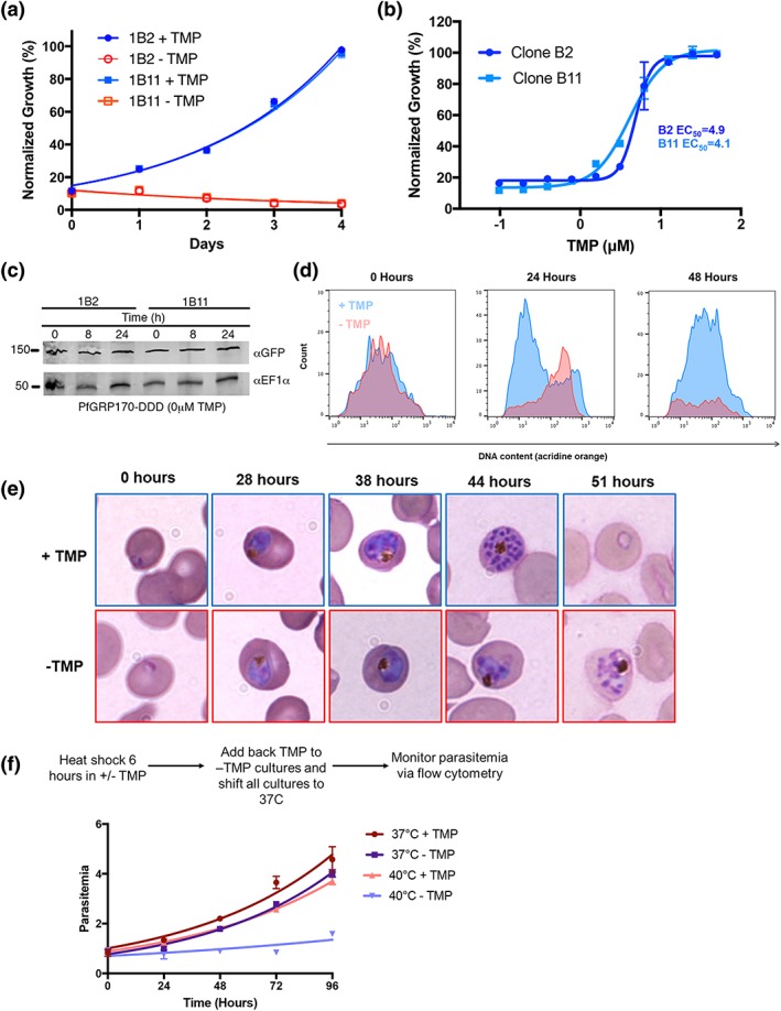
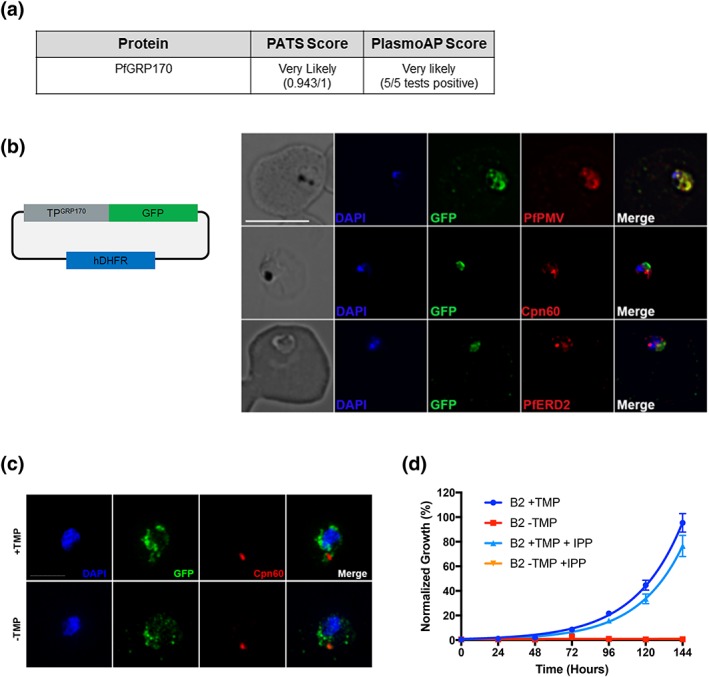
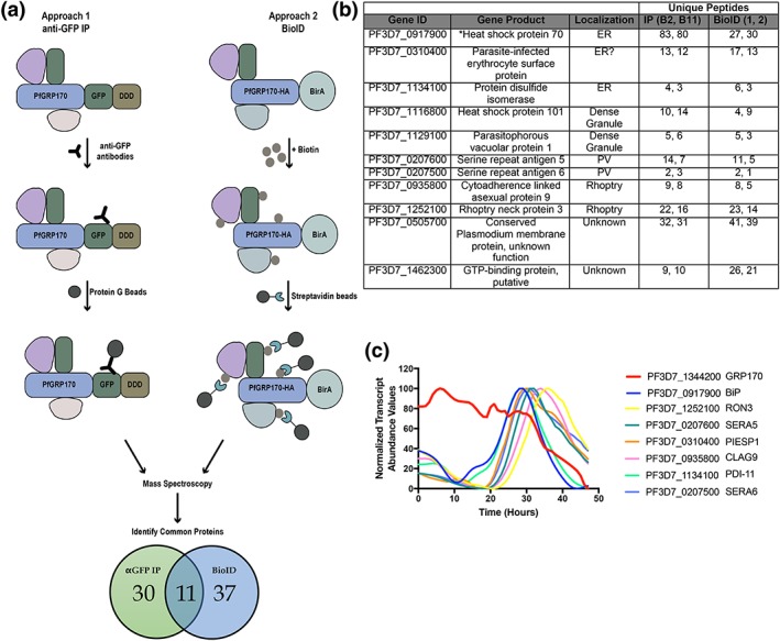
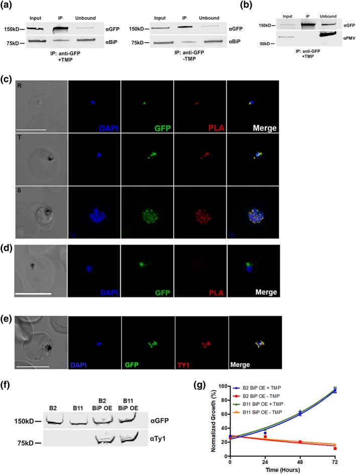
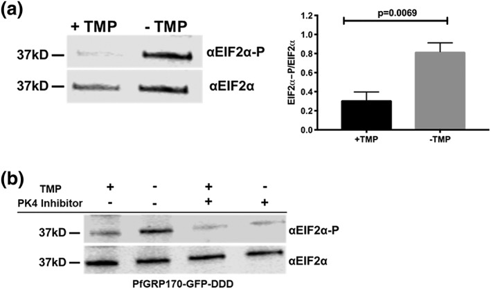
The vast majority of malaria mortality is attributed to one parasite species: Plasmodium falciparum. Asexual replication of the parasite within the red blood cell is responsible for the pathology of the disease. In Plasmodium, the endoplasmic reticulum (ER) is a central hub for protein folding and trafficking as well as stress response pathways. In this study, we tested the role of an uncharacterised ER protein, PfGRP170, in regulating these key functions by generating conditional mutants. Our data show that PfGRP170 localises to the ER and is essential for asexual growth, specifically required for proper development of schizonts. PfGRP170 is essential for surviving heat shock, suggesting a critical role in cellular stress response. The data demonstrate that PfGRP170 interacts with the Plasmodium orthologue of the ER chaperone, BiP. Finally, we found that loss of PfGRP170 function leads to the activation of the Plasmodium eIF2α kinase, PK4, suggesting a specific role for this protein in this parasite stress response pathway.

Keywords: Plasmodium falciparum; endoplasmic reticulum; malaria; parasitology.


Figures

Figure 1
Figure 1
Generation of PfGRP170‐GFP‐DD Parasites. (a) Schematic detailing the putative domain boundaries of PfGRP170 (PF3D7_1344200) based on the yeast homologue, Lhs1: signal peptide (SP), nucleotide binding domain (NBD), substrate‐binding domain (SBD), extended C‐terminus region (783–928), and an ER retention signal (KDEL). (b) Schematic diagram demonstrating the conditional inhibition of PfGRP170. Conditional inhibition of PfGRP170 is achieved by the removal of trimethoprim (TMP), which results in the unfolding of the destabilisation (DDD). The chaperone recognises and binds the unfolded DDD and is inhibited from interacting with client proteins. (c) (Top) Schematic diagram of the PfGRP170 locus in the parental line (PM1KO) and the modified locus where PfGRP170 is endogenously tagged with GFP and DDD. Primers used for integration test and control PCR are indicated by arrows. The relative positions of Primer 1 (blue) and Primer 2 (grey) on the PfGRP170 locus are shown. These two primers will amplify PfGRP170 in parental and transfected parasites. Primer 3 (Red) recognises the GFP sequence. Primers 1 and 3 were used to screen for proper integration into the PfGRP170 locus. (Bottom) PCR integration test and control PCRs on gDNA isolated from the PM1KO (parental), the original transfection of the pPfGRP170‐GFP‐DDD plasmid after three rounds of blasticidin (BSD) drug selection (transfection), the PfGRP170‐GFP‐DDD transfected parasite lines after two rounds of enrichment for GFP positive cells (S3 enrichment), and PfGRP170‐GFP‐DDD clones 1B2 and 1B11 after MoFlo XDP flow sorting. The first five lanes are control PCRs using primers to amplify the PfGRP170 locus. The last five lanes are integration PCRs that only amplify if the GFP‐DDD has been integrated into the genome. (d) (Left) MoFlo XDP flow data demonstrating the percentage of GFP positive parasites in transfected PfGRP170‐GFP‐DDD parasites following three rounds of drug selection with blasticidin (BSD) and two rounds of enrichment with an S3 cell sorter. Using the MoFlo, single GFP positive cells were cloned into a 96‐well plate. Two clones, 1B2 and 1B11, were isolated using this method. (Right) 1B2 and 1B11 parasites were observed using live fluorescence microscopy. (e) Southern blot analysis of PfGRP170‐GFP‐DDD clones 1B2 and 1B11, PM1KO (parental control), and the PfGRP170‐GDB plasmid is shown. Mfe1 restriction sites, the probe used to detect the DNA fragments, and the expected sizes are denoted in the schematic (left). Expected sizes for PfGRP170‐GFP‐DDD clones (blue), parental DNA (red), and plasmid (grey) were observed (right). Parental and plasmid bands were absent from the PfGRP170‐GFP‐DDD clonal cell lines. (f) Western blot analysis of protein lysates from PM1KO (parental) and PfGRP170‐GFP‐DDD clonal cell lines 1B2 and 1B11 is shown. Lysates were probed with anti‐GFP to visualise PfGRP170 and anti‐PfEF1α as a loading control. (g) Asynchronous PfGRP170‐GFP‐DDD parasites were paraformaldehyde fixed and stained with anti‐GFP, anti‐PfGRP78 (BiP), and DAPI to visualise the nucleus. Images were taken as a Z‐stack using super resolution microscopy and SIM processing was performed on the Z‐stacks. Images are displayed as a maximum intensity projection. The scale bar is 2 μm
Figure 2
Figure 2
PfGRP170 is essential and required for surviving a heat shock. (a) Growth of asynchronous PfGRP170‐GFP‐DDD clonal cell lines 1B2 and 1B11, in the presence or absence of 20 μM TMP, was observed using flow cytometry over 4 days. One hundred percent growth is defined as the highest parasitemia in samples with TMP, on the final day of the experiment. Data were fit to an exponential growth curve equation. Each data point is representative of the mean of three replicates ± SEM. (b) Asynchronous PfGRP170‐GFP‐DDD clonal cell lines 1B2 and 1B11 were grown in a range of TMP concentrations for 48 hr. After 48 hr, parasitemia was observed using flow cytometry. One hundred percent growth is defined as the highest parasitemia in the presence of TMP on the final day of the experiment. Data were fit to a dose–response equation. Each data point is representative of the mean of three replicates ± SEM. (c) Western blot analysis of PfGRP170‐GFP‐DDD lysates at 0, 8, and 24 hr following the removal of TMP is shown. Lysates were probed with anti‐GFP to visualise PfGRP170 and anti‐PfEF1α as a loading control. (d) Flow cytometric analysis of asynchronous PfGRP170‐GFP‐DDD parasites, incubated with (blue) and without TMP (red), and stained with acridine orange. Data at 0, 24, and 48 hr after the removal of TMP are shown. (e) TMP was removed from tightly synchronised PfGRP170‐GFP‐DDD ring stage parasites and their growth and development through the life cycle was monitored by Hema 3 stained thin blood smears. Representative images are shown from the parasite culture at the designated times. (f) PfGRP170‐GFP‐DDD clones 1B2 and 1B11 were incubated with and without TMP for 6 hr at either 37°C or 40°C. Following the incubation, TMP was added back to all cultures and parasites were shifted back to 37°C. Parasitemia was then observed over 96 hr via flow cytometry. Data were fit to an exponential growth curve equation. Each data point shows the mean of three replicates ± SEM
Figure 3
Figure 3
Putative PfGRP170 apicoplast transit peptide localises to the ER and conditional inhibition of PfGRP170 does not affect trafficking of apicoplast proteins. (a) Analysis of PfGRP170's protein sequence using two apicoplast transit peptide prediction programmes: prediction of apicoplast‐targeted sequences (PATS) and PlasmoAP. (b) PfGRP170's putative apicoplast transit peptide was fused to GFP and transfected into 3D7 parasites. Parasites were fixed with acetone and stained with DAPI, anti‐GFP (to label the PfGRP170 putative transit peptide) and either anti‐PfPMV (ER), anti‐PfERD2 (Golgi), or anti‐Cpn60 (apicoplast) to determine subcellular localisation. The images were taken with Delta Vision II, deconvolved, and are displayed as a maximum intensity projection. The scale bar is 5 μm. (c) Synchronised ring stage PfGRP170 parasites were incubated for 24 hr with and without TMP. Following the incubation, the parasites were fixed with paraformaldehyde and stained with DAPI, anti‐GFP (PfGRP170), and anti‐Cpn60 (apicoplast). Images were taken as a Z‐stack using super resolution microscopy and SIM processing was performed on the Z‐stacks. Images are displayed as a maximum intensity projection. The scale bar is 2 μm. (d) Asynchronous PfGRP170‐GFP‐DDD parasites were incubated with and without TMP and in the presence or absence of 200 μM IPP. Parasitemia was monitored using flow cytometry for 144 hr. One hundred percent growth is defined as the highest parasitemia in the presence of TMP, on the final day of the experiment. Data were fit to an exponential growth curve equation. Each data point is representative of the mean of three replicates ± SEM
Figure 4
Figure 4
PfGRP170 interacting partners. (a) Schematic diagram illustrating the two independent methods used to identify potential interacting partners of PfGRP170: anti‐GFP IP using lysates from PfGRP170‐GFP‐DDD parasites and streptavidin IP of PfGRP170‐BirA parasites incubated with biotin for 24 hr followed by mass spectroscopy. The proteins identified from each IP were filtered to include only proteins that had a signal peptide and/or transmembrane domain using PlasmoDB. Proteins found in the respective control IP's (excluding PfBiP) were also removed from further analysis (data in Table S1). (b) The 11 proteins identified in both independent mass spectroscopy approaches (see Figure 4a; Table S1). The PlasmoDB gene ID, gene product, putative subcellular localisation, and number of unique peptides identified for each protein in each independent experiment are listed. (c) The relative transcript abundance of interacting proteins, with peak expression around the time the PfGRP170‐GFP‐DDD parasites die (36–44 hr), are plotted using genome‐wide real‐time transcript data (Painter et al., 2018)
Figure 5
Figure 5
PfGRP170 Interacts with BiP. (a) Synchronised ring stage PfGRP170‐GFP‐DDD parasites were incubated with and without TMP for 24 hr. Following this incubation, an anti‐GFP IP was performed, and input, IP, and unbound fractions were analysed using a western blot. The blot was probed using anti‐GFP and anti‐BiP. (b) Western blot analysis of an anti‐GFP IP performed on asynchronous PfGRP170‐GFP‐DDD parasites. Input, IP, and unbound fractions are shown. The blot was probed using anti‐GFP and anti‐PfPMV. (c) In vivo interaction of PfGRP170 and BiP. PfGRP170‐GFP‐DDD parasites were paraformaldehyde fixed and stained with anti‐GFP and anti‐BiP. A proximity ligation assay (PLA) was then performed. The scale bar is 5 μm. A negative control using anti‐GFP and anti‐PfPMV is shown in (d). (e) Asynchronous PfGRP170‐GFP‐DDD parasites overexpressing PfBiP‐Ty1 were paraformaldehyde fixed and stained with anti‐GFP (PfGRP170), anti‐Ty1 (PfBiP‐Ty1‐KDEL), and DAPI to visualise the nucleus. The images were taken with Delta Vision II, deconvolved, and are displayed as a maximum intensity projection. The scale bar is 5 μm. (f) Western blot analysis of protein lysates from parental 1B2 and 1B11 parasites as well as 1B2 and 1B11 parasites overexpressing the PfBiP‐Ty1fusion protein. Lysates were probed with anti‐GFP to visualise PfGRP170 and anti‐Ty1 to visualise PfBiP‐Ty1‐KDEL. (g) Parasitemia of asynchronous PfGRP170‐GFP‐DDD parasites expressing PfBiP‐Ty1‐KDEL, in the presence or absence of 20 μM TMP, was observed using flow cytometry over 3 days. One hundred percent growth is defined as the highest parasitemia on the final day of the experiment. Data were fit to an exponential growth curve equation. Each data point is representative of the mean of three replicates ± SEM
Figure 6
Figure 6
Loss of PfGRP170 function activates the PK4 stress pathway. (a) (Left) Synchronised ring stage PfGRP170‐GFP‐DDD parasites were incubated with and without TMP for 24 hr. Protein was isolated from these samples and analysed via western blot, probing for anti‐eIF2α and anti‐Phospho‐eIF2α. (Right) The ratio of phosphorylated EIF2α over total EIF2α for PfGRP170‐GFP‐DDD parasites incubated with and without TMP is shown. Western blot band intensities were calculated using ImageJ software (NIH), and the significance was calculated using an unpaired t test. Data are representative of four biological replicates ± SEM. (b) Synchronised ring stage PfGRP170‐GFP‐DDD parasites were incubated with and without TMP and in the presence and absence of 2 μM PK4 inhibitor GSK2606414 for 24 hr. Protein was isolated from these samples and analysed via western blot by probing for anti‐eIF2a and anti‐Phospho‐eIF2α

References

    1. Amaratunga, C. , Witkowski, B. , Khim, N. , Menard, D. , & Fairhurst, R. M. (2014). Artemisinin resistance in Plasmodium falciparum . The Lancet Infectious Disease, 14, 449–450. 10.1016/S1473-3099(14)70777-7 - DOI - PMC - PubMed
    1. Andreasson, C. , Rampelt, H. , Fiaux, J. , Druffel‐Augustin, S. , & Bukau, B. (2010). The endoplasmic reticulum Grp170 acts as a nucleotide exchange factor of Hsp70 via a mechanism similar to that of the cytosolic Hsp110. The Journal of Biological Chemistry, 285, 12445–12453. 10.1074/jbc.M109.096735 - DOI - PMC - PubMed
    1. Araki, K. , & Nagata, K. (2011). Protein folding and quality control in the ER. Cold Spring Harbor Perspectives in Biology, 3, a007526 10.1101/cshperspect.a007526 - DOI - PMC - PubMed
    1. Babbitt, S. E. , Altenhofen, L. , Cobbold, S. A. , Istvan, E. S. , Fennell, C. , Doerig, C. , … Goldberg, D. E. (2012). Plasmodium falciparum responds to amino acid starvation by entering into a hibernatory state. Proceedings of the National Academy of Sciences of the United States of America, 109, E3278–E3287. 10.1073/pnas.1209823109 - DOI - PMC - PubMed
    1. Balu, B. , Shoue, D. A. , Fraser, M. J. Jr. , & Adams, J. H. (2005). High‐efficiency transformation of Plasmodium falciparum by the lepidopteran transposable element piggyBac. Proceedings of the National Academy of Sciences of the United States of America, 102, 16391–16396. 10.1073/pnas.0504679102 - DOI - PMC - PubMed

Publication types

MeSH terms