Review
. 2019 May 31;9(5):190036.
doi: 10.1098/rsob.190036.

Tissue tropism in parasitic diseases

Affiliations
Review

Tissue tropism in parasitic diseases

Sara Silva Pereira et al. Open Biol. .

Erratum in

  • Correction to 'Tissue tropism in parasitic diseases'.
    Silva Pereira S, Trindade S, De Niz M, Figueiredo LM. Silva Pereira S, et al. Open Biol. 2019 Jun 28;9(6):190124. doi: 10.1098/rsob.190124. Epub 2019 Jun 26. Open Biol. 2019. PMID: 31238821 Free PMC article. No abstract available.

Abstract

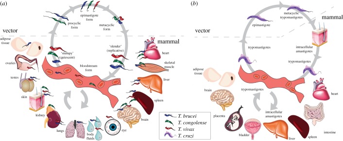
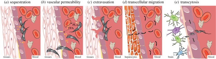
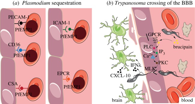
Parasitic diseases, such as sleeping sickness, Chagas disease and malaria, remain a major cause of morbidity and mortality worldwide, but particularly in tropical, developing countries. Controlling these diseases requires a better understanding of host-parasite interactions, including a deep appreciation of parasite distribution in the host. The preferred accumulation of parasites in some tissues of the host has been known for many years, but recent technical advances have allowed a more systematic analysis and quantifications of such tissue tropisms. The functional consequences of tissue tropism remain poorly studied, although it has been associated with important aspects of disease, including transmission enhancement, treatment failure, relapse and clinical outcome. Here, we discuss current knowledge of tissue tropism in Trypanosoma infections in mammals, describe potential mechanisms of tissue entry, comparatively discuss relevant findings from other parasitology fields where tissue tropism has been extensively investigated, and reflect on new questions raised by recent discoveries and their potential impact on clinical treatment and disease control strategies.

Keywords: nagana; parasites; sleeping sickness; tissue tropism; trypanosomes.


Conflict of interest statement

We declare we have no competing interests.

Figures

Figure 1.
Figure 1.
Life cycle of trypanosomes. (a) African trypanosomes (T. brucei, T. congolense and T. vivax): a tsetse takes a bloodmeal on an infected mammal and becomes a vector of African trypanosomiasis. Procyclic forms establish in the midgut by clonal expansion. The parasites travel to the proventriculus, salivary glands and/or proboscis, where they become epimastigotes and then infective metacyclics. In the following bloodmeal, the fly injects some of these parasites into the mammalian host, through its saliva. Parasites in the tissues (dermis, hypodermis) enter the bloodstream as metacyclic trypomastigotes and differentiate to bloodstream forms. Tissues affected by each parasite species are also depicted in the figure. T. vivax bloodstream forms can also be mechanically transmitted by non-tsetse vectors to new mammalian hosts, without biological differentiation. (b) Trypanosoma cruzi: a triatomine bug feeds on an infected mammalian host and becomes a vector of Chagas disease. Trypomastigotes establish in the midgut, where they differentiate into epimastigotes and multiply. Epimastigotes travel to the hindgut and differentiate into infective metacyclic trypomastigotes. In the following bloodmeal, the triatomine releases the infective metacyclic trypomastigotes in its faeces in the skin near the bite site. Trypomastigotes enter the mammalian host via mucosal membranes and invade cells, where they differentiate into intracellular amastigotes. These intracellular forms continue to multiply until they differentiate back into trypomastigotes, which burst out of the cell and are released into the bloodstream, reaching a variety of tissues. This figure was modified from Servier Medical Art, licensed under a Creative Commons Attribution 3.0 Generic License. https://smart.servier.com.
Figure 2.
Figure 2.
Potential mechanisms of tissue tropism in African trypanosome and Plasmodium spp. (a) Sequestration. Trypanosomes can adhere to the endothelial cells through membrane receptors, potentially sequestering to particular tissues. Sequestration is also done by RBCs infected with Plasmodium spp. (b) Vascular permeability. Trypanosomes secrete molecules, including phospholipase A (PLA), that cause lysis of the RBCs, resulting in the release of free fatty acids (FFAs) and other cell contents to the bloodstream. These molecules increase vascular permeability, which may facilitate migration of parasites through the vascular endothelium into the underlying tissues. (c) Extravasation. Attachment of trypanosomes to RBCs and/or endothelial cells can cause cell damage, promoting endothelial tissue rupture and necrosis, followed by extravasation of blood cells and parasites. (d) Transcellular migration. Plasmodium sporozoites can invade tissues by crossing the endothelial cell layer, in a process called transcellular migration. (e) Transcytosis. Trypanosome invasion of the cerebral parenchyma may occur by transcytosis, in a process where endothelial cells uptake parasites and, inside a vacuole, they are transported and released in the abluminal side of the vessel, into the brain parenchyma. This figure was modified from Servier Medical Art, licensed under a Creative Commons Attribution 3.0 Generic License. https://smart.servier.com.
Figure 3.
Figure 3.
Molecular mechanisms of tissue tropism. (a) Plasmodium sequestration. P. falciparum erythrocyte membrane protein 1 (PfEMP1) is the main mediator of parasite sequestration. Despite being always the same surface protein, the different expressed domains determine affinity to different tissues. PfEMP1 domains include DBL-5, CIDR1α, VAR2CSA, DC4 and DC8/13, which bind to endothelial cell receptors PECAM-1, CD36, CSA, ICAM-1 and EPCR, respectively. (b) Trypanosoma crossing of the BBB. Brain invasion can happen through opening of the tight junctions of the endothelial cells at the BBB. T. brucei, in particular, secretes brucipain, which acts on G-protein-coupled receptors (GPCRs) that activate phospholipase C (PLC). PLC activation results in increased inositol triphosphate (IP3) levels that cause an increase in cytosolic calcium (Ca2+). Increased calcium levels activate phosphokinase C (PKC), which acts on myosin light chain kinases to open the tight junctions, thus allowing parasite crossing. The release of interferon γ (IFNγ) by the parasite is also thought to induce astrocytes to release chemokine ligand 10 (CXCL-10), which may facilitate parasite movement to the brain parenchyma. This figure was modified from Servier Medical Art, licensed under a Creative Commons Attribution 3.0 Generic License. https://smart.servier.com/.

References

    1. Liscum E, Askinosie SK, Leuchtman DL, Morrow J, Willenburg KT, Coats DR. 2014. Phototropism: growing towards an understanding of plant movement. Plant Cell 26, 38–55. (10.1105/tpc.113.119727) - DOI - PMC - PubMed
    1. Prudêncio M, Rodriguez A, Mota MM. 2006. The silent path to thousands of merozoites: the Plasmodium liver stage. Nat. Rev. Microbiol. 4, 849–856. (10.1038/nrmicro1529) - DOI - PubMed
    1. Lopes AH. 2010. Trypanosomatids: odd organisms, devastating diseases. Open Parasitol. J. 4, 30–59. (10.2174/1874421401004010030) - DOI
    1. Stanaway JD, Roth G. 2015. The burden of Chagas disease. Glob. Heart 10, 139–144. (10.1016/j.gheart.2015.06.001) - DOI - PubMed
    1. World Health Organization. 2018. Trypanosomiasis, human African (sleeping sickness). Fact Sheet. See https://www.who.int/news-room/fact-sheets/detail/trypanosomiasis-human-a...) (accessed on 19 March 2019).

Publication types

LinkOut - more resources