. 2019 May 14;12(1):225.
doi: 10.1186/s13071-019-3463-2.

Shared species of crocodilian trypanosomes carried by tabanid flies in Africa and South America, including the description of a new species from caimans, Trypanosoma kaiowa n. sp

Affiliations

Shared species of crocodilian trypanosomes carried by tabanid flies in Africa and South America, including the description of a new species from caimans, Trypanosoma kaiowa n. sp

Bruno R Fermino et al. Parasit Vectors. .

Abstract

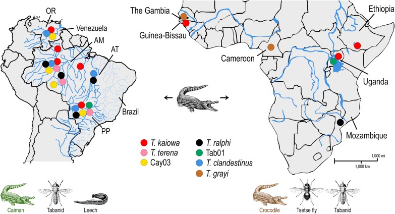
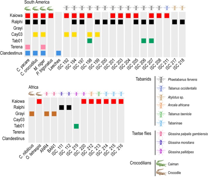
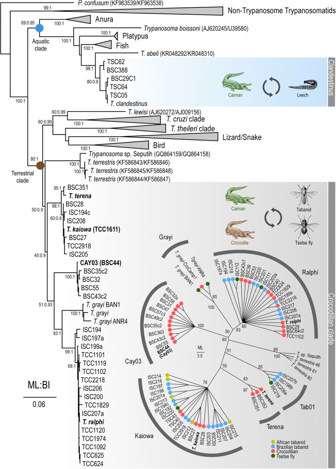
Background: The genus Trypanosoma Gruby, 1843 is constituted by terrestrial and aquatic phylogenetic lineages both harboring understudied trypanosomes from reptiles including an increasing diversity of crocodilian trypanosomes. Trypanosoma clandestinus Teixeira & Camargo, 2016 of the aquatic lineage is transmitted by leeches to caimans. Trypanosoma grayi Novy, 1906 of the terrestrial lineage is transmitted by tsetse flies to crocodiles in Africa, but the vectors of Neotropical caiman trypanosomes nested in this lineage remain unknown.

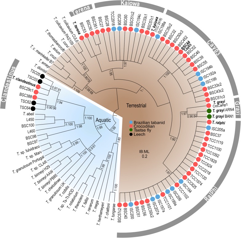
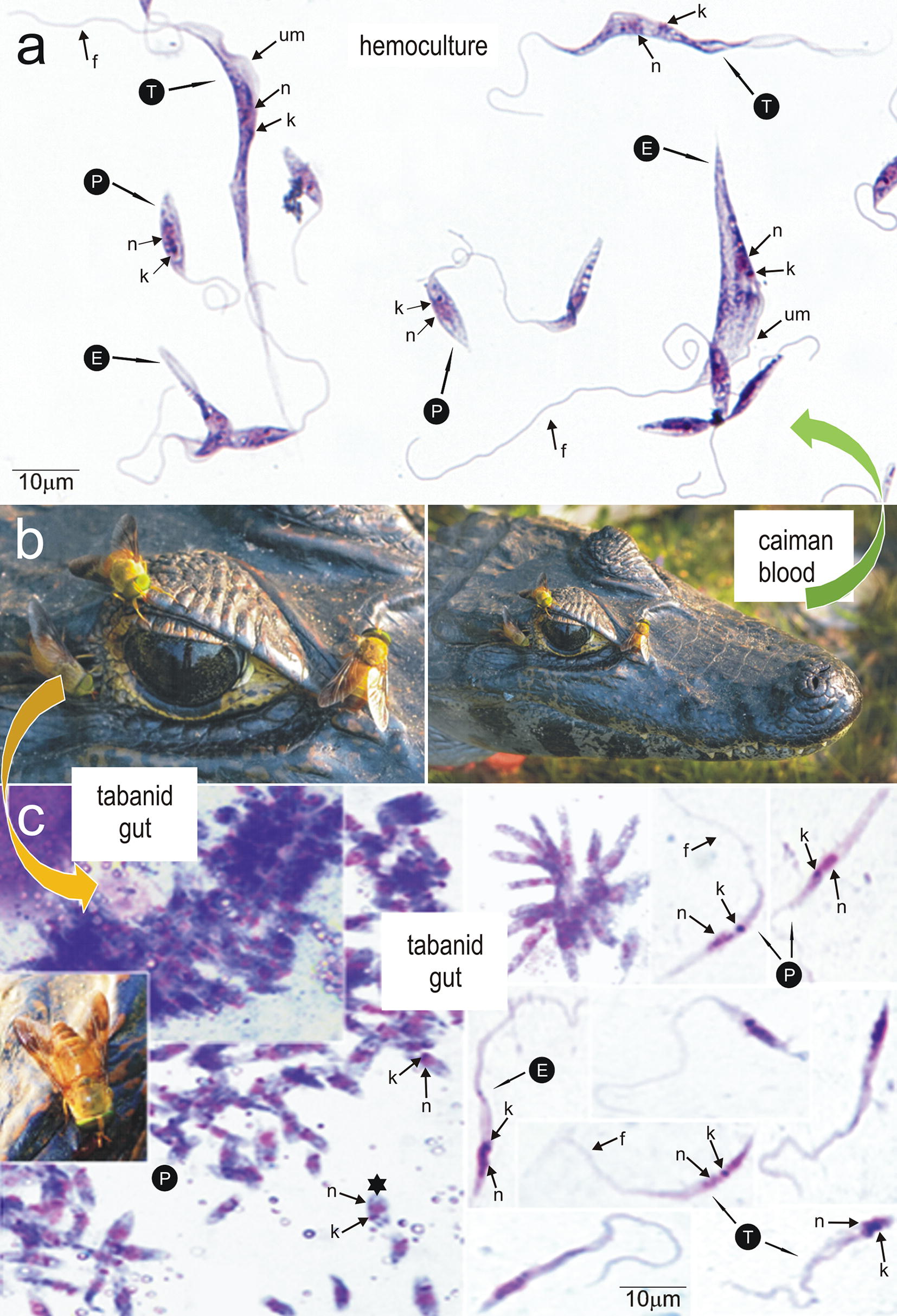
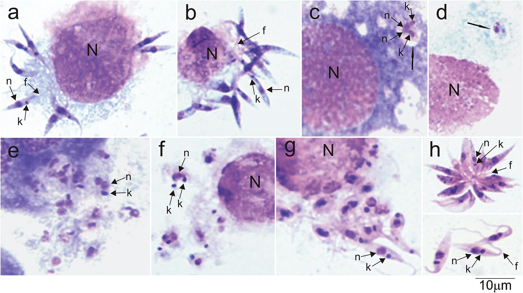
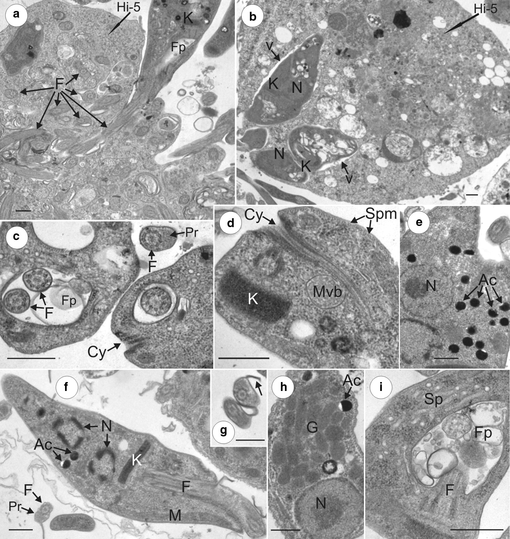
Results: Our phylogenetic analyses uncovered crocodilian trypanosomes in tabanids from South America and Africa, and trypanosomes other than T. grayi in tsetse flies. All trypanosomes found in tabanids clustered in the crocodilian clade (terrestrial lineage) forming six clades: Grayi (African trypanosomes from crocodiles and tsetse flies); Ralphi (trypanosomes from caimans, African and Brazilian tabanids and tsetse flies); Terena (caimans); Cay03 (caimans and Brazilian tabanids); and two new clades, Tab01 (Brazilian tabanid and tsetse flies) and Kaiowa. The clade Kaiowa comprises Trypanosoma kaiowa n. sp. and trypanosomes from African and Brazilian tabanids, caimans, tsetse flies and the African dwarf crocodile. Trypanosoma kaiowa n. sp. heavily colonises tabanid guts and differs remarkably in morphology from other caiman trypanosomes. This species multiplied predominantly as promastigotes on log-phase cultures showing scarce epimastigotes and exhibited very long flagellates in old cultures. Analyses of growth behavior revealed that insect cells allow the intracellular development of Trypanosoma kaiowa n. sp.

Conclusions: Prior to this description of Trypanosoma kaiowa n. sp., no crocodilian trypanosome parasitic in tabanid flies had been cultured, morphologically examined by light, scanning and transmission microscopy, and phylogenetically compared with other crocodilian trypanosomes. Additionally, trypanosomes thought to be restricted to caimans were identified in Brazilian and African tabanids, tsetse flies and the dwarf crocodile. Similar repertoires of trypanosomes found in South American caimans, African crocodiles and tabanids from both continents support the recent diversification of these transcontinental trypanosomes. Our findings are consistent with trypanosome host-switching likely mediated by tabanid flies between caimans and transoceanic migrant crocodiles co-inhabiting South American wetlands at the Miocene.

Keywords: Caiman; Crocodile; Evolution; Morphology; Tabanids; Taxonomy; Transoceanic dispersal; Tsetse flies.


Conflict of interest statement

The authors declare that they have no competing interests.

Figures

Fig. 1
Fig. 1
Geographical origin of crocodilian trypanosomes in South America and Africa. Trypanosomes were obtained from blood of caiman and crocodiles, and guts of tabanid and tsetse flies. Samples of T. kaiowa n. sp., T. ralphi and Trypanosoma sp. Tab01 were from Africa and South America; T. terena, T. clandestinus and Cay03 from South America; T. grayi from Africa. Hydrographic basins: OR, Orinoco; AM, Amazonas; AT, Araguaya-Tocantins; PP, Paraná-Paraguay
Fig. 2
Fig. 2
Trypanosomes in crocodilians, tabanids and tsetse flies in South America and Africa. Single and mixed infections identified by V7V8 SSU rRNA and/or gGAPDH barcodes of trypanosomes from the clades Kaiowa, Ralphi, Grayi, Cay03, Tab01, Terena and Clandestinus in individual blood samples from caimans (Caiman yacare, Caiman crocodilus, Melanonosuchus niger and Paleosuchus trigonatus), leeches (Haementeria sp.) and crocodiles (Crocodylus niloticus and Osteolaemus tetrapsis), and guts of tabanids (ISC 212-216) and tsetse flies (ANR4, BAN1, and ISC111, 112, 219, 220). In South America, T. kaiowa, T. ralphi and Trypanosoma sp. Cay03 were detected in tabanids and caimans, T. terena exclusively in caimans, Trypanosoma sp. Tab01 in tabanids, and T. clandestinus in caimans and leeches. In Africa, T. kaiowa n. sp. was identified in tabanids, tsetse flies and crocodiles, T. ralphi and Trypanosoma sp. Tab01 in tsetse flies, and T. grayi in tsetse flies and crocodiles
Fig. 3
Fig. 3
Phylogenetic analysis based on V7V8 SSU rRNA gene sequences of trypanosomes from caimans, crocodiles, tabanids and tsetse flies. Maximum Likelihood inference (903 characters, Ln = −4371.770725) supported the clades Ralphi, Kaiowa, Terena, Cay03 and Grayi in the terrestrial lineage. Trypanosomes of the aquatic lineages, including T. clandestinus, were used as outgroups. Numbers at the nodes (Bayesian Inference/Maximum Likelihood) represent the posterior probability > 0.8 and bootstrap support > 60%, respectively, derived from 500 replicates
Fig. 4
Fig. 4
Phylogenetic tree (ML) inferred using concatenated SSU rRNA and gGAPDH gene sequences of trypanosomes from caimans, crocodiles, tabanids, and tsetse flies. The analyses were inferred by Maximum Likelihood (ML, 1720 characters, Ln = −17593.067085) and Bayesian Inference (BI). Trypanosomes of the crocodilian clade were distributed in the clades Ralphi, Terena, Cay03, Tab01, Kaiowa, and Grayi. Trypanosomes of both terrestrial and aquatic lineages were used as outgroups. Numbers at nodes (ML/BI) are bootstrap support > 50%, and Bayesian posterior probability > 0.8 derived from 500 replicates. Insert: phylogram (ML) using gGAPDH sequences. Numbers at nodes are bootstrap values derived from 500 replicates
Fig. 5
Fig. 5
Trypanosoma kaiowa n. sp. predicted life-cycle. a Light microscopy of Giemsa-stained preparations of early hemocultures (BALB/LIT medium) from Caiman yacare naturally infected with T. kaiowa n. sp. showing large trypomastigote (T) and epimastigote (E) forms with noticeable undulating membrane, and small forms resembling promastigotes (P). b Caiman yacare and the tabanid Phaeotabanus fervens in the Pantanal, Brazil. c Clumps of flagellates adhered to a fragment of tabanid wall gut (yellow arrow), slim promastigotes (P), epimastigotes (E), and trypomastigotes (T) from gut contents of tabanids. Abbreviations: n, nucleus; k, kinetoplast; f, flagellum. Scale-bars: 10 µm
Fig. 6
Fig. 6
Growth behavioral of Trypanosoma kaiowa n. sp. co-cultivated with Hi-5 insect cells at 25 °C. Light microscopy of Giemsa-stained preparations showing: (a, b) flagellates adhered to Hi-5 cells by their flagella; (c, d) rounded flagellates within vacuoles (black arrows) in the cytoplasm of the insect cells; (e, f) agglomerates of dividing rounded forms; (g) rounded and promastigote forms, apparently released by host cells; (h) rosette and free short promastigotes in the supernatant of Hi-5 cells. Abbreviations: n, nucleus; k, kinetoplast; f, flagellum. Scale-bar: 10 µm
Fig. 7
Fig. 7
Development of Trypanosoma kaiowa n. sp. cultivated in LIT medium. a Rosettes of flagellates united by the flagella in log-phase cultures (3 days). b, c Detached free-swimming promastigotes (5–7 days). df Epimastigotes with undeveloped undulating membrane. e Rosette of large, irregular and pointed flagellates with a dividing flagellate still attached by the flagellum (10 days). g Long flagellates from stationary cultures varying in size and shape with long free flagellum. h, i Large flagellates with a long and pointed anterior extremity from old (15–20 days) cultures. Abbreviations: P, promastigote; E, epimastigote; n, nucleus; k, kinetoplast; f, flagellum; um, undulating membrane. Black crosses indicate dividing flagellates. Scale-bar: 10 µm
Fig. 8
Fig. 8
Scanning electron microscopy of Trypanosoma kaiowa n. sp. a, b Flagellates adhered to the membrane of Hi-5 insect cell (white arrow) cultivated in TC100 medium at 25 °C. b Flagellates apparently invading a Hi-5 cell via flagellum. c Promastigotes from culture supernatant. d, e Rosettes of promastigotes. f Promastigote (P) and epimastigote (E). g A clump of slender promastigotes exhibiting long flagella. h Epimastigote with an inconspicuous undulating membrane. Scale-bars: 10 µm
Fig. 9
Fig. 9
Ultrastructural features of T. kaiowa n. sp. revealed by TEM microscopy. a Flagellate invading Hi-5 cells via flagellum, and many transverse sections of the flagellum inside cell cytoplasm. b Small flagellates within tight vacuoles (indicated by black arrows) in the cytoplasm. c Transverse section of the flagellar pocket showing the typical structure of flagella, the absence of paraflagellar structure inside the flagellar pocket, prominent paraflagellar structure in free flagellum, and a small portion of the cytostome. d Longitudinal section of flagellate showing a deeply invaginated cytostome, compacted kinetoplast, and a multivesicular body probably containing viral particles. e Many acidocalcisomes in the cytoplasm. f Longitudinal sections of a dividing flagellate showing two nuclei. g Transverse section of flagella showing noticeable paraflagellar structure and lamella (black arrow). h Multiple glycosomes near the nucleus. i Network of tubules forming the spongiome adjacent to the flagellar pocket containing many vesicles. Abbreviations: N, nucleus; K, kinetoplast; Fp, flagellar pocket; F, flagellum; Cy, Cytostome; Pr, paraflagellar structure; Ac, acidocalcisomes; Spm, subpellicular microtubules; Mvb, multivesicular bodies; M, mitochondria; Sp, spongiome; v, vacuoles. Scale-bars: 0.5 µm

References

    1. Hoare C. The trypanosomes of mammals: a zoological monograph. Oxford: Blackwell Scientific Publications; 1972.
    1. Telford S., Jr . Parasitic protozoa. In: Kreir JP, editor. The kinetoplastid hemoflagellates of reptiles. New York: Academic Press; 1995. pp. 161–223.
    1. Stevens J, Noyes H, Schofield C, Gibson W. The molecular evolution of trypanosomatidae. Adv Parasitol. 2001;48:1–53. doi: 10.1016/S0065-308X(01)48003-1. - DOI - PubMed
    1. Hamilton P, Gibson W, Stevens J. Patterns of co-evolution between trypanosomes and their hosts deduced from ribosomal RNA and protein-coding gene phylogenies. Mol Phylogen Evol. 2007;44:15–25. doi: 10.1016/j.ympev.2007.03.023. - DOI - PubMed
    1. Hoare C. Studies on Trypanosoma grayi II. Experimental transmission to the crocodile. Trans R Soc Trop Med Hyg. 1929;23:39–56. doi: 10.1016/S0035-9203(29)90831-2. - DOI

LinkOut - more resources