. 2019 May 14;10(3):e00330-19.
doi: 10.1128/mBio.00330-19.

Langerhans Cells Sense Staphylococcus aureus Wall Teichoic Acid through Langerin To Induce Inflammatory Responses

Affiliations

Langerhans Cells Sense Staphylococcus aureus Wall Teichoic Acid through Langerin To Induce Inflammatory Responses

Rob van Dalen et al. mBio. .

Abstract

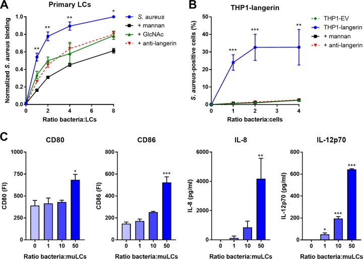
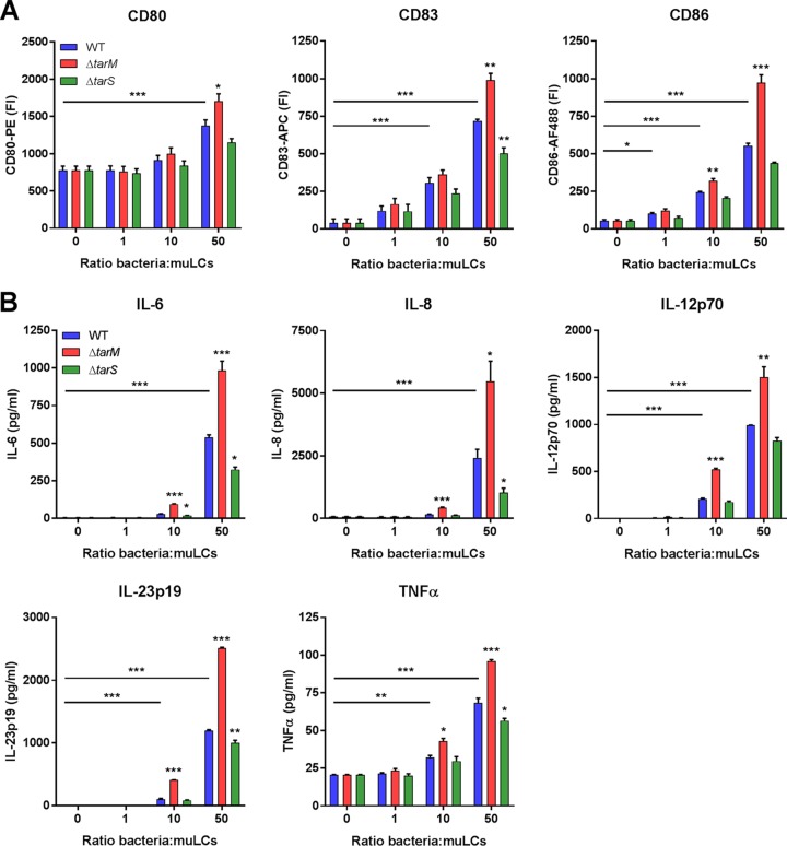
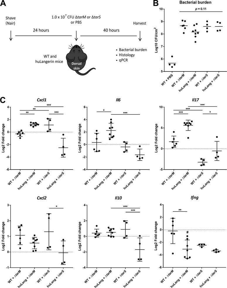
Staphylococcus aureus is a major cause of skin and soft tissue infections and aggravator of the inflammatory skin disease atopic dermatitis (AD [eczema]). Epicutaneous exposure to S. aureus induces Th17 responses through skin Langerhans cells (LCs), which paradoxically contribute to host defense but also to AD pathogenesis. The molecular mechanisms underlying the interaction between S. aureus and LCs are poorly understood. Here we demonstrate that human LCs directly interact with S. aureus through the pattern recognition receptor langerin (CD207). Human, but not mouse, langerin interacts with S. aureus through the conserved β-N-acetylglucosamine (GlcNAc) modifications on wall teichoic acid (WTA), thereby discriminating S. aureus from other staphylococcal species. Importantly, the specific S. aureus WTA glycoprofile strongly influences the level of proinflammatory cytokines that are produced by in vitro-generated LCs. Finally, in a murine epicutaneous infection model, S. aureus strongly upregulated transcripts of Cxcl1, Il6, and Il17, which required the presence of both human langerin and WTA β-GlcNAc. Our findings provide molecular insight into the unique proinflammatory capacities of S. aureus in relation to skin inflammation.IMPORTANCE The bacterium Staphylococcus aureus is an important cause of skin infections and is also associated with the occurrence and severity of eczema. Langerhans cells (LCs), a specific subset of skin immune cells, participate in the immune response to S. aureus, but it is yet unclear how LCs recognize S. aureus Therefore, we investigated the molecular mechanism underlying the interaction between LCs and S. aureus We identified that wall teichoic acid, an abundant polymer on the S. aureus surface, is recognized by langerin, a receptor unique to LCs. This interaction allows LCs to discriminate S. aureus from other related staphylococcal species and initiates a proinflammatory response similar to that observed in patients with eczema. Our data therefore provide important new insights into the relationship between S. aureus, LCs, and eczema.

Keywords: Langerhans cell; Staphylococcus aureus; atopic dermatitis; glycosylation; langerin; wall teichoic acid.


Figures

FIG 1
FIG 1
Langerin is a receptor for S. aureus on human LCs. (A) Binding of GFP-expressing S. aureus Newman to isolated primary human LCs from four different donors. The interaction was blocked by addition of mannan (n = 4), GlcNAc (n = 4), or anti-langerin blocking antibody (n = 2; using GFP-expressing S. aureus Newman Δspa Δsbi). Data were pooled and normalized to the maximum binding level observed in each donor (ratio 8) and are presented as mean ± standard error of mean (SEM). Indicated statistical differences refer to the blocking conditions compared to the nonblocked control. (B) Binding of S. aureus to THP1-langerin cells. Human langerin-transduced or empty vector (EV)-transduced THP1 cells were incubated with different amounts of GFP-expressing S. aureus Newman Δspa Δsbi. The interaction was blocked by addition of mannan or anti-langerin blocking antibody. Data are presented as percentage GFP-positive cells ± SEM from three independent experiments. (C) Expression of costimulatory molecules CD80 and CD86 and production of cytokines IL-8 and IL-12p70 by muLCs after stimulation with gamma-irradiated S. aureus USA300. Mean concentrations ± SEM from three independent experiments are shown. *, P < 0.05; **, P < 0.01; ***, P < 0.001.
FIG 2
FIG 2
Langerin specifically recognizes S. aureus in a tarS-dependent manner through the conserved WTA β-GlcNAc epitope. Shown is binding of recombinant human langerin-FITC to (A) 18 wild-type S. aureus strains (11 different clonal complexes, indicated above the bars and by different colors) and a selection of coagulase-negative staphylococcal species (CoNS), (B) the S. aureus USA300 wild-type (WT) and WTA biosynthesis ΔtarMS, ΔtarM, ΔtarS, ΔtarMS ptarM, and ΔtarMS ptarS strains, and (C) two representative S. aureus isolates (82086 and PS66) that naturally lack tarM and their isogenic ΔtarS mutants. Data are presented as geometric mean fluorescence intensity ± SEM from three independent experiments. *, P < 0.05; **, P < 0.01; ***, P < 0.001.
FIG 3
FIG 3
The S. aureus WTA glycoprofile affects the proinflammatory cytokine response of LCs. (A) Expression of costimulatory molecules CD80 and CD86 and maturation marker CD83 and (B) production of cytokines IL-6, IL-8, IL12p70, IL23p19, and TNF-α by muLCs upon incubation with gamma-irradiated S. aureus USA300 wild-type (WT) or ΔtarM or ΔtarS mutant cells. muLCs stimulated with WT S. aureus were compared to unstimulated controls, and muLCs stimulated with the ΔtarM and ΔtarS mutants were compared to their respective WT controls within the same ratio. IL-4, IL-10, and IFN-γ concentrations were assessed, but were below the detection limit. Data are presented as geometric mean fluorescence intensity or mean concentration ± SEM from three independent experiments. *, P < 0.05; **, P < 0.01; ***, P < 0.001.
FIG 4
FIG 4
Epicutaneous infection with S. aureus induces skin inflammation that requires both human langerin and WTA β-GlcNAc expression. (A) Experimental design for epicutaneous infection of WT mice with the S. aureus ΔtarM mutant (n = 6), huLangerin mice with the S. aureus ΔtarM mutant (n = 8), WT mice with the S. aureus ΔtarS mutant (n = 4), huLangerin mice with the S. aureus ΔtarS mutant (n = 4), and WT mice with PBS controls (n = 4). (B) Bacterial burden of the lesions 40 h postinoculation, presented as log10-transformed CFU/cm2 ± SD. (C) Transcript abundance of Cxcl1, Cxcl2, Il6, Il17, Il10, and Ifng in whole-skin homogenates 40 h post-epicutaneous inoculation. Data are presented as log2-transformed fold change ± standard deviation (SD) relative to Gapdh and normalized to the mean value of the group WT mice plus PBS. *, P < 0.05; **, P < 0.01; ***, P < 0.001. #, no CT value was reached for Ifng in one sample.

References

    1. Nutten S. 2015. Atopic dermatitis: global epidemiology and risk factors. Ann Nutr Metab 66(Suppl 1):8–16. doi:10.1159/000370220. - DOI - PubMed
    1. Totte JE, van der Feltz WT, Hennekam M, van Belkum A, van Zuuren EJ, Pasmans SG. 2016. Prevalence and odds of Staphylococcus aureus carriage in atopic dermatitis: a systematic review and meta-analysis. Br J Dermatol 175:687–695. doi:10.1111/bjd.14566. - DOI - PubMed
    1. Nakatsuji T, Chen TH, Narala S, Chun KA, Two AM, Yun T, Shafiq F, Kotol PF, Bouslimani A, Melnik AV, Latif H, Kim JN, Lockhart A, Artis K, David G, Taylor P, Streib J, Dorrestein PC, Grier A, Gill SR, Zengler K, Hata TR, Leung DY, Gallo RL. 2017. Antimicrobials from human skin commensal bacteria protect against Staphylococcus aureus and are deficient in atopic dermatitis. Sci Transl Med 9:eaah4680. doi:10.1126/scitranslmed.aah4680. - DOI - PMC - PubMed
    1. Kong HH, Oh J, Deming C, Conlan S, Grice EA, Beatson MA, Nomicos E, Polley EC, Komarow HD, NISC Comparative Sequence Program, Murray PR, Turner ML, Segre JA. 2012. Temporal shifts in the skin microbiome associated with disease flares and treatment in children with atopic dermatitis. Genome Res 22:850–859. doi:10.1101/gr.131029.111. - DOI - PMC - PubMed
    1. DeLeo FR, Otto M, Kreiswirth BN, Chambers HF. 2010. Community-associated meticillin-resistant Staphylococcus aureus. Lancet 375:1557–1568. doi:10.1016/S0140-6736(09)61999-1. - DOI - PMC - PubMed

Publication types

MeSH terms