. 2019 May 14;10(1):2153.
doi: 10.1038/s41467-019-09884-6.

Bacteroides fragilis polysaccharide A induces IL-10 secreting B and T cells that prevent viral encephalitis

Affiliations

Bacteroides fragilis polysaccharide A induces IL-10 secreting B and T cells that prevent viral encephalitis

Chandran Ramakrishna et al. Nat Commun. .

Abstract

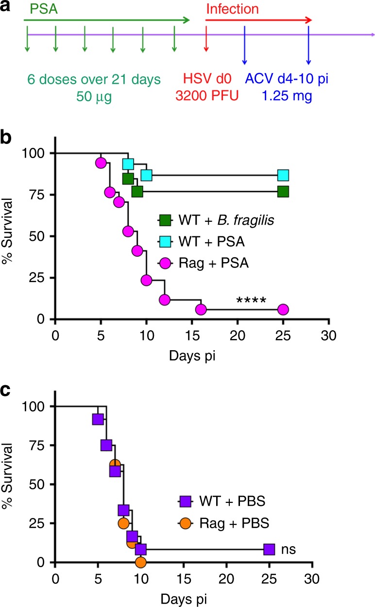
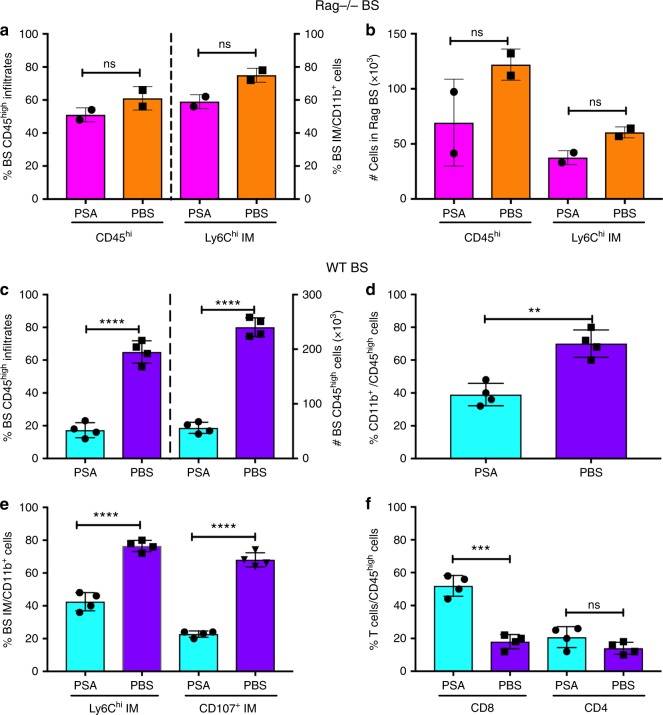
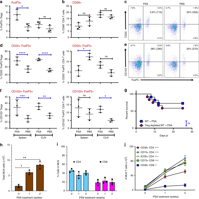
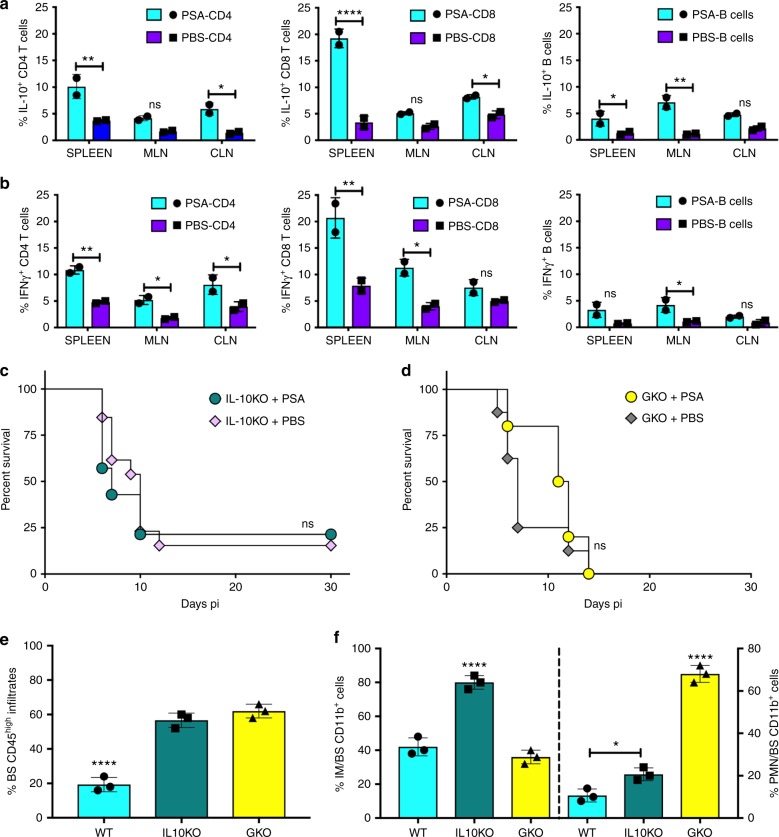
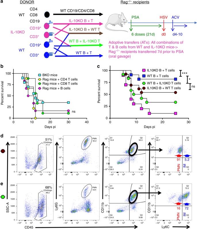
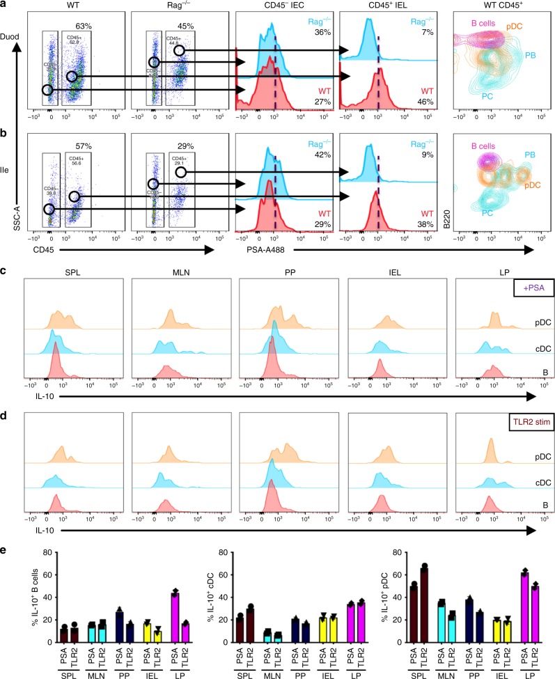
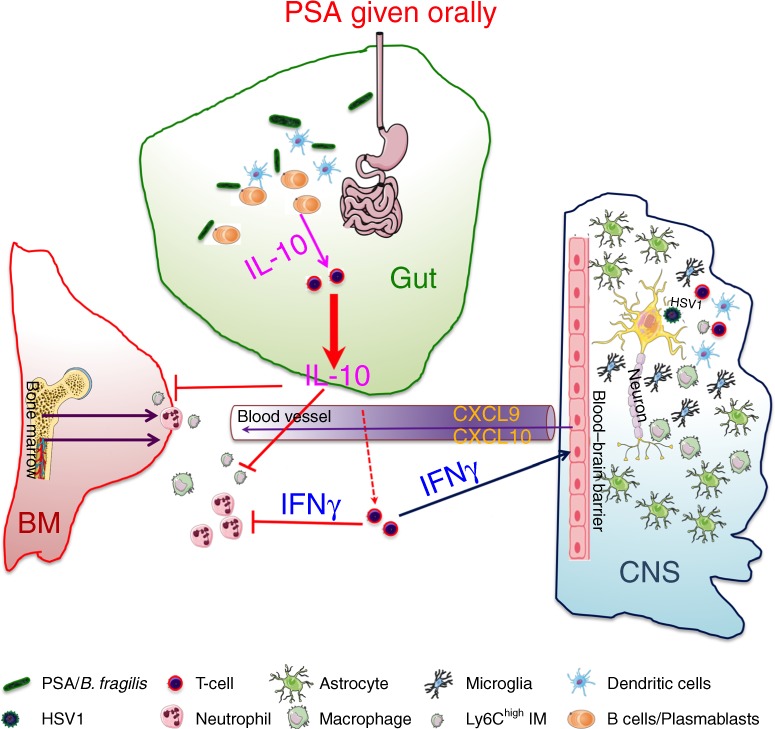
The gut commensal Bacteroides fragilis or its capsular polysaccharide A (PSA) can prevent various peripheral and CNS sterile inflammatory disorders. Fatal herpes simplex encephalitis (HSE) results from immune pathology caused by uncontrolled invasion of the brainstem by inflammatory monocytes and neutrophils. Here we assess the immunomodulatory potential of PSA in HSE by infecting PSA or PBS treated 129S6 mice with HSV1, followed by delayed Acyclovir (ACV) treatment as often occurs in the clinical setting. Only PSA-treated mice survived, with dramatically reduced brainstem inflammation and altered cytokine and chemokine profiles. Importantly, PSA binding by B cells is essential for induction of regulatory CD4+ and CD8+ T cells secreting IL-10 to control innate inflammatory responses, consistent with the lack of PSA mediated protection in Rag-/-, B cell- and IL-10-deficient mice. Our data reveal the translational potential of PSA as an immunomodulatory symbiosis factor to orchestrate robust protective anti-inflammatory responses during viral infections.


Conflict of interest statement

E.M.C., C.R. and S.K.M. are inventors of patent application: PCT/US2016/036803, which describes use of PSA as a treatment for viral inflammatory diseases. The remaining authors declare no competing interests.

Figures

Fig. 1
Fig. 1
B. fragilis PSA protects against HSE. a Experimental regimen: In all experiments, PSA (six doses, 50 μg/mouse) or PBS was given orally before HSV infection on day 0 and thereafter daily ip injections of ACV from day 4 pi for 7 days. Survival of wildtype (WT) or Rag mice pre-treated with b B. fragilis, PSA or c PBS, (n = 8–17 mice/group). ****p < 0.001, WT + B. fragilis/PSA vs. Rag + PSA; ns: not significant, WT + PBS vs. Rag + PBS as determined by log rank (Mantel–Cox) test
Fig. 2
Fig. 2
PSA reduces CNS inflammation in HSV-infected WT but not Rag mice. a % and b total numbers (#) of CD45high leukocytes and CD45high Ly6Chigh inflammatory monocytes (IM) infiltrating the brainstem (BS) of Rag mice. c % (left y-axis) and # (right y-axis) infiltrating CD45high leukocytes in the BS of WT mice. d % CD11b+ cells within BS infiltrating CD45high cells; e % Ly6Chigh and CD107+ IM within the CD11b+ population; f % CD4+ and CD8+ T cells within CD45high cells in the BS of WT mice. Data compiled from 2 to 4 experiments with n = 6–8/group at day 6 pi. All data show mean ± SEM. ***p < 0.0005, ****p < 0.0001, ns: not significant, as determined by two-tailed Student's t-test
Fig. 3
Fig. 3
PSA protection from HSE is independent of induced Tregs. a % FoxP3+ CD4 Tregs and b CD69+ CD4 T cells in spleen and CLN of PSA or PBS-treated WT mice at day 6 pi. c CD25 expression within FoxP3+ CD4 Tregs in WT mice at day 6 pi, % and mean fluorescence intensity (MFI) in () shown in right top quadrant. d % CD25 within FoxP3+ Tregs (left plot) and FoxP3 CD4+ T cells (right plot), e CD103 expression within FoxP3+ Tregs in WT mice at day 6 pi; % and MFI in () shown in right top quadrant. f CD103 within FoxP3+ Tregs (left plot) and FoxP3 CD4+ T cells (right plot) in the spleen or CLN of WT mice at day 6 pi. Data from three experiments shown. g PSA-treated Treg depleted and control WT mice were monitored for survival after HSV infection and ACV treatment as in Fig. 1a, ns: not significant determined by log rank Mantel–Cox test (n = 11–12 mice). After administration of three (1 week) or six doses (2 weeks) of PSA, MLN in uninfected WT mice were monitored for h cellularity, i % CD4 and CD8 T cells, and j # ICOS+, CD39+, and CD73+ CD4 and CD8 T cells (n = 3 mice); ****p < 0.0001, **p < 0.01 as determined by two-way ANOVA or one-way ANOVA with Sidaks or Turkeys correction, respectively, for multiple comparisons tests. All data show mean ± SEM
Fig. 4
Fig. 4
PSA increases IL-10 and IFNγ-secreting T cells. CD4 and CD8 T cells and B cells in spleens, mesenteric lymph nodes (MLN), and cervical lymph nodes (CLN) of PSA or PBS-treated WT mice at day 6 pi were analyzed for a IL-10 and b IFNγ secretion, n = 2 experiments; *p < 0.05, **p < 0.01, ****p < 0.0001, as determined by two-way ANOVA with Sidak’s multiple comparisons test. Survival of PSA or PBS treated c IL-10KO mice or d IFN-GKO mice (n = 8–16 mice); ns: not significant. Bar plots show e % CD45high leukocytes, f (left y-axis) % Ly6Chigh IM and (right y-axis) % Ly6G+ neutrophils (PMN) within CD45high CD11b+ cells infiltrating the BS of PSA treated 129 WT, IL10KO, and GKO mice at day 6 pi, n = 3 experiments with 2–3 BS/group; *p < 0.05, ****p < 0.0001 as determined by ordinary one-way ANOVA with Turkey’s multiple comparisons tests
Fig. 5
Fig. 5
PSA protection against HSE requires B and T cells secreting IL-10. a Experimental design for experiments in b and c Donor WT (In black text): Naïve Rag mice were transferred with WT CD4+ or CD8+ T cells or CD19+ B cells 7 days before PSA treatment. Donor IL-10KO (magenta text) and WT (Blue text): four groups of naïve Rag mice were transferred with combinations of donor WT B and T cells, IL-10KO B and T cells, WT B and IL-10KO T cells, IL-10KO B and WT T cells 7-days before PSA treatment. All Rag recipients received six doses of PSA before HSV infection and ACV treatment. b Survival of B cell-depleted mice (BKO, n = 20 mice) and Rag recipients of WT single cell subsets (n = 6–9 mice/group). B cell depletion in WT mice was initiated 10 days prior to PSA treatment and continued throughout infection, ns: not significant. c Survival of Rag recipients of WT and IL-10KO combination of T and B cells (n = 10–13/group). ***p < 0.001, *p < 0.05, ns: not significant as determined by log rank (Mantel–Cox) test. FACS plots of BS CD45high cells (left), Ly6G+ PMN (left middle), CD11b+ cells within CD45high cells (right middle), and Ly6Chigh IM and Ly6Cint CD11b+ PMN within CD45high CD11b+ cells (right) were analyzed at day 6 pi in the BS of Rag recipients of d IL-10KO B + WT T cells (brown circle) and e WT B + IL-10KO T cells (green circle)
Fig. 6
Fig. 6
TLR2+ macrophages, pDCs, and PB in the small intestine bind PSA and induce IL-10 secretion. Gating strategy for mononuclear cells isolated from a duodenum (Duod) and b ileum (Ile) of WT and Rag mice analyzed for binding of fluorescent A488-conjugated PSA (left two plots). CD45 intra-epithelial cells (CD45 IEC: middle histogram) and CD45+ gated intra-epithelial leukocytes (CD45+ IEL: second right histogram) isolated from the a Duod and b Ile of WT (red) and Rag (blue) mice were analyzed for reactivity to PSA-A488. CD45+CD11cB220+ B cells, PDCA1+ B220+CD11c+ pDCs and CD138+B220+ PB and B220low PC isolated from a Duod and b Ile of WT mice were analyzed for PSA reactivity (right histogram). Flow cytometry plots show CD11c+PDCA1 cDC, PDCA1+B220+ pDC, and B220+CD19+ B cells isolated from spleen, MLN, PP, IEL, or LP of WT mice were stimulated with c PSA or d LTA-SA (TLR2 agonist) and analyzed for IL-10 expression. e Plots summarize data from c and d and show % IL-10+ B cells (left), cDCs (middle), and pDCs (right) from spleen, MLN, PP, IEL, and LP stimulated with PSA or TLR2 (LTA-SA) (n = 3 mice). All data show mean ± SEM
Fig. 7
Fig. 7
Role of the bacterial symbiosis factor PSA in preventing viral encephalitis. HSV infection of susceptible 129 WT mice provokes excessive production of neutrophils (PMN) and Ly6Chigh inflammatory monocytes (IM) in the bone marrow that invade the brainstem in massive numbers resulting in fatal HSV encephalitis (HSE), despite antiviral treatment from day 4 pi. The bacterial symbiosis factor, PSA given orally is bound by B cells/CD138+ plasmablasts (PB) in the small intestine, which induces IL-10 and IFNγ production by regulatory CD4 and CD8 T cells resulting in the suppression of pathogenic inflammatory myeloid cells concomitant with the induction of IFNγ inducible chemokines in the BS. This novel study reveals the immunomodulatory potential of PSA in protecting from lethal viral infections of the CNS in combination with an antiviral. Cells involved in this protective mechanism are shown in the key. Inhibitory pathways indicated by red blocking arrows

References

    1. Sender R, Fuchs S, Milo R. Revised estimates for the number of human and bacteria cells in the body. PLoS Biol. 2016;14:e1002533. doi: 10.1371/journal.pbio.1002533. - DOI - PMC - PubMed
    1. Lynch SV, Pedersen O. The human intestinal microbiome in health and disease. New Engl. J. Med. 2016;375:2369–2379. doi: 10.1056/NEJMra1600266. - DOI - PubMed
    1. Bashiardes S, Zilberman-Schapira G, Elinav E. Use of metatranscriptomics in microbiome research. Bioinform Biol. Insights. 2016;10:19–25. doi: 10.4137/BBI.S34610. - DOI - PMC - PubMed
    1. Blander JM, Longman RS, Iliev ID, Sonnenberg GF, Artis D. Regulation of inflammation by microbiota interactions with the host. Nat. Immunol. 2017;18:851–860. doi: 10.1038/ni.3780. - DOI - PMC - PubMed
    1. Thomas S, et al. The host microbiome regulates and maintains human health: a primer and perspective for non-microbiologists. Cancer Res. 2017;77:1783–1812. doi: 10.1158/0008-5472.CAN-16-2929. - DOI - PMC - PubMed

Publication types

MeSH terms