. 2019 May 14;9(1):7354.
doi: 10.1038/s41598-019-43669-7.

Quantified forces between HepG2 hepatocarcinoma and WA07 pluripotent stem cells with natural biomaterials correlate with in vitro cell behavior

Affiliations

Quantified forces between HepG2 hepatocarcinoma and WA07 pluripotent stem cells with natural biomaterials correlate with in vitro cell behavior

Riina Harjumäki et al. Sci Rep. .

Erratum in

Abstract

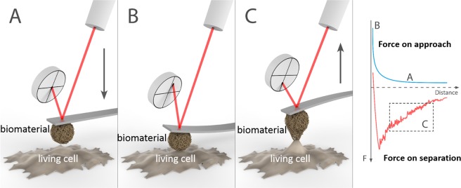
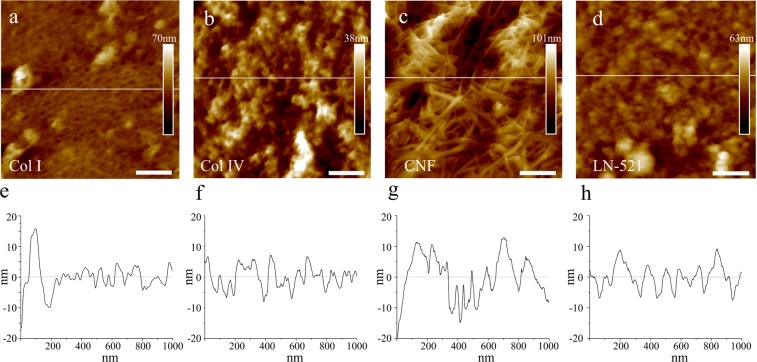
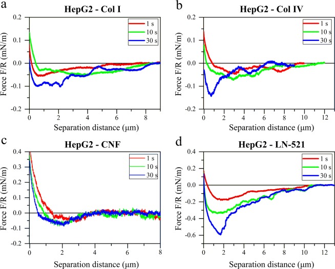
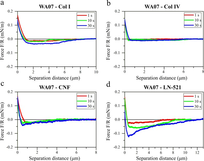
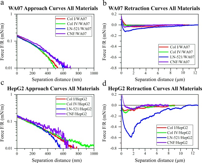
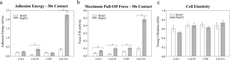
In vitro cell culture or tissue models that mimic in vivo cellular response have potential in tissue engineering and regenerative medicine, and are a more economical and accurate option for drug toxicity tests than animal experimentation. The design of in vivo-like cell culture models should take into account how the cells interact with the surrounding materials and how these interactions affect the cell behavior. Cell-material interactions are furthermore important in cancer metastasis and tumor progression, so deeper understanding of them can support the development of new cancer treatments. Herein, the colloidal probe microscopy technique was used to quantify the interactions of two cell lines (human pluripotent stem cell line WA07 and human hepatocellular carcinoma cell line HepG2) with natural, xeno-free biomaterials of different chemistry, morphology, and origin. Key components of extracellular matrices -human collagens I and IV, and human recombinant laminin-521-, as well as wood-derived, cellulose nanofibrils -with evidenced potential for 3D cell culture and tissue engineering- were analysed. Both strength of adhesion and force curve profiles depended on biomaterial nature and cell characteristics. The successful growth of the cells on a particular biomaterial required cell-biomaterial adhesion energies above 0.23 nJ/m. The information obtained in this work supports the development of new materials or hybrid scaffolds with tuned cell adhesion properties for tissue engineering, and provides a better understanding of the interactions of normal and cancerous cells with biomaterials in the human body.


Conflict of interest statement

The authors declare no competing interests.

Figures

Figure 1
Figure 1
Schematic representation of the measurement of cell-biomaterial interaction forces by colloidal probe microscopy. A biomaterial-coated colloidal probe and a substrate with living cells are approached each other (A) until contact (B), and then they are retracted (C) until detachment. The interaction forces are quantified from the deflection of the cantilever, which is monitored with a laser and a photodetector. Figure prepared by Joel Wolff.
Figure 2
Figure 2
Representative AFM height images of biomaterial substrates formed by adsorption of the biomaterials on colloidal glass probes. The biomaterials used were (a) collagen I, (b) collagen IV, (c) CNF, (d) laminin-521 (scale bar is 200 nm). (eh) Show the height topographic profiles corresponding to the lines marked in figures (ad), respectively.
Figure 3
Figure 3
Retraction force curves between HepG2 cells and colloidal probes coated with: (a) collagen I, (b) collagen IV, (c) cellulose nanofibrils, and (d) laminin-521. Representative force curves are presented normalized by the probe radius R (Table S2) after different cells-probes contact times (1 s, 10 s, and 30 s).
Figure 4
Figure 4
Retraction force curves between WA07 cells and colloidal probes coated with: (a) collagen I, (b) collagen IV, (c) cellulose nanofibrils, and (d) laminin-521. Representative force curves are presented normalized by the probe radius R (Table S2) after different cells-probes contact times (1 s, 10 s, and 30 s).
Figure 5
Figure 5
Representative normalized force curves between different biomaterial-coated probes and living cells: (a) approach curves in logarithmic scale and (b) retraction curves on WA07 cells; (c) approach curves in logarithmic scale and (d) retraction curves on HepG2 cells. The retraction curves were recorded after 30 s contact time between cells and probes. Force values were normalized by the probe radius R (Table S2).
Figure 6
Figure 6
Comparison of adhesion energies (a), maximum pull-off forces (b), and cell elasticity (c) for HepG2 and WA07 cell interactions with collagen I (Col I), collagen IV (Col IV), cellulose nanofibrils (CNF), and laminin-521 (LN-521) at contact time of 30 s. Error bars are standard errors of mean and significant differences of p ≤ 0.05 are marked with *. Values were normalized by the probe radius R.
Figure 7
Figure 7
Representative images of HepG2 and WA07 cells before, during and after the force experiments. The cell viability after the experiments was controlled with Trypan Blue exclusion test (scale bars are 200 µm).
Figure 8
Figure 8
HepG2 and WA07 cells after 20 hours culture on a control matrix (plastic and Matrigel, respectively) and on test materials: collagen I, collagen IV, laminin-521 and cellulose nanofibrils (scale bars are 200 μm).

References

    1. Schlie-Wolter S, Ngezahayo A, Chichkov BN. The selective role of ECM components on cell adhesion, morphology, proliferation and communication in vitro. Exp. Cell Res. 2013;319:1553–1561. doi: 10.1016/j.yexcr.2013.03.016. - DOI - PubMed
    1. Inman JL, Robertson C, Mott JD, Bissell MJ. Mammary gland development: cell fate specification, stem cells and the microenvironment. Development. 2015;142:1028–1042. doi: 10.1242/dev.087643. - DOI - PubMed
    1. Kaukonen R, et al. Normal stroma suppresses cancer cell proliferation via mechanosensitive regulation of JMJD1a-mediated transcription. Nat. Commun. 2016;7:12237. doi: 10.1038/ncomms12237. - DOI - PMC - PubMed
    1. Lee EY, Parry G, Bissell MJ. Modulation of secreted proteins of mouse mammary epithelial cells by the collagenous substrata. J. Cell Biol. 1984;98:146–155. doi: 10.1083/jcb.98.1.146. - DOI - PMC - PubMed
    1. Heydarkhan-Hagvall S, et al. Three-dimensional electrospun ECM-based hybrid scaffolds for cardiovascular tissue engineering. Biomaterials. 2008;29:2907–2914. doi: 10.1016/j.biomaterials.2008.03.034. - DOI - PMC - PubMed

Publication types