. 2019 Nov;56(11):7631-7650.
doi: 10.1007/s12035-019-1604-4. Epub 2019 May 14.

Transcriptional Response and Morphological Features of the Neurovascular Unit and Associated Extracellular Matrix After Experimental Stroke in Mice

Affiliations

Transcriptional Response and Morphological Features of the Neurovascular Unit and Associated Extracellular Matrix After Experimental Stroke in Mice

Susanne Aleithe et al. Mol Neurobiol. 2019 Nov.

Abstract

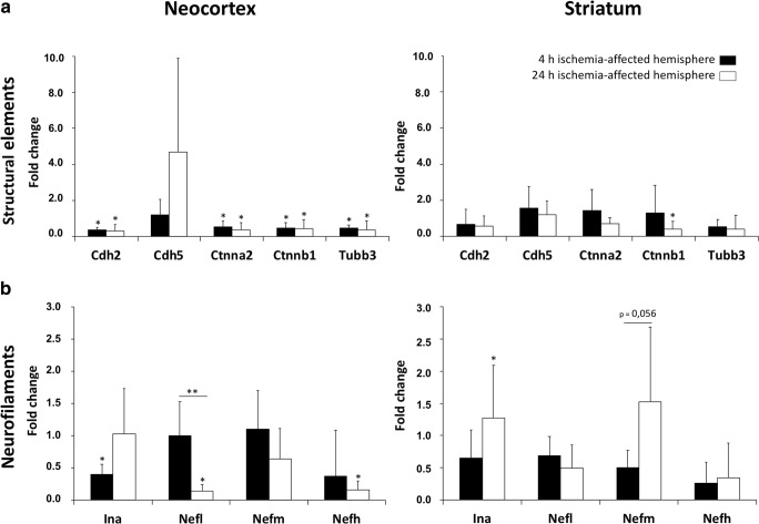
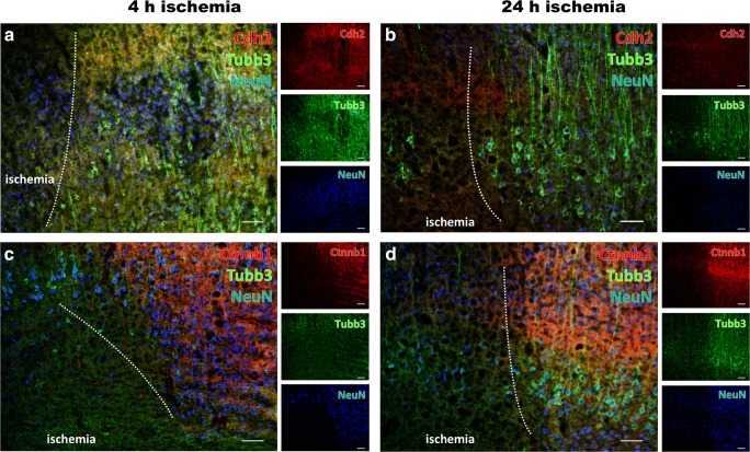
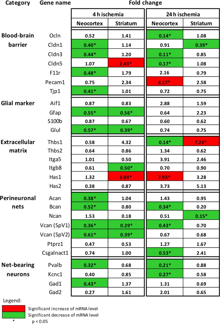
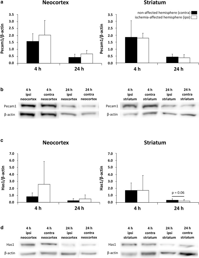
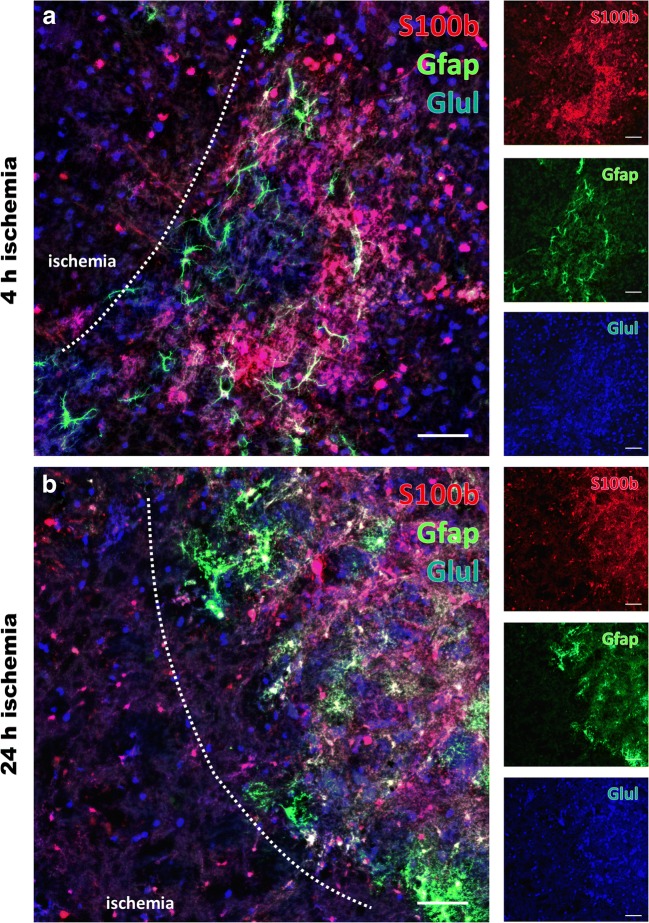
Experimental stroke studies yielded insights into single reactions of the neurovascular unit (NVU) and associated extracellular matrix (ECM). However, the extent of simultaneous processes caused by ischemia and their underlying transcriptional changes are still poorly understood. Strictly following the NVU and ECM concept, this study explored transcriptional responses of cellular and non-cellular components as well as their morphological characteristics following ischemia. Mice were subjected to 4 or 24 h of unilateral middle cerebral artery occlusion. In the neocortex and the striatum, cytoskeletal and glial elements as well as blood-brain barrier and ECM components were analyzed using real-time PCR. Western blot analyses allowed characterization of protein levels and multiple immunofluorescence labeling enabled morphological assessment. Out of 37 genes analyzed, the majority exhibited decreased mRNA levels in ischemic areas, while changes occurred as early as 4 h after ischemia. Down-regulated mRNA levels were predominantly localized in the neocortex, such as the structural elements α-catenin 2, N-cadherin, β-catenin 1, and βIII-tubulin, consistently decreasing 4 and 24 h after ischemia. However, a few genes, e.g., claudin-5 and Pcam1, exhibited increased mRNA levels after ischemia. For several components such as βIII-tubulin, N-cadherin, and β-catenin 1, matching transcriptional and immunofluorescence signals were obtained, whereas a few markers including neurofilaments exhibited opposite directions. In conclusion, the variety in gene regulation emphasizes the complexity of interactions within the ischemia-affected NVU and ECM. These data might help to focus future research on a set of highly sensitive elements, which might prospectively facilitate neuroprotective strategies beyond the traditional single target perspective.

Keywords: Extracellular matrix; Focal cerebral ischemia; Gene expression; Neurovascular unit; Stroke.


Conflict of interest statement

The authors declare that there is no conflict of interest.

Figures

Fig. 1
Fig. 1
Immunolabeling of the neurofilament-L (Nefl) combined with astroglial Gfap and neuronal NeuN as well as microglia/macrophages (Aif1) and endothelial Pecam1 in the neocortex of mice affected by 24 h of focal ischemia. Figure 1a provides a general overview of the infarcted neocortex in the superior part of a forebrain section. Here, the ischemic area is delineated by an increased immunosignal of Nefl and a concomitantly decreased immunosignal of NeuN. The rectangle indicates the ischemic border zone, shown in b and c at higher magnification to explore regional arrangements. Here, a significant increase of Nefl-immunoreactivity (red, b′, c′), apparently fragmented astrocytes illustrated by immunolabeling of Gfap (green, b″), decreased NeuN immunosignals (b‴), ameboid Aif1-positive microglia/macrophages (c″), and a slightly decreased Pecam1 immunoreactivity (c‴) are visible in the ischemic area. Corresponding contralateral control areas show homogeneous distribution patterns for NeuN and Gfap (d), an only weak immunosignal of Nefl (d, e) and an evenly distributed Pecam1 immunoreactivity. Scale bars: a 300 μm; be 100 μm
Fig. 2
Fig. 2
Comparative quantification of mRNA levels obtained from real-time PCR analyses of structural elements (a) and neurofilaments (b) after 4 and 24 h of ischemia in the neocortex and the striatum. Data are given as fold changes, calculated by an inter-hemispheric relation considering mRNA levels from the ischemic hemisphere (neocortex and striatum) and the corresponding contralateral, i.e., non-affected, regions. Bars represent means and added lines the standard deviations. Significance levels at the top of added lines are related to the inter-hemispheric comparison, while added horizontal lines with significance levels are related to potential temporal changes. *p < 0.05; **p < 0.01; sample size: n = 5 for each time point
Fig. 3
Fig. 3
Immunofluorescence labeling of structural elements in the neocortex affected by 4 and 24 h of focal ischemia. Diminished immunosignals of N-cadherin (Cdh2, red, a, b) and β-catenin 1 (Ctnnb1, red, c, d) as well as βIII-tubulin (Tubb3, green, ad), together with the neuronal marker neuronal nuclei (NeuN, blue, ad), indicate morphological changes in terms of degenerated structural elements due to ischemia. While considering the duration of ischemia, the degree of altered immunosignals for structural elements indicates a more severe affection towards the later time point, i.e., 24 h of ischemia (b, d). Scale bars: ad 100 μm
Fig. 4
Fig. 4
Changes in mRNA levels revealed by real-time PCR analyses of different markers of the blood-brain barrier, the neurovascular unit, and associated extracellular matrix constituents 4 and 24 h after ischemia onset in the neocortex and the striatum. The given values represent fold changes, calculated as the ratio of the mRNA levels between the ischemic hemisphere (neocortex and striatum) and the corresponding contralateral, i.e., non-affected, regions. Thereby, significant changes are color-coded: A green background represents a down-regulation, a red background stands for an up-regulation, whereas a white background indicates no relevant changes. *p < 0.05; sample size: n = 5 for each time point
Fig. 5
Fig. 5
Comparative quantifications of protein levels of selected blood-brain barrier- and extracellular matrix-related markers obtained from Western blot analyses after 4 and 24 h of ischemia in the neocortex and the striatum. a The Pecam1 protein levels normalized by β-actin remain statistically unaltered after ischemia, but tend to increase in the ischemia-affected neocortex 4 and 24 h after ischemia. c The neocortical and striatal protein levels of hyaluronan synthase 1 (Has1) normalized by β-actin similarly show a non-significant increase in the ischemia-affected neocortex 4 and 24 h after ischemia. However, in the striatum the protein level tends to decrease 24 h after ischemia, barely missing statistical significance. b, d Representative protein expression bands of Pecam1 (b) and Has1 (d) with corresponding bands of β-actin (b, d) in samples from neocortex and striatum 4 and 24 h after ischemia. Bars represent means and added lines the standard deviations. Sample size: n = 5 for each time point
Fig. 6
Fig. 6
Glial alterations following 4 and 24 h of focal cerebral ischemia. Multiple immunofluorescence labeling applied to the ischemia-affected striatum reveals significantly altered signals of the astroglial markers S100β (S100b, red, a, b) and Gfap (green, a, b) as well as glutamine synthetase (Glul, blue, a, b) decreasing towards ischemic regions. When considering potential time-dependent effects, the degree of affection seems to be pronounced towards the later time point, i.e., 24 h after ischemia (b). Remarkably, directly at the ischemic border zone, slightly increased immunosignals become visible for Gfap and S100b with a carpet-like appearance. Scale bars: ad 100 μm
Fig. 7
Fig. 7
Alterations of perineuronal nets and net-bearing neurons in the neocortex affected by 4 and 24 h of focal ischemia. Multiple immunofluorescence labeling of aggrecan (Acan, red, a, b), combined with the visualization of parvalbumin (Pvalb, green, a, b) and the voltage-gated potassium channel subfamily C member 1 (Kcnc1, blue, a, b) as markers for a large subset of net-bearing neurons indicate significantly decreased immunosignals for Acan and Pvalb after 24 h of ischemia (b), while the respective signals after 4 h of ischemia (a) appear largely stable. Additional time-dependent changes are revealed by triple fluorescence labeling of enzyme glutamate decarboxylase 1 (Gad1, green, c, d), Pvalb (blue, c, d) and binding sites for the lectin Wisteria floribunda agglutinin (WFA, red, c, d). Notably, the labelling of all 3 markers is strongly decreased strongly decreased in the ischemic zone after 24 h of ischemia (d). Although to a much lesser degree, a degradation of net-like structures becomes already visible after 4 h of ischemia (c) in terms of a weakened signal for WFA in ischemic areas. Scale bars: ad 100 μm

References

    1. Kolominsky-Rabas PL, Sarti C, Heuschmann PU, et al. A prospective community-based study of stroke in Germany-the Erlangen Stroke Project (ESPro): incidence and case fatality at 1, 3, and 12 months. Stroke. 1998;29:2501–2506. doi: 10.1161/01.STR.29.12.2501. - DOI - PubMed
    1. Benjamin EJ, Blaha MJ, Chiuve SE, et al. Heart Disease and Stroke Statistics-2017 update: a report from the American Heart Association. Circulation. 2017;135:e146–e603. doi: 10.1161/CIR.0000000000000485. - DOI - PMC - PubMed
    1. Donnan GA, Fisher M, Macleod M, et al. Stroke. Lancet. 2008;371:1612–1323. doi: 10.1016/S0140-6736(08)60694-7. - DOI - PubMed
    1. Hacke W, Kaste M, Bluhmki E, et al. Thrombolysis with alteplase 3 to 4.5 hours after acute ischemic stroke. N Engl J Med. 2008;359:1317–1329. doi: 10.1056/NEJMoa0804656. - DOI - PubMed
    1. Berkhemer OA, Fransen PS, Beumer D, et al. A randomized trial of intraarterial treatment for acute ischemic stroke. N Engl J Med. 2015;372:11–20. doi: 10.1056/NEJMoa1411587. - DOI - PubMed