. 2019 Dec;70(6):2123-2141.
doi: 10.1002/hep.30766. Epub 2019 Jun 24.

Dysregulated Autophagy and Lysosome Function Are Linked to Exosome Production by Micro-RNA 155 in Alcoholic Liver Disease

Affiliations

Dysregulated Autophagy and Lysosome Function Are Linked to Exosome Production by Micro-RNA 155 in Alcoholic Liver Disease

Mrigya Babuta et al. Hepatology. 2019 Dec.

Abstract

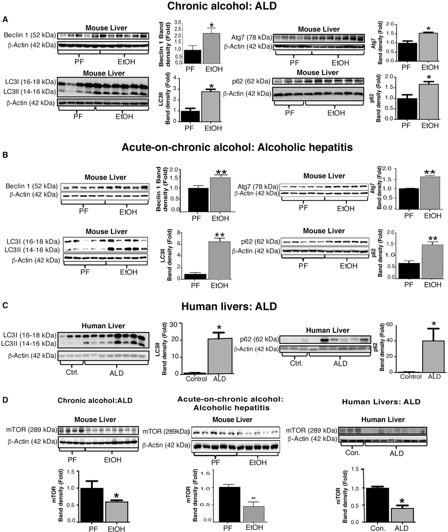
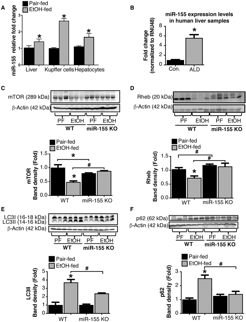
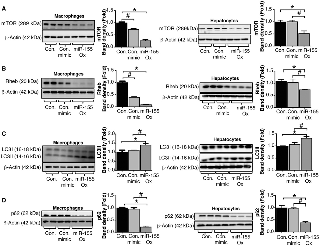
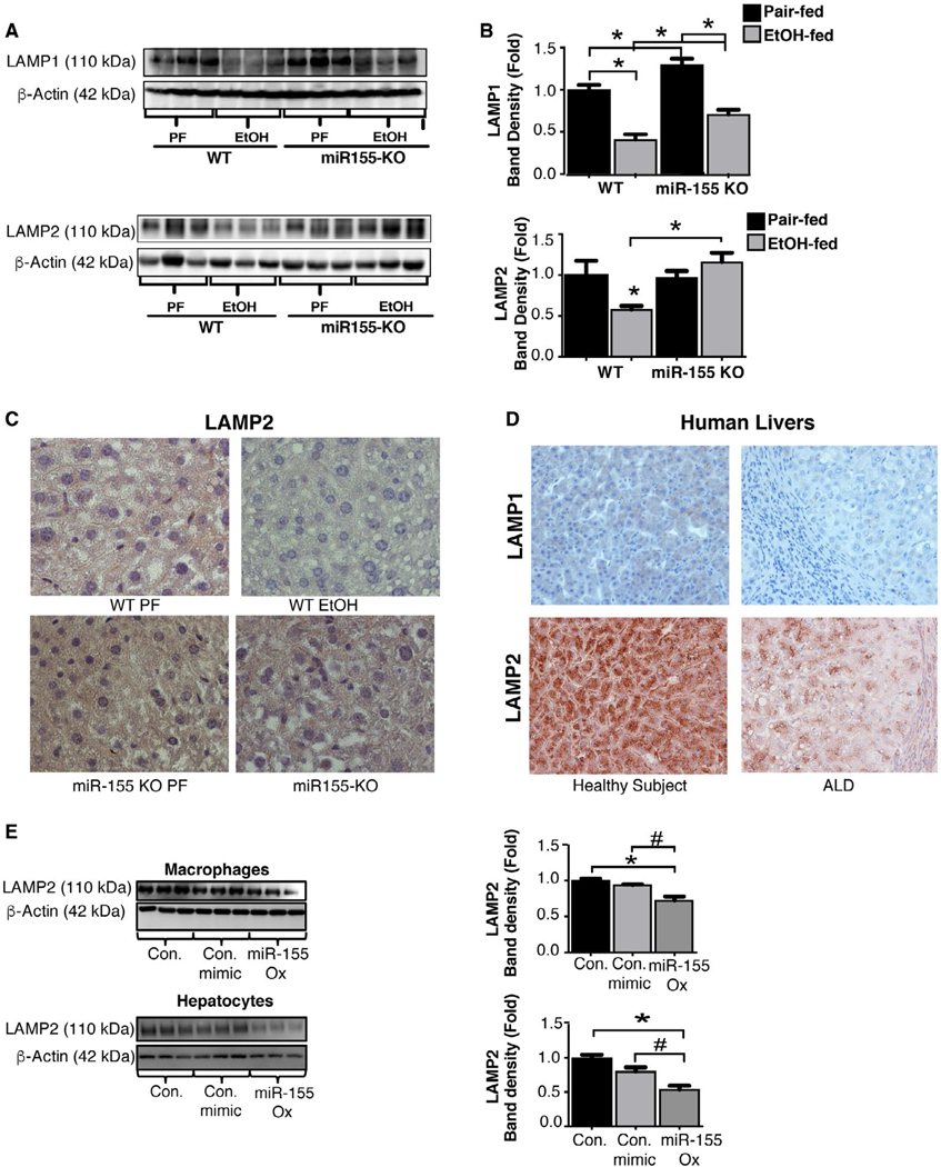
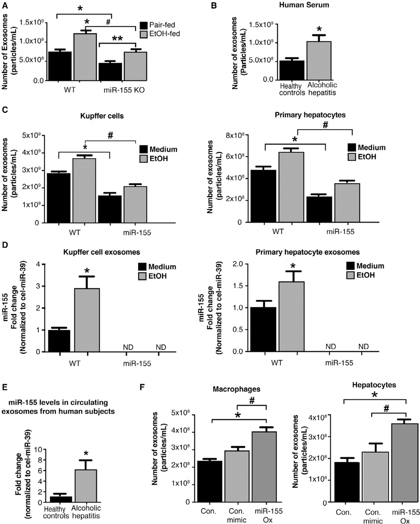
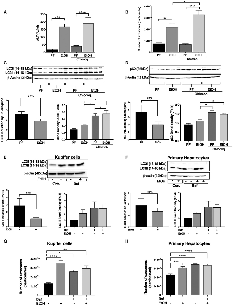
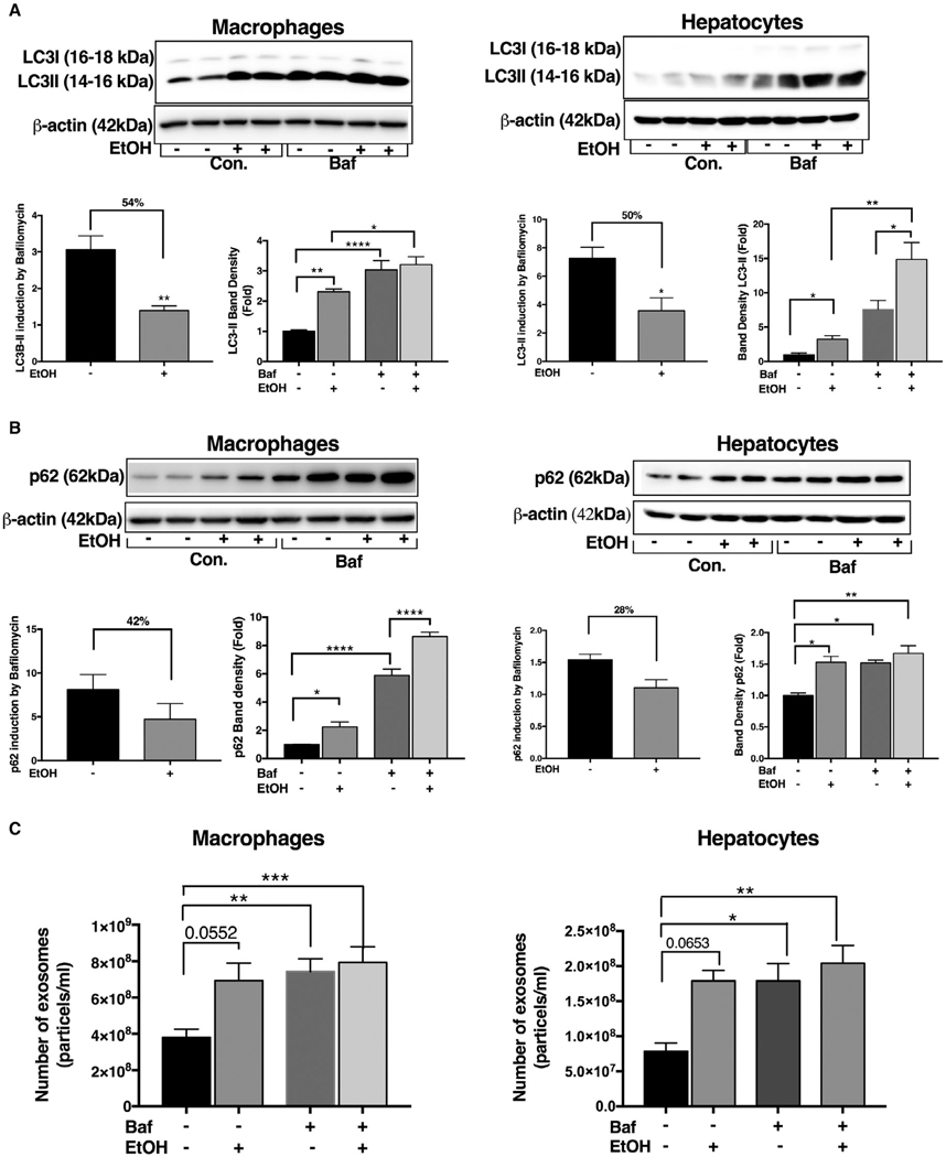
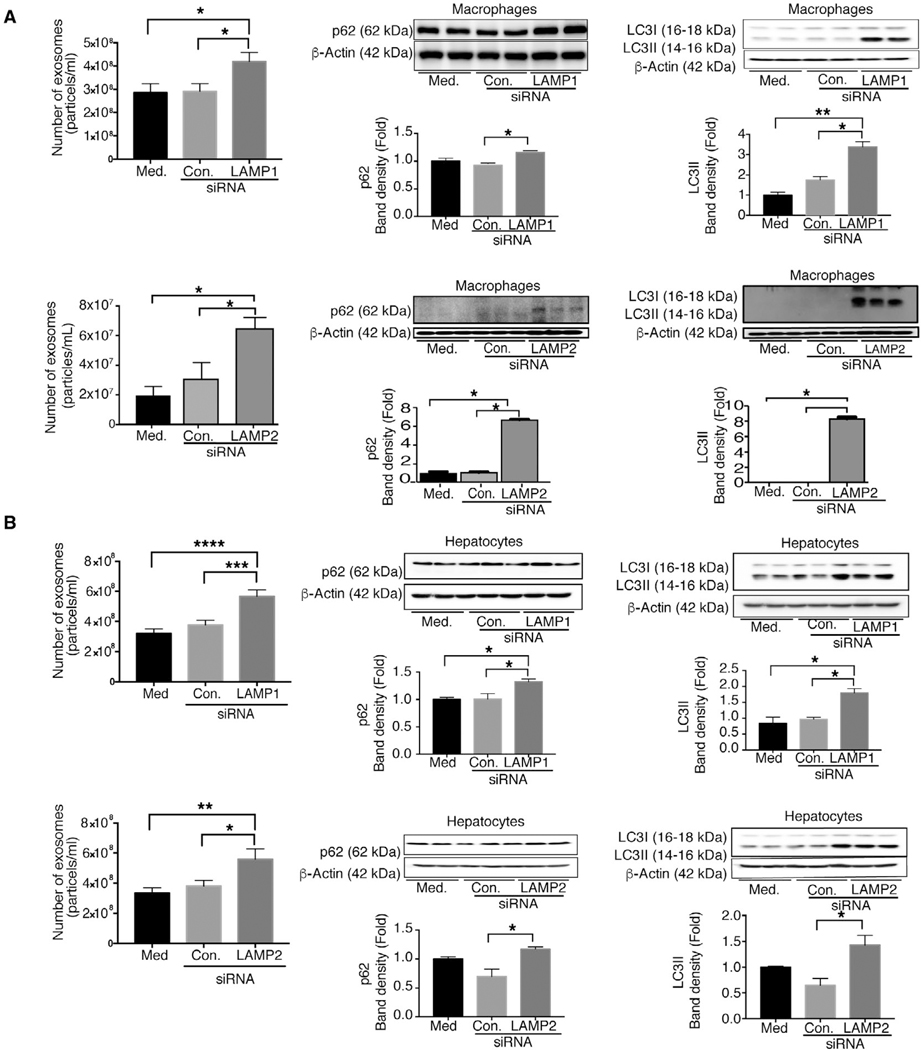
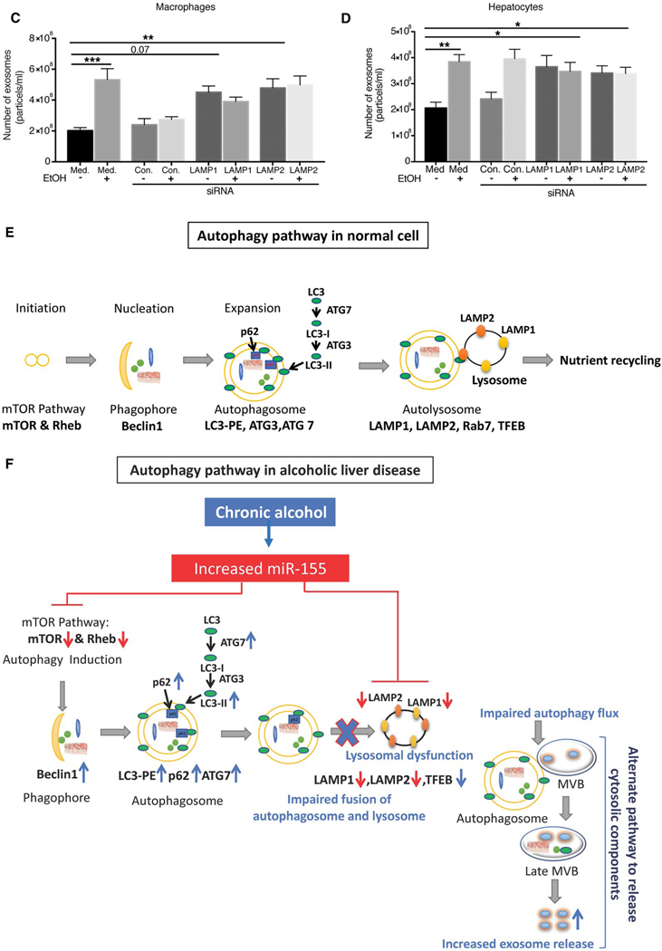
Cellular homeostais, that is normally maintained through autophagy, is disrupted in alcoholic liver disease (ALD). Because autophagy and exosome biogenesis share common elements, we hypothesized that increased exosome production in ALD may be linked to disruption of autophagic function. We found impaired autophagy both in ALD and alcoholic hepatitis (AH) mouse models and human livers with ALD as indicated by increased hepatic p62 and LC3-II levels. Alcohol reduced autophagy flux in vivo in chloroquine-treated mice as well as in vitro in hepatocytes and macrophages treated with bafilomycin A. Our results revealed that alcohol targets multiple steps in the autophagy pathway. Alcohol-related decrease in mechanistic target of rapamycin (mTOR) and Ras homolog enriched in brain (Rheb), that initiate autophagy, correlated with increased Beclin1 and autophagy-related protein 7 (Atg7), proteins involved in phagophore-autophagosome formation, in ALD. We found that alcohol disrupted autophagy function at the lysosomal level through decreased lysosomal-associated membrane protein 1 (LAMP1) and lysosomal-associated membrane protein 2 (LAMP2) in livers with ALD. We identified that micro-RNA 155 (miR-155), that is increased by alcohol, targets mTOR, Rheb, LAMP1, and LAMP2 in the authophagy pathway. Consistent with this, miR-155-deficient mice were protected from alcohol-induced disruption of autophagy and showed attenuated exosome production. Mechanistically, down-regulation of LAMP1 or LAMP2 increased exosome release in hepatocytes and macrophages in the presence and absence of alcohol. These results suggested that the alcohol-induced increase in exosome production was linked to disruption of autophagy and impaired autophagosome and lysosome function. Conclusion: Alcohol affects multiple genes in the autophagy pathway and impairs autophagic flux at the lysosome level in ALD. Inhibition of LAMP1 and LAMP2 promotes exosome release in ALD. We identified miR-155 as a mediator of alcohol-related regulation of autophagy and exosome production in hepatocytes and macrophages.


Conflict of interest statement

Potential conflict of interest: Dr. Szabo consults for and received grants from Allergan. She consultsfor Terra Firma, Glympse Bio, Quest, Arrow, GLG, Salix, and Tobira. She received grants from Gilead, Genfit, Intercept, Verlyx, Novartis, SignaBlok, and Shire. She holds intellectual property rights with Up to Date.

Figures

FIG. 1.
FIG. 1.
ALD in mice and humans is associated with impaired autophagic flux. (A,B) Total liver proteins were extracted from pair-fed (PF), chronic alcohol diet (EtOH), and acute-on-chronic alcohol (EtOH) fed mice (n = 8–10/group) and analyzed by western blotting using β-actin as a loading control. Protein levels of Beclinl, Atg7, LC3-I, LC3-II, and p62 in chronic alcohol (A) and in acute-on-chronic alcohol-fed (B) mice. Western blotting analysis for LC3-I, LC3-II, and p62 from livers of control subjects and ALD patients (C). mTOR protein levels in mouse livers in chronic alcohol- and in acute-on-chronic alcohol-fed mice and in ALD patients (D). The densitometry analysis is shown as bar diagrams. Human liver sample data are representative of 3 control donors and 6–8 patients with cirrhosis and superimposed AH. *P < 0.05; **P < 0.01. Abbreviations: Con., control; ctrl., control.
FIG. 2.
FIG. 2.
miR-155 deficiency prevents alcohol-induced impairment in autophagic flux in mice. Total RNA from livers, KCs, and hepatocytes from mice as well as liver samples from ALD patients (n = 8–10) analyzed for miR-155 expression by RT-qPCR (A,B). qCT values were normalized to snoRNA-202 for mouse samples and to RNU48 for human samples. Total liver protein was extracted from pair-fed (PF) or chronic alcohol-fed (EtOH) WT and miR-155 KO mice (n = 8–10) and analyzed by western blotting using β-actin as a loading control. Immunoblottings were probed with antibodies for mTOR (C), Rheb (D), LC3-I and LC3-II (E), and p62 (F). The densitometry analysis is shown as bar diagrams. *,#P < 0.05. Abbreviation: Con., control.
FIG. 3.
FIG. 3.
miR-155 regulates autophagy in macrophages and hepatocytes by targeting the mTOR pathway. Macrophages (RAW 264.7) or hepatocytes (Hepa1–6) were untreated or transfected with either a miR-155 mimic or a control mimic. Protein levels from untreated, control mimic, and miR-155 mimic transfected macrophages and hepatocytes were analyzed by western blotting for mTOR (A), Rheb (B), LC3-I and LC3-II (C), and p62 (D) using β-actin as a loading control (n = 7). The densitometry analysis is shown as bar diagrams. *,#P < 0.05. Abbreviations: Con., control; ox, overexpression.
FIG. 4.
FIG. 4.
Chronic alcohol-induced increase in miR-155 regulates autophagy at the autophagosome formation stage by targeting lysosome-associated membrane proteins. Total liver protein was extracted from pair-fed (PF) or chronic alcohol-fed (EtOH) WT and miR-155 KO mice (n = 8–10) and analyzed for LAMP2 and LAMP1 (A,B) by western blotting using β-actin as a loading control. Representative IHC images of LAMP2 from WT and miR-155 KO mice in livers (C). LAMP1 and LAMP2 IHC images from livers of healthy subjects and from patients with ALD (n = 64) (D). Macrophages (RAW 264.7) or hepatocytes (Hepa1–6) were untreated or transfected with either a miR-155 mimic or a control mimic and the LAMP2 protein levels determined by western blotting (E). *,#P < 0.05. Abbreviations: Con., control; ox, overexpression.
FIG. 5.
FIG. 5.
Alcohol-induced increased miR-155 regulates the cellular release of exosomes. NTA on the number of exosomes from sera of WT and miR-155 KO mice (n = 19; A) and the number of serum exosomes from AH patients (n = 8; B). NTA quantification of the number of exosomes in supernatants from KCs and hepatocytes (n = 15) after alcohol (50 mM, 24 hours) treatment (C). qPCR on miR-155 expression levels in exosomes isolated from KCs and primary hepatocytes (D) and in exosomes from sera of healthy subjects and patients with AH (n = 5–7; E); miR-155 levels were normalized to a spiked-in synthetic Cel-miR-39 used as an internal control. Macrophages (RAW 264.7) or hepatocytes (Hepa1–6) were untreated or transfected with either a miR-155 mimic or a control mimic and the number of exosomes in the supernatants quantified by NTA (n = 9) (F). *,#P < 0.05; **P < 0.01. Abbreviations: Con., control; ND, not detected; ox, overexpression.
FIG. 6.
FIG. 6.
Functional block of autophagy at the autophagosome-lysosome fusion step results in increased liver injury and exosome production in acute-on-chronic alcohol feeding. Serum ALT levels were used to determine the level of liver damage in acute-on-chronic alcohol feeding (A). The number of exosomes from sera of pair-fed (PF)- and acute-on-chronic alcohol-fed (EtOH)-fed mice with or without chloroquine injection was quantified by NTA (n = 8; B). Total liver protein was extracted from PF or EtOH mice (n = 6–8) with or without 65 mg/kg of chloroquine injected intraperitonealy and analyzed by western blotting using β-actin as a loading control. Immunoblottings were probed with antibodies for LC3-I and LC3-II (C) and p62 (D). The densitometry analysis is shown as bar diagrams. The LC3-II and P62 ratio in chloroquine treated to that of untreated alcohol-fed and pair-fed animals was calculated. (C,D) KCs and primary hepatocytes were treated with and without 50 mM of alcohol for 24 hours, and bafilomycin was added (in last 12 hours) as indicated. After 24 hours, the protein levels from KCs and primary hepatocytes were analyzed by western blotting for LC3-I andLC3-II (E,F) using β-actin as a loading control. The densitometry analysis is shown as bar diagrams. The LC3-II ratio was calculated (E,F). The number of exosomes from KCs and primary hepatocytes supernatants after 24 hours was quantified by NTA (G,H). *P < 0.05; **P < 0.01; ***P < 0.001; ****P < 0.0001. Abbreviations: Baf, bafilomycin; Chloroq., chloroquine; Con., control.
FIG. 7.
FIG. 7.
Inhibition of autophagy at the lysosomal level results in increased exosome production in macrophages and hepatocytes. Macrophages (RAW 264.7) and hepatocytes (Hepal-6) were treated with or without 50 mM of alcohol (EtOH) for 24 hours, and bafilomycin A (100 nM) was added in the last 12 hours of the experiment. After 24 hours, protein levels from macrophages and hepatocytes were analyzed by western blotting for LC3-I and L3-II (A) and p62 (B) using β-actin as a loading control. The densitometry analysis is shown as bar diagrams. The LC3-II and p62 ratio in bafilomycin-treated macrophages and hepatocytes to that of untreated macrophages and hepatocytes was calculated to show reduction in autophagic flux (B). The number of exosomes from macrophages and hepatocytes supernatants after 24 hours was quantified by NTA (C; n = 12). *P < 0.05; **P < 0.01; ***P < 0.001; ****P < 0.0001. Abbreviations: Baf, bafilomycin; Con., control.
FIG. 8.
FIG. 8.
Knockdown of LAMP1 and LAMP2 increases exosome production in macrophages and hepatocytes. Macrophages (RAW 264.7) and hepatocytes (HEPA 1.6) were untreated or transfected with control or LAMP1 or LAMP2 siRNA, and the number of exosomes from supernatants of LAMP1/LAMP2 siRNA-treated macrophages and hepatocytes was quantified by NTA (A,B; n = 6). p62 (A,B) and LC3-I and LC3-II (A,B) were analyzed by western blotting using β-actin as a loading control (n = 6). The densitometry analysis is shown as bar diagrams. The number of exosomes from supernatants of LAMP1 and LAMP2 siRNA-treated macrophages and hepatocytes in the presence and absence of alcohol was quantified by NTA (n = 9; C,D). *P < 0.05; **P < 0.01; ***P < 0.001; ****P < 0.0001. Schematic representation of miR-155-autophagy-exosome axis in ALD (E,F). Chronic alcohol induces miR-155, which then decreases mTOR and Rheb, thus inhibiting the mTOR pathway (E,F). Chronic alcohol increases Beclin1, Atg7, and LC3II expression. However, alcohol impairs autophagic flux by affecting LAMPs (LAMP1 and LAMP2) through miR-155. miR-155 regulates lysosome function by targeting LAMP1 and LAMP2. Because of impaired lysosome function, autophagosomes merge with MVBs to release their components outside the cell, and, as a consequence, there is increased exosomes secretion. Abbreviation: Med, medium.
FIG. 8.
FIG. 8.
Knockdown of LAMP1 and LAMP2 increases exosome production in macrophages and hepatocytes. Macrophages (RAW 264.7) and hepatocytes (HEPA 1.6) were untreated or transfected with control or LAMP1 or LAMP2 siRNA, and the number of exosomes from supernatants of LAMP1/LAMP2 siRNA-treated macrophages and hepatocytes was quantified by NTA (A,B; n = 6). p62 (A,B) and LC3-I and LC3-II (A,B) were analyzed by western blotting using β-actin as a loading control (n = 6). The densitometry analysis is shown as bar diagrams. The number of exosomes from supernatants of LAMP1 and LAMP2 siRNA-treated macrophages and hepatocytes in the presence and absence of alcohol was quantified by NTA (n = 9; C,D). *P < 0.05; **P < 0.01; ***P < 0.001; ****P < 0.0001. Schematic representation of miR-155-autophagy-exosome axis in ALD (E,F). Chronic alcohol induces miR-155, which then decreases mTOR and Rheb, thus inhibiting the mTOR pathway (E,F). Chronic alcohol increases Beclin1, Atg7, and LC3II expression. However, alcohol impairs autophagic flux by affecting LAMPs (LAMP1 and LAMP2) through miR-155. miR-155 regulates lysosome function by targeting LAMP1 and LAMP2. Because of impaired lysosome function, autophagosomes merge with MVBs to release their components outside the cell, and, as a consequence, there is increased exosomes secretion. Abbreviation: Med, medium.

References

    1. Szabo G, Bala S. Alcoholic liver disease and the gut-liver axis. World J Gastroenterol 2010;16:1321–1329. - PMC - PubMed
    1. Nagy LE. The role of innate immunity in alcoholic liver disease. Alcohol Res 2015;37:237–250. - PMC - PubMed
    1. Ding WX, Li M, Chen X, Ni HM, Lin CW, Gao W, et al. Autophagy reduces acute ethanol-induced hepatotoxicity and steatosis in mice. Gastroenterology 2010;139:1740–1752. - PMC - PubMed
    1. Lin CW, Zhang H, Li M, Xiong X, Chen X, Chen X, et al. Pharmacological promotion of autophagy alleviates steatosis and injury in alcoholic and non-alcoholic fatty liver conditions in mice. J Hepatol 2013;58:993–999. - PMC - PubMed
    1. Cho HI, Choi JW, Lee SM. Impairment of autophagosome- lysosome fusion contributes to chronic ethanol-induced liver injury. Alcohol 2014;48:717–725. - PubMed

Publication types

MeSH terms

Substances