. 2019 May 15;13(5):e0007397.
doi: 10.1371/journal.pntd.0007397. eCollection 2019 May.

Interplay between proinflammatory cytokines, miRNA, and tissue lesions in Anisakis-infected Sprague-Dawley rats

Affiliations

Interplay between proinflammatory cytokines, miRNA, and tissue lesions in Anisakis-infected Sprague-Dawley rats

Jerko Hrabar et al. PLoS Negl Trop Dis. .

Abstract

Background: Anisakiasis is an emerging public health problem, caused by Anisakis spp. nematode larvae. Anisakiasis presents as variable and unspecific gastrointestinal and/or allergic clinical symptoms, which accounts for the high rate of misdiagnosed cases.

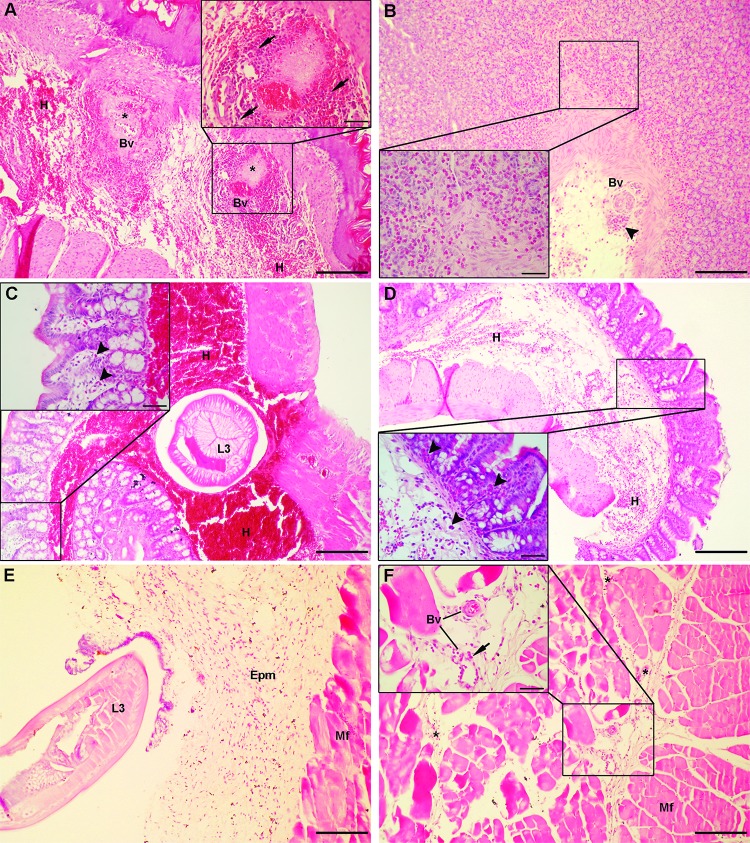
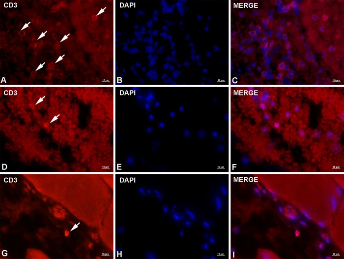
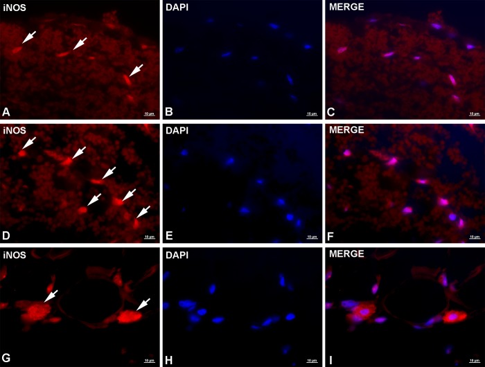
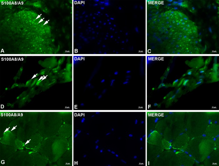
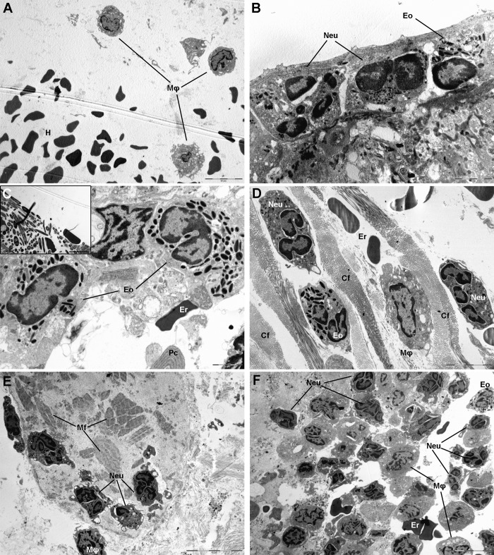
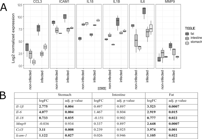
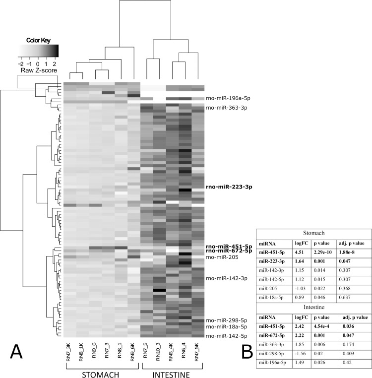
Methodology/principal findings: The aim of this study was to characterize the early cellular (6-72 h p.i.) and molecular (6 h p.i.) immune response and general underlying regulatory mechanism in Anisakis infected rats. Each Sprague-Dawley rat was infected with 10 Anisakis spp. larvae by gastric intubation. Tissues with visible lesions were processed for: i) classic histopathology (HE), immunofluorescence (CD3, iNOS, S100A8/A9), and transmission electron microscopy (TEM); ii) target genes (Il1b, Il6, Il18, Ccl3, Icam1, Mmp9) and microRNA (Rat Immunopathology MIRN-104ZF plate, Quiagen) expression analysis; and iii) global DNA methylation. Histopathology revealed that Anisakis larval migration caused moderate to extensive hemorrhages in submucosal and epimysial/perimysial connective tissue. In stomach and muscle, moderate to abundant mixed inflammatory infiltrate was present, dominated by neutrophils and macrophages, while only mild infiltration was seen in intestine. Lesions were characterized by the presence of CD3+, iNOS+, and S100A8/A9+ cells. The greatest number of iNOS+ and S100A8/A9+ cells was seen in muscle. Il6, Il1b, and Ccl3 showed particularly strong expression in stomach and visceral adipose tissues, but the order of expression differed between tissues. In total, three miRNAs were differentially expressed, two in stomach (miRNA-451 and miRNA-223) and two in intestine (miRNA-451 and miRNA-672). No changes in global DNA methylation were observed in infected tissues relative to controls.

Conclusions/significance: Anisakis infection induces strong immune responses in infected rats with marked induction of specific proinflammatory cytokines and miRNA expression. Deciphering the functional role of these cytokines and miRNAs will help in understanding the anisakiasis pathology and controversies surrounding Anisakis infection in humans.


Conflict of interest statement

The authors have declared that no competing interests exist.

Figures

Fig 1
Fig 1. Histopathological findings in rats infected with Anisakis spp. L3.
(A) Severe submucosal hemorrhage (H) in stomach at the stomach-esophageal junction, adjacent to the site of larval penetration with two necrotic foci (asterisks) around large blood vessels (Bv) (HE, 100x); (a) inset: high-power magnification of a necrotic focus showing vascular and perivascular necrosis, surrounded by abundant neutrophil and macrophage infiltrate (arrows) (400x); (B) Stomach of a rat euthanized 24 h p.i. with abundant eosinophil infiltrate in lamina propria and around blood vessels (Bv) (arrowhead) indicating recent extravasation (HE, 100x); (b) inset: detail of abundant eosinophil infiltrate (400x); (C) Severe submucosal hemorrhage (H) in intestine with a cross-section of Anisakis spp. larva (L3) (HE, 100x); (c) inset: detail of mucosa with rare eosinophils in lamina propria (arrowheads) (HE, 400x); (D) Extensive submucosal hemorrhage (H) in caecum (HE, 100x); (d) inset: detail of mucosa with abundant eosinophil infiltration (arrowheads) in lamina propria (HE, 400x); (E) Migration of Anisakis spp. larvae (L3) through epimysial connective tissue (Epm) causing moderate to extensive hemorrhage accompanied by mixed inflammatory infiltrate, composed mostly of neutrophils and macrophages (Mf = muscle fibers) (HE, 100x); (F) Muscle tissue adjacent to site of larval migration showing mild perimysial hemorrhage (asterisks) and focal necrosis of muscle fibers (Mf) (HE, 100x); (f) inset: detail of perimysial connective tissue with two blood vessels (Bv) surrounded by moderate neutrophil infiltrate (arrow) (400x). Scale bar (A, B, C, D, E, F) = 200 μm, scale bar (inset) = 50 μm.
Fig 2
Fig 2. Representative micrographs of CD3 immunolabeling in rats experimentally infected with Anisakis spp. third-stage larvae.
(A) Moderate CD3 expression on a large number of T lymphocytes (arrows) in stomach adjacent to blood vessel; (B) DAPI-stained cell nuclei; (C) Merge of (A) + (B); (D) Two lymphocytes in the intestinal submucosa showing strong CD3 expression (arrows); (E) DAPI-stained cell nuclei; (F) Merge of (D) + (E); (G) Single T lymphocyte with very strong CD3 expression in perimysial connective tissue (arrow); (H) DAPI-stained nuclei; (I) Merge of (G) + (H). Immunofluorescence, scale bar = 10 μm.
Fig 3
Fig 3. Representative micrographs of iNOS immunolabeling in rats experimentally infected with Anisakis spp. third-stage larvae.
(A) Moderate to strong granular cytoplasmic expression of iNOS in a low number of cells in stomach (arrows); (B) DAPI-stained cell nuclei; (C) Merge of (A) + (B); (D) Strong iNOS expression in intestinal cells adjacent to migrating Anisakis larvae (arrows); (E) DAPI-stained cell nuclei; (F) Merge of (D) + (E); (G) Very strong iNOS expression in perimysial connective tissue in cells most likely belonging to the mononuclear phagocyte system, adjacent to blood vessel (arrows); (H) DAPI-stained nuclei; (I) Merge of (G) + (H). Immunofluorescence, scale bar = 10 μm.
Fig 4
Fig 4. Representative micrographs of S100A8/A9 (MRP8+MRP14) immunolabeling in rats experimentally infected with Anisakis spp. third-stage larvae.
(A) Strong diffuse cytoplasmic labeling of S100A8/A9 in a low number of cells in the inner layer of the muscularis externa of rat stomach (arrows); (B) DAPI-stained cell nuclei; (C) Merge of (A) + (B); (D) Moderate diffuse cytoplasmic expression of S100A8/A9 in endothelial cells in intestinal submucosa; (E) DAPI-stained cell nuclei; (F) Merge of (D) + (E); (G) Very strong sarcoplasmic expression of S100A8/A9 in abdominal muscle myocytes (arrows); (H) DAPI-stained nuclei; (I) Merge of (G) + (H). Immunofluorescence, scale bar = 10 μm.
Fig 5
Fig 5. Transmission electron micrographs of lesions in rats experimentally infected with Anisakis spp. third-stage larvae.
(A) Hemorrhage (H) in lamina propria of stomach with three macrophages (Mφ); (B) Rat stomach adjacent to site of larval migration through stomach wall with neutrophil (Neu) and eosinophil (Eo) infiltration; (C) Rat intestine adjacent to site of larval migration through intestinal wall with a single erythrocyte (Er) indicative of hemorrhage and eosinophil (Eo) and macrophage (Mφ) infiltration; bottom of picture shows elaborate endoplasmic reticulum indicative of a plasma cell (Pc). Inset: numerous elongated fungus-like organisms with electron lucent intracytoplasmic granules; (D) Mixed inflammatory infiltrate of rat caecum composed of neutrophils (Neu), eosinophils (Eo) and macrophages (Mφ) interspersed between collagen fibers (Cf) and mixed with non-nucleated erythrocytes (Er) indicating hemorrhage; (E) Necrotic myocytes of abdominal muscle with fragmented muscle fibers (Mf); few neutrophils with outspread pseudopodia migrating over muscle fibers (Mf) and a single macrophage (Mφ) are visible; (F) Mixed inflammatory infiltrate of rat muscle dominated by neutrophils (Neu), and few eosinophils (Eo), macrophages (Mφ), and erythrocytes (Er), indicating hemorrhage. Osmium tetroxide/lead citrate, scale bar: (A, E, F) = 10 μm, scale bar (B, C) = 2 μm, scale bar (D, C inset) = 5 μm.
Fig 6
Fig 6. Expression of target genes in tissues of rats experimentally infected with Anisakis spp. third-stage larvae.
(A) Distribution of target gene expression in stomach, intestine, and visceral adipose tissues grouped according to state (infected or uninfected). Box plots represent distribution of log2-normalized expression; (B) Log2-transformed fold changes of target gene expression with respective adjusted p-values.
Fig 7
Fig 7. miRNAs expression in stomach and intestine of rats experimentally infected with Anisakis spp. third-stage larvae.
(A) Two-dimensional hierarchical clustering and heatmap of log2-normalized miRNA expression (Manhattan distance), grouped according to their expression profiles, tissue of origin (stomach and intestine), and tissue state (infected or uninfected). Color key ranges from white to light grey for downregulated (−2 to 0, Z-scores) and from light grey to black for upregulated (0–2, Z-scores) miRNAs. Only differentially expressed miRNAs (p < 0.05) in stomach and intestine are listed on the right (adjusted p-value < 0.05 in bold). (RN7_3, RN8_1, RN9_6: stomach, infected; RN7_3K, RN8_1K, RN9_6K: stomach, uninfected; RN6_4, RN7_5, RN10_3: intestine, infected; RN6_4K, RN7_5K: intestine, uninfected); (B) Log2-transformed fold changes with respective p-values and adjusted p-values of differentially expressed miRNAs in stomach and intestine.

References

    1. Broglia A, Kapel C. Changing dietary habits in a changing world: Emerging drivers for the transmission of foodborne parasitic zoonoses. Vet Parasitol. Elsevier B.V.; 2011;182: 2–13. 10.1016/j.vetpar.2011.07.011 - DOI - PubMed
    1. Chai J-Y, Murrell KD, Lymbery AJ. Fish-borne parasitic zoonoses: Status and issues. Int J Parasitol. 2005;35: 1233–1254. 10.1016/j.ijpara.2005.07.013 - DOI - PubMed
    1. WHO. First formal meeting of the Foodborne Disease Burden Epidemiology Reference Group (FERG). 2007;
    1. Torgerson PR, de Silva NR, Fèvre EM, Kasuga F, Rokni MB, Zhou X-N, et al. The global burden of foodborne parasitic diseases: an update. Trends Parasitol. 2014;30: 20–26. 10.1016/j.pt.2013.11.002 - DOI - PubMed
    1. EFSA Panel on Biological Hazards. Scientific opinion on risk assessment of parasites in fishery products. EFSA J. 2010;8: 1543.

Publication types

MeSH terms