. 2019 May 14;27(7):2029-2035.e5.
doi: 10.1016/j.celrep.2019.04.074.

Mesenchymal Stromal Cells Are Required for Regeneration and Homeostatic Maintenance of Skeletal Muscle

Affiliations

Mesenchymal Stromal Cells Are Required for Regeneration and Homeostatic Maintenance of Skeletal Muscle

Michael N Wosczyna et al. Cell Rep. .

Abstract

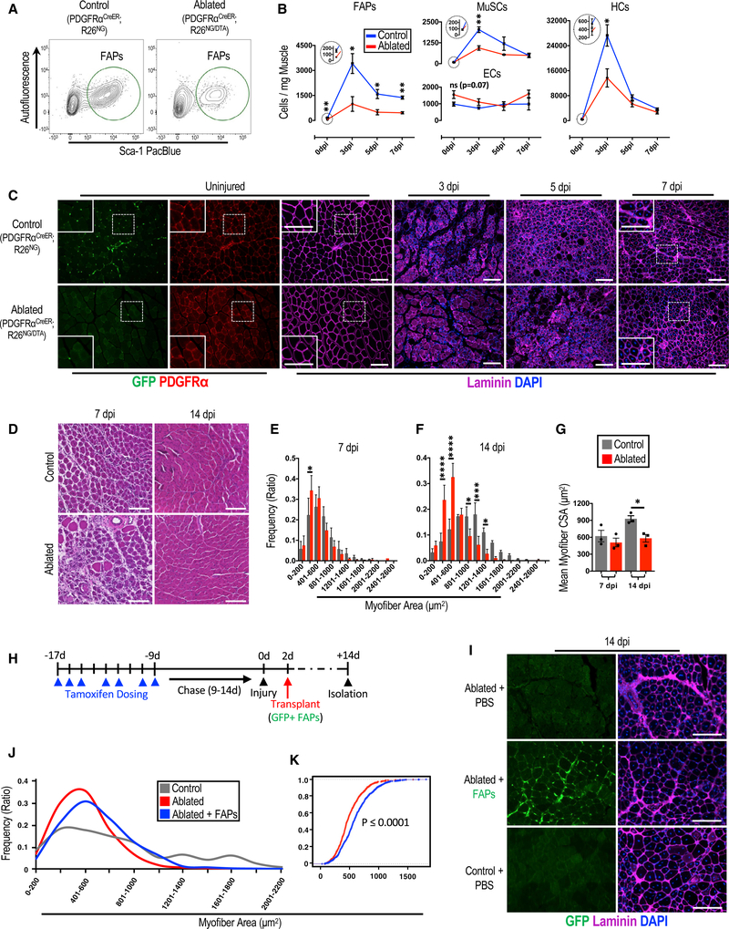
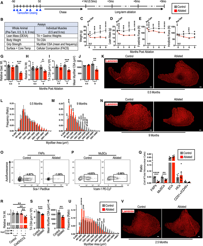
The necessity of mesenchymal stromal cells, called fibroadipogenic progenitors (FAPs), in skeletal muscle regeneration and maintenance remains unestablished. We report the generation of a PDGFRαCreER knockin mouse model that provides a specific means of labeling and targeting FAPs. Depletion of FAPs using Cre-dependent diphtheria toxin expression results in loss of expansion of muscle stem cells (MuSCs) and CD45+ hematopoietic cells after injury and impaired skeletal muscle regeneration. Furthermore, FAP-depleted mice under homeostatic conditions exhibit muscle atrophy and loss of MuSCs, revealing that FAPs are required for the maintenance of both skeletal muscle and the MuSC pool. We also report that local tamoxifen metabolite delivery to target CreER activity in a single muscle, removing potentially confounding systemic effects of ablating PDGFRα+ cells distantly, also causes muscle atrophy. These data establish a critical role of FAPs in skeletal muscle regeneration and maintenance.

Keywords: FAP; Mesenchymal; PDGFRα; fibroadipogenic progenitor; local recombination; muscle; niche; satellite cell; stem cell; stromal.


Figures

Figure 1.
Figure 1.. FAPs Are Required for Efficient Skeletal Muscle Regeneration
(A-C) The effects of depletion of FAPs on skeletal muscle. (A) FACS plots showing the efficiency of ablating FAPs. (B) A 7-day time course examining the influence of FAP ablation on the noted cellular fractions following skeletal muscle injury in ablated (PDGFRαCreER/+; R26NG/DTA or +/DTA) and control mice (n = 5–6 [0 dpi], 4–5 [3 dpi], and 3 [5 and 7 dpi]). The large circles are magnified views of areas in the small circles at 0 dpi for each respective graph. (C) Immunofluorescence images of TA muscle cross sections from ablated and control mice corresponding to the time points in (B). Note the decrease in PDGFRα+ cells (punctate interstitial staining) in uninjured tissue from ablated mice and impaired myofiber necrosis and regeneration in the ablated tissue at 3 dpi and 7 dpi, respectively. (D-G) Histological analyses of skeletal muscle regeneration from ablated and control mice. (D) H&E staining of TA muscle cross sections from ablated and control mice at 7 and 14 dpi. (E-G) Myofiber cross-sectional area (CSA) plotted as discrete sizes (E, 7 dpi; and F, 14 dpi) and overall means (G) (n = 3). (H-K) Transplantation of FAPs into skeletal muscle of FAP-ablated mice to rescue regeneration impairment. (H) Experimental design of the transplantation experiments. (I) Immunofluorescent images showing GFP+ FAPs transplanted into the TA muscle. (J) Quantification of myofiber CSAs from each experimental group, shown as a line histogram (n = 3). (K) An empirical cumulative distribution function graph of all myofiber CSAs from the noted groups, with significance determined by a KS test (n = 681 [ablated] and 857 [ablated + FAPs]). Scale bars, 100 μm. Error bars represent ± SEM. *p ≤ 0.05, **p ≤ 0.01, ***p ≤ 0.001, ****p ≤ 0.0001.
Figure 2.
Figure 2.. FAPs Are Necessary for Long-Term Maintenance and Normal Growth of Skeletal Muscle
(A) Experimental design of the long-term FAP depletion experiments. (B) Assays used to test the effect of FAP depletion on the whole animal and the noted skeletal muscles. (C-F) Assessments of animal and muscle parameters (C, weight; D, lean mass; E, forelimb force; and F, hindlimb force) overtime before and after FAP depletion (n = 20–26 [pre-(Tam)], 17–24 [0.5 months], 6–9 [3 months], 5–9 [6 months], and 5–8 [9 months]). (G-N) Assessment of muscle parameters 0.5 and 9 months after FAP depletion. Wt., weight; Gast., gastrocnemius. n = 4 (G), n = 4–9 (H), n = 3–6 (I and J), and n = 3–5 (L and M). (K and N) Representative immunofluorescence images (K, 0.5 months; and N, 9 months) of TA cross-sections analyzed in bar graphs of (I)-(N). (O-Q) The consequence of FAP ablation on cellular fractions of skeletal muscle. (O and P) Representative FACS plots of (O) MuSCs and (P) FAPs 9 months after FAP ablation (quantified in Q). (Q) Quantification of mononuclear cell composition of skeletal muscle 9 months after FAP ablation (n = 4–6). (R-U) Assessment of muscle parameters (R, TA weight; S, TACSA; T, mean my ofiber CSA; and U, my ofiber CSA distribution)following localized FAP depletion in TA muscles. Impl, implant; Ctrl, control-PCL implants; Edx, endoxifen-PCL implants; CreER;DTA, PDGFRαCreER;R26 NG/DTA or +/DTA mice. n = 5 (R-T) and n = 7 (U). (V) Representative immunofluorescent images of TA muscle cross-sections analyzed in (R)-(U). Ablated and control notations in (S)-(V) refer to TA muscles targeted with Edx-PCL from CreER;DTA mice and control mice, respectively. Scale bars, 200 μm. Error bars represent ± SEM. *p ≤ 0.05, **p ≤ 0.01,***p ≤ 0.001, ****p ≤ 0.0001.

References

    1. Butler A, Hoffman P, Smibert P, Papalexi E, and Satija R. (2018). Integrating single-cell transcriptomic data across different conditions, technologies, and species. Nat. Biotechnol 36, 411–420. - PMC - PubMed
    1. Carlson BM, and Faulkner JA (1989). Muscle transplantation between young and old rats: age of host determines recovery. Am. J. Physiol 256, C1262–C1266. - PubMed
    1. Chakkalakal JV, Jones KM, Basson MA, and Brack AS (2012). The aged niche disrupts muscle stem cell quiescence. Nature 490, 355–360. - PMC - PubMed
    1. Farley FW, Soriano P, Steffen LS, and Dymecki SM (2000). Widespread recombinase expression using FLPeR (flipper) mice. Genesis 28, 106–110. - PubMed
    1. Frontera WR, and Ochala J. (2015). Skeletal muscle: a brief review of structure and function. Calcif. Tissue Int 96, 183–195. - PubMed

Publication types