

Graphene oxide containing self-assembling peptide hybrid hydrogels as a potential 3D injectable cell delivery platform for intervertebral disc repair applications
- PMID: 31091473
- PMCID: PMC6582688
- DOI: 10.1016/j.actbio.2019.05.004
Graphene oxide containing self-assembling peptide hybrid hydrogels as a potential 3D injectable cell delivery platform for intervertebral disc repair applications
Abstract
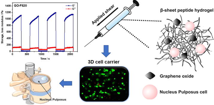
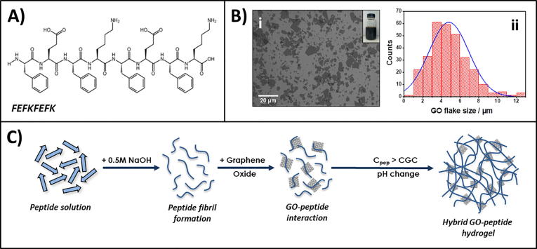
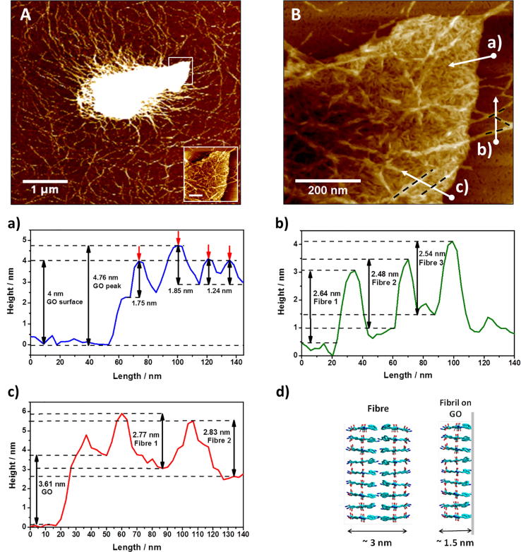
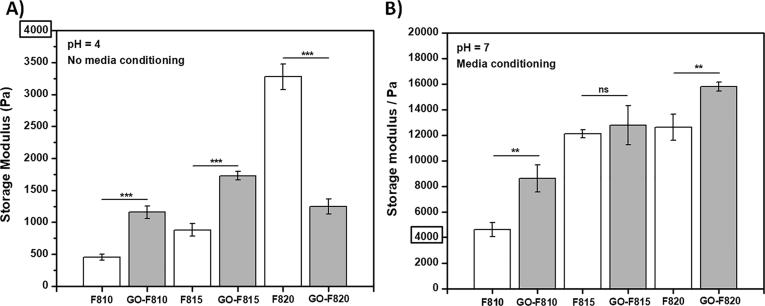
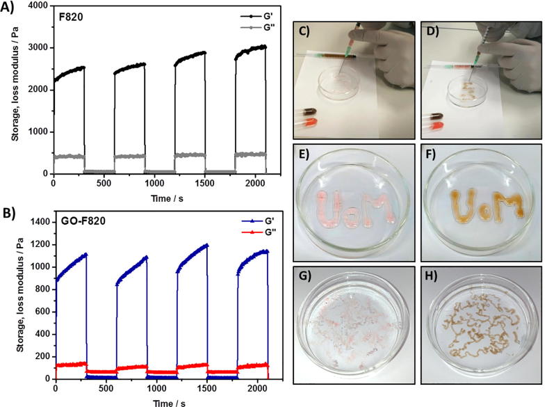
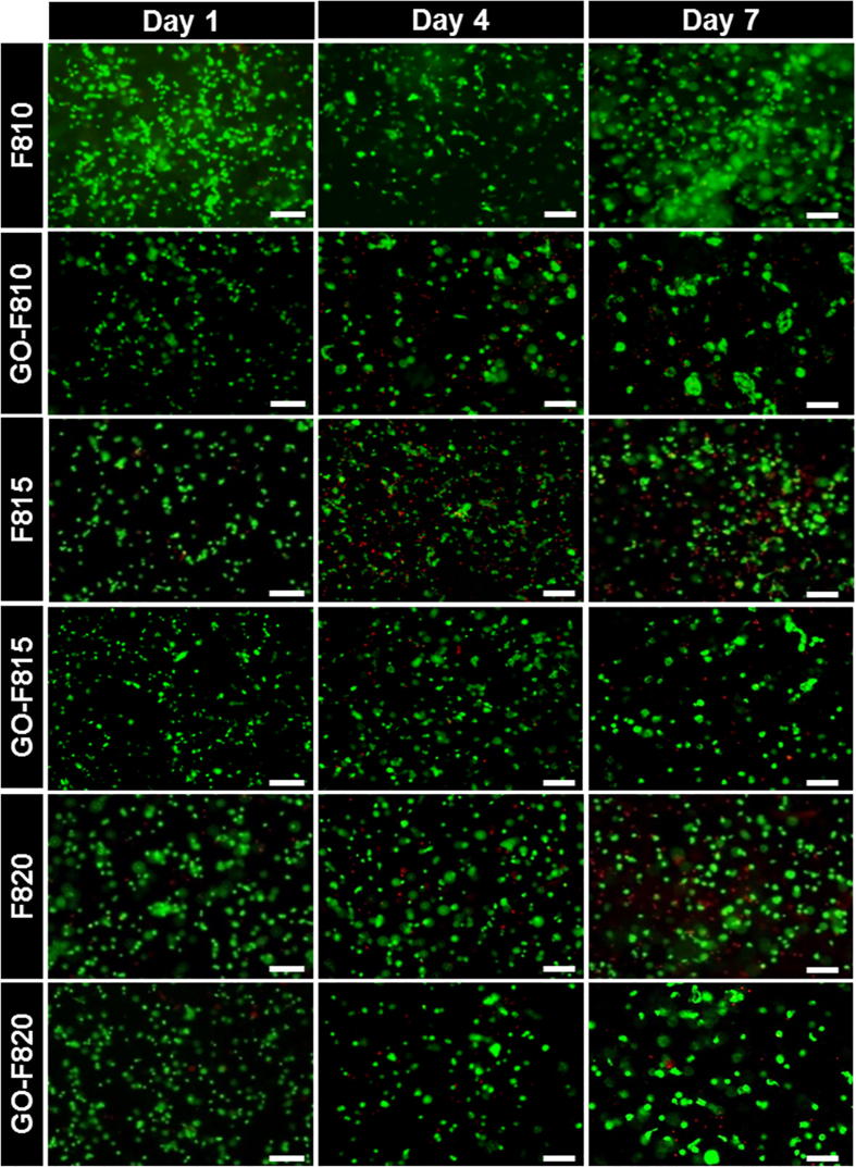
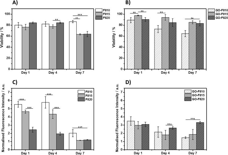
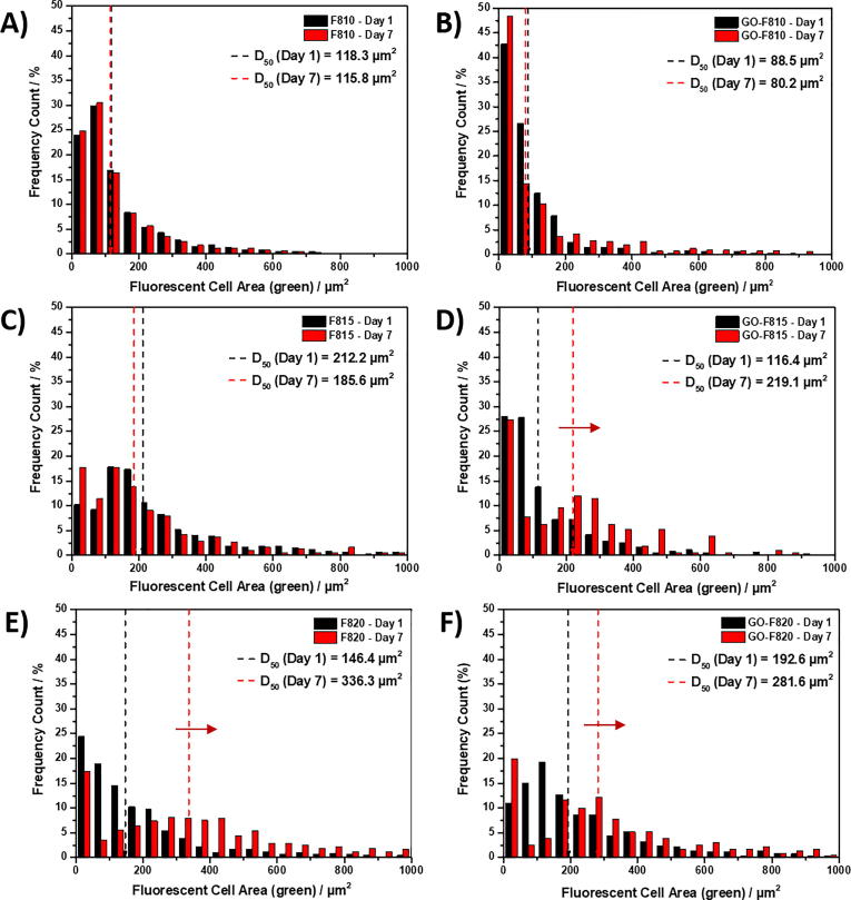
Cell-based therapies have shown significant promise in tissue engineering with one key challenge being the delivery and retention of cells. As a result, significant efforts have been made in the past decade to design injectable biomaterials to host and deliver cells at injury sites. Intervertebral disc (IVD) degeneration, a major cause of back pain, is a particularly relevant example where a minimally-invasive cellular therapy could bring significant benefits specifically at the early stages of the disease, when a cell-driven process starts in the gelatinous core of the IVD, the nucleus pulposus (NP). In this present study we explore the use of graphene oxide (GO) as nano-filler for the reinforcement of FEFKFEFK (β-sheet forming self-assembling peptide) hydrogels. Our results confirm the presence of strong interactions between FEFKFEFK and GO flakes with the peptide coating and forming short thin fibrils on the surface of the flakes. These strong interactions were found to affect the bulk properties of hybrid hydrogels. At pH 4 electrostatic interactions between the peptide fibres and the peptide-coated GO flakes are thought to govern the final bulk properties of the hydrogels while at pH 7, after conditioning with cell culture media, electrostatic interactions are removed leaving the hydrophobic interactions to govern hydrogel final properties. The GO-F820 hybrid hydrogel, with mechanical properties similar to the NP, was shown to promote high cell viability and retained cell metabolic activity in 3D over the 7 days of culture and therefore shown to harbour significant potential as an injectable hydrogel scaffold for the in-vivo delivery of NP cells. STATEMENT OF SIGNIFICANCE: Short self-assembling peptide hydrogels (SAPHs) have attracted significant interest in recent years as they can mimic the natural extra-cellular matrix, holding significant promise for the ab initio design of cells' microenvironments. Recently the design of hybrid hydrogels for biomedical applications has been explored through the incorporation of specific nanofillers. In this study we exploited graphene oxide (GO) as nanofiller to design hybrid injectable 3Dscaffolds for the delivery of nucleus pulposus cells (NPCs) for intervertebral disc regeneration. Our work clearly shows the presence of strong interactions between peptide and GO, mimicking the mechanical properties of the NP tissue and promoting high cell viability and metabolic activity. These hybrid hydrogels therefore harbour significant potential as injectable scaffolds for the in vivo delivery of NPCs.
Keywords: Cell culture; Cell delivery; Graphene oxide; Hydrogels; Injectable; Nucleus pulposus; Peptide; Tissue engineering.
Copyright © 2019 Acta Materialia Inc. Published by Elsevier Ltd. All rights reserved.
Figures


References
-
- Buzhor E., Leshansky L., Blumenthal J., Barash H., Warshawsky D., Mazor Y., Shtrichman R. Cell-based therapy approaches: the hope for incurable diseases. Regen. Med. 2014;9:649–672. - PubMed
- E. Buzhor, L. Leshansky, J. Blumenthal, H. Barash, D. Warshawsky, Y. Mazor, R. Shtrichman, Cell-based therapy approaches: the hope for incurable diseases., Regen. Med. 9 (2014) 649–72. doi:10.2217/rme.14.35. - PubMed
-
- Cheung K.M.C., Karppinen J., Chan D., Ho D.W.H., Song Y.-Q., Sham P., Cheah K.S.E., Leong J.C.Y., Luk K.D.K. Prevalence and pattern of lumbar magnetic resonance imaging changes in a population study of one thousand forty-three individuals. Spine (Phila. Pa. 1976) 2009;34:934–940. - PubMed
- K.M.C. Cheung, J. Karppinen, D. Chan, D.W.H. Ho, Y.-Q. Song, P. Sham, K.S.E. Cheah, J.C.Y. Leong, K.D.K. Luk, Prevalence and pattern of lumbar magnetic resonance imaging changes in a population study of one thousand forty-three individuals., Spine (Phila. Pa. 1976). 34 (2009) 934–940. doi:10.1097/BRS.0b013e3181a01b3f. - PubMed
-
- Richardson S.M., Freemont A.J., Hoyland J.A. Pathogenesis of intervertebral disc degeneration. In: Shapiro I.M., Risbud M.V., editors. Intervertebral Disc Mol. Struct. Stud. Disc Heal. Dis. Springer; Vienna, Vienna: 2014. pp. 177–200.
- S.M. Richardson, A.J. Freemont, J.A. Hoyland, Pathogenesis of Intervertebral Disc Degeneration, in: I.M. Shapiro, M. V Risbud (Eds.), Intervertebral Disc Mol. Struct. Stud. Disc Heal. Dis., Springer Vienna, Vienna, 2014: pp. 177–200. doi:10.1007/978-3-7091-1535-0_11.
Publication types
MeSH terms
Substances
Grants and funding
LinkOut - more resources
Full Text Sources
Research Materials
Miscellaneous
