Review
. 2019 May 14;8(5):681.
doi: 10.3390/jcm8050681.

Immuno-Imaging to Predict Treatment Response in Infection, Inflammation and Oncology

Affiliations
Review

Immuno-Imaging to Predict Treatment Response in Infection, Inflammation and Oncology

Alberto Signore et al. J Clin Med. .

Abstract

Background: Molecular nuclear medicine plays a pivotal role for diagnosis in a preclinical phase, in genetically susceptible patients, for radio-guided surgery, for disease relapse evaluation, and for therapy decision-making and follow-up. This is possible thanks to the development of new radiopharmaceuticals to target specific biomarkers of infection, inflammation and tumour immunology.

Methods: In this review, we describe the use of specific radiopharmaceuticals for infectious and inflammatory diseases with the aim of fast and accurate diagnosis and treatment follow-up. Furthermore, we focus on specific oncological indications with an emphasis on tumour immunology and visualizing the tumour environment.

Results: Molecular nuclear medicine imaging techniques get a foothold in the diagnosis of a variety of infectious and inflammatory diseases, such as bacterial and fungal infections, rheumatoid arthritis, and large vessel vasculitis, but also for treatment response in cancer immunotherapy.

Conclusion: Several specific radiopharmaceuticals can be used to improve diagnosis and staging, but also for therapy decision-making and follow-up in infectious, inflammatory and oncological diseases where immune cells are involved. The identification of these cell subpopulations by nuclear medicine techniques would provide personalized medicine for these patients, avoiding side effects and improving therapeutic approaches.

Keywords: infection; inflammation; personalised medicine; therapy follow-up; tumour immunology; tumour microenvironment.


Conflict of interest statement

The authors declare no conflict of interest.

Figures

Figure 1
Figure 1
38-year-old male, known with acute lymphatic leukaemia. Because of fever and bacteraemia he was treated with meropenem (antibiotics). High-resolution computed tomography (HR-CT) (left figure): several lung densities with irregular borders, not typical for bacterial infections, but typical for angio-invasive aspergillosis, because of the halo sign. 18F-fluorodeoxyglucose positron emission tomography/computed tomography (18F-FDG-PET) (right figure): not only lung involvement but also infectious lesions in the liver, spleen and in muscles throughout the body.
Figure 2
Figure 2
20-year-old female, known with recurrent acute lymphatic leukaemia. After first chemotherapy cycle fever and neutropenia. Blood cultures: candidia. Left scan: infectious foci around catheter in the oesophagus and heterogeneous uptake in the kidneys. Scan 2, 6 weeks after treatment with fluconazole: progressive disease with now involvement of the lungs, spleen and kidneys. Biopsy of the parenchyma of the right kidney confirmed renal candidiasis. Caspofungin was added to the antifungal treatment. Scan 3, 3 months after treatment with fluconazole and caspofungin: improvement, but still involvement of spleen and kidneys. Treatment continued. Scan 4, after 6 months treatment: no active infection anymore.
Figure 3
Figure 3
Scintigraphy with 99mTc-infliximab before (a) and 4 months after (b) intra-articular administration of infliximab. Red colour represents uptake of the 99mTc-infliximab in scintigraphy (from Conti, F.; et al. Arthritis Rheum, 2005, 52, 1224–1226 [68]).
Figure 4
Figure 4
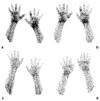
Scintigraphic images of wrists of a rheumatoid arthritis (RA) patient injected with (99mTc)-adalimumab (anti-TNFα mAb) before (a and b; dorsal images after 6 and 20 h p.i., respectively) and 3 months after systemic therapy with adalimumab (c and d; dorsal images after 6 h and 20 h p.i., respectively), (from Malviya, G.; et al. Q. J. Nucl. Med. Mol. Imaging 2008, 52, 13–14) [71].
Figure 5
Figure 5
Female, 64 years-old; (18F)-FDG-PET/CT MIP in giant cell arteritis (GCA) before (A) and after (B) immuno-suppressive treatment; ascending and descending aorta (white arrows), before (C1) and after (C2) treatment; aortic arc (yellow arrow), before (D1) and after (D2) treatment; abdominal aorta (white arrow-head), before (E1) and after (E2) treatment.

References

    1. Signore A., Anzola K.L., Auletta S., Varani M., Petitti A., Pacilio M., Galli F., Lauri C. Current status of molecular imaging in inflammatory disorders. Curr. Pharm. Des. 2018;24:743–753. doi: 10.2174/1381612824666180130115153. - DOI - PubMed
    1. Glaudemans A.W., Prandini N., Di Girolamo M., Argento G., Lauri C., Lazzeri E., Muto M., Sconfienza L.M., Signore A. Hybrid imaging of musculoskeletal infections. Q. J. Nucl. Med. Mol. Imaging. 2018;62:3–13. - PubMed
    1. Chianelli M., Boerman O.C., Malviya G., Galli F., Oyen W.J., Signore A. Receptor binding ligands to image infection. Curr. Pharm. Des. 2008;14:3316–3325. doi: 10.2174/138161208786549416. - DOI - PubMed
    1. Ruf J., Oeser C., Amthauer H. Clinical role of anti-granulocyte MoAb versus radiolabeled white blood cells. Q. J. Nucl. Med. Mol. Imaging. 2010;54:599–616. - PubMed
    1. Malherbe C., Dupont A.C., Maia S., Venel Y., Erra B., Santiago-Ribeiro M.J., Arlicot N. Estimation of the added value of 99mTc-HMPAO labelled white blood cells scintigraphy for the diagnosis of infectious foci. Q. J. Nucl. Med. Mol. Imaging. 2017;61 doi: 10.23736/S1824-4785.17.02964-8. - DOI - PubMed

LinkOut - more resources