. 2019 Jun 3;218(6):2051-2070.
doi: 10.1083/jcb.201902017. Epub 2019 May 15.

Proteome of the central apparatus of a ciliary axoneme

Affiliations

Proteome of the central apparatus of a ciliary axoneme

Lei Zhao et al. J Cell Biol. .

Abstract

Nearly all motile cilia have a "9+2" axoneme containing a central apparatus (CA), consisting of two central microtubules with projections, that is essential for motility. To date, only 22 proteins are known to be CA components. To identify new candidate CA proteins, we used mass spectrometry to compare axonemes of wild-type Chlamydomonas and a CA-less mutant. We identified 44 novel candidate CA proteins, of which 13 are conserved in humans. Five of the latter were studied more closely, and all five localized to the CA; therefore, most of the other candidates are likely to also be CA components. Our results reveal that the CA is far more compositionally complex than previously recognized and provide a greatly expanded knowledge base for studies to understand the architecture of the CA and how it functions. The discovery of the new conserved CA proteins will facilitate genetic screening to identify patients with a form of primary ciliary dyskinesia that has been difficult to diagnose.


Figures

Figure 1.
Figure 1.
Structural characterization of WT and pf18 axonemes used for MS analysis. (A and B) Representative TEMs of isolated axonemes of WT (A) and pf18 (B). Nearly all WT axonemes contained a CA, whereas all pf18 axonemes lacked the CA (see Fig. S1 for quantitation). The axonemes of both preparations were highly pure, with little apparent cell body contamination. Insets: Higher-magnification images confirming integrity of the 9+2 structure in WT axonemes and the 9+0 structure in pf18 axonemes. The lower inset in B shows that the electron-dense material in the lumens of some pf18 axonemes is not the CA. Bars, 0.5 µm (A and B); 0.1 µm (insets).
Figure 2.
Figure 2.
pf18:WT ratios for select flagellar proteins, abundances for known CA proteins, and criteria used to screen for candidate novel CA proteins. (A) pf18:WT ratios for axonemal and IFT proteins (in this example, the ratios are based on IBAQ scores from replicate 1; see Table S2). Ratios for subunits such as LC7 and LC8, which are present in more than one axonemal structure, are repeated for each structure with which they are associated. The four CA proteins with the highest ratios (calmodulin, enolase, HSP70, and PP1c, in order of decreasing ratios) are located both in the CA and elsewhere in the flagellum. NAP, an unconventional actin previously shown to be a subunit of some inner arm dyneins in the absence of conventional actin, is grouped separately. Our first criterion for considering an uncharacterized protein as a potentially novel CA component was that it had to have a pf18:WT ratio ≤0.2 (red line), ensuring that it is reduced in pf18 axonemes at least as much as PP1c and the known CA-specific proteins. (B) IBAQ values, representing protein abundance, for known CA proteins (values from replicate 1 used as example here). Based on both IBAQ and Top3 values, the least abundant of the known CA proteins in replicate 1 was FAP297. Therefore, our second criterion for considering an uncharacterized protein as a potentially novel CA component was that it had to have an abundance based on IBAQ or Top3 score ≥0.8 of the FAP297 value (red line).
Figure 3.
Figure 3.
Assignment of novel candidate CA proteins to the C1 or C2 microtubule. The results of the two different approaches described in the text were combined to predict association with either the C1 or the C2 microtubule. Each approach was repeated twice to provide two biological replicates (Rep. 1 and Rep. 2), and each replicate was analyzed using IBAQ and Top3 methods. Proteins with similar ratios were then grouped manually into the clusters shown. The soluble:insoluble ratios for replicates 1 and 2 are indicated by green and blue bars, respectively; the pf16:WT ratios for replicates 1 and 2 are indicated by purple and red bars, respectively. For each replicate, the ratios as reported by IBAQ and Top3 are shown in that order using the same color. To the left of each bar graph, known C1 proteins are indicated by a light-red background; known C2 proteins have a light-green background. FAP174, indicated by a light-blue background, previously was assigned to C2 (Rao et al., 2016), but this assignment does not fully agree with our other results (see Some candidate proteins can be assigned to specific CA projections). Candidate CA proteins in Cluster 1 are predicted to be C1 proteins, whereas those in Cluster 3 are predicted to be C2 proteins. The locations of candidate CA proteins in the other clusters are less certain.
Figure 4.
Figure 4.
Phenotypic analysis of insertional mutants and rescued strains. (A) Flagellar length of WT, the fap47-1, fap76-1, fap99-1, fap196-1, fap246-1, and dpy30-1 insertional mutants, and the mutants following transformation with constructs designed to express WT HA-tagged versions of the proteins defective in each. n = number of flagella scored; error bars indicate standard deviation; *, significant difference from WT (Student’s t test, P ≤ 0.05). (B) Swimming speed of the same strains as in A. n = number of cells scored; error bars indicate standard deviation; *, significant difference (Student’s t test, P ≤ 0.05) between WT and mutant or mutant and rescued strains as indicated. (C) Swimming paths of WT, the fap76-1 mutant, and the mutant following transformation with the construct expressing FAP76-HA. The exposure time was 1 s; bar, 50 µm. (D) Swimming paths of WT, the fap47-1 mutant, and the mutant following transformation with the construct expressing FAP47-HA, in the absence of photostimulation (top panels) and in the presence of photostimulation coming from the left (arrow; lower panels). The large dot in each swimming path indicates the end of the track. fap47-1 has a slow-swimming phenotype that was partially rescued by FAP47-HA; the phototaxis defect was not noticeably rescued. The exposure time was 0.5 s; bar, 50 µm.
Figure 5.
Figure 5.
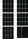
Localization of selected candidate CA proteins to the CA. (A–E) SIM images of isolated axonemes from mutant cells rescued with FAP47-HA (A), FAP76-HA (B), FAP99-HA (C), FAP246-HA (D), and DPY30-HA (E). Each set of images shows control intact axonemes from the nonrescued mutant (subpanels A, A′, A″), intact axonemes from the rescued mutant (subpanels B, B′, B″), and axonemes from the rescued mutant after treatment with ATP to induce partial (subpanels C, C′, C″) or complete (subpanels D, D′, D″) extrusion of the CA. Axonemes were double labeled with antibodies to acetylated tubulin (red; A–D) and the HA tag (green; A′–D′); merged images are shown in A″–D″. There was no anti-HA signal from the control axonemes. In the intact rescued axonemes, the CA can be seen as a thin anti-HA–labeled structure centered in and extending the length of the axonemes. In the samples of rescued axonemes with partially or fully extruded CAs, the CAs appear, respectively, as thin microtubular structures projecting (arrows in C″) or completely separated (arrows in D″) from the axonemes. In the merged images, regions of overlap between the CA and outer doublet microtubules appear yellow. (F) (A) Enlargement of B″ from B, and (B) enlargement of C″ from E. Bar, 5 µm.
Figure 6.
Figure 6.
Summary of CA proteins and their predicted locations in the C1 and C2 microtubules. Diagram of cross section of the Chlamydomonas CA (Carbajal-González et al., 2013) showing predicted locations of previously known CA proteins (normal font) and new candidate or confirmed CA proteins (bold font). Earlier biochemical and structural studies assigned many known proteins to the C1a, C1b, and C2c projections, but more recent cryo-ET data indicate that these structures should be divided into C1a and C1e, C1b and C1f, and C2c and C2d projections, respectively (Carbajal-González et al., 2013). Hence these proteins and new candidate CA proteins that interact with them may be located in either member of these pairs of projections, as indicated by the curved brackets. The FAP47 complex (box, upper right) includes FAP49 and so likely is associated with C2, but where in C2 has not been determined. The question mark indicates proteins whose locations in the CA are not yet known.

References

    1. Adams, G.M., Huang B., Piperno G., and Luck D.J.. 1981. Central-pair microtubular complex of Chlamydomonas flagella: polypeptide composition as revealed by analysis of mutants. J. Cell Biol. 91:69–76. 10.1083/jcb.91.1.69 - DOI - PMC - PubMed
    1. Ahmed, N.T., Gao C., Lucker B.F., Cole D.G., and Mitchell D.R.. 2008. ODA16 aids axonemal outer row dynein assembly through an interaction with the intraflagellar transport machinery. J. Cell Biol. 183:313–322. 10.1083/jcb.200802025 - DOI - PMC - PubMed
    1. Awata, J., Song K., Lin J., King S.M., Sanderson M.J., Nicastro D., and Witman G.B.. 2015. DRC3 connects the N-DRC to dynein g to regulate flagellar waveform. Mol. Biol. Cell. 26:2788–2800. 10.1091/mbc.E15-01-0018 - DOI - PMC - PubMed
    1. Bernstein, M., Beech P.L., Katz S.G., and Rosenbaum J.L.. 1994. A new kinesin-like protein (Klp1) localized to a single microtubule of the Chlamydomonas flagellum. J. Cell Biol. 125:1313–1326. 10.1083/jcb.125.6.1313 - DOI - PMC - PubMed
    1. Berthold, P., Schmitt R., and Mages W.. 2002. An engineered Streptomyces hygroscopicus aph 7” gene mediates dominant resistance against hygromycin B in Chlamydomonas reinhardtii. Protist. 153:401–412. 10.1078/14344610260450136 - DOI - PubMed

Publication types

Associated data