. 2019 May 15;11(492):eaav4523.
doi: 10.1126/scitranslmed.aav4523.

Intein-mediated protein trans-splicing expands adeno-associated virus transfer capacity in the retina

Affiliations

Intein-mediated protein trans-splicing expands adeno-associated virus transfer capacity in the retina

Patrizia Tornabene et al. Sci Transl Med. .

Abstract

Retinal gene therapy with adeno-associated viral (AAV) vectors holds promises for treating inherited and noninherited diseases of the eye. Although clinical data suggest that retinal gene therapy is safe and effective, delivery of large genes is hindered by the limited AAV cargo capacity. Protein trans-splicing mediated by split inteins is used by single-cell organisms to reconstitute proteins. Here, we show that delivery of multiple AAV vectors each encoding one of the fragments of target proteins flanked by short split inteins results in protein trans-splicing and full-length protein reconstitution in the retina of mice and pigs and in human retinal organoids. The reconstitution of large therapeutic proteins using this approach improved the phenotype of two mouse models of inherited retinal diseases. Our data support the use of split intein-mediated protein trans-splicing in combination with AAV subretinal delivery for gene therapy of inherited blindness due to mutations in large genes.


Conflict of interest statement

Competing interests: A.A., P. Tornabene and I.T. are co-inventors on patent application number EP 18200490.3 entitled “Intein proteins and uses thereof”. The other authors declare that they have no competing interests.

Figures

Figure 1
Figure 1. AAV intein reconstitute EGFP both in vitro and in mouse and pig retina at levels that are higher than dual AAV and up to those achieved with a single AAV.
(A) Schematic representation of AAV intein-mediated protein trans-splicing. ITR: AAV2 inverted terminal repeats; CDS: coding sequence;formula image
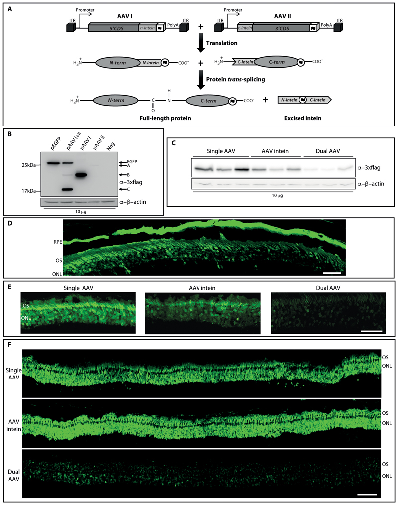
: 3xflag tag; PolyA: polyadenylation signal. (B) Western blot (WB) analysis of lysates from HEK293 transfected with either full-length or AAV intein CMV-EGFP plasmids. pEGFP: full-length EGFP plasmid; pAAV I+II: AAV-EGFP I+II intein plasmids; pAAV I: single AAV-EGFP I intein plasmid; pAAV II: single AAV-EGFP II intein plasmid; Neg: untransfected cells. The arrows indicate both the full-length EGFP protein (EGFP), the N- and C-terminal halves of the EGFP protein (B and A, respectively), and the reconstituted intein excised from the full-length EGFP protein (C). The WB is representative of N=3 independent experiments. (C) WB analysis of lysates from HEK293 infected with either single, intein or dual AAV2/2-CMV-EGFP vectors. The WB is representative of N=5 independent experiments. (D) Retinal cryosection from C57BL/6J mice injected subretinally with AAV2/8-CMV-EGFP intein vectors. Scale bar: 50 μm. RPE: retinal pigment epithelium; OS: outer segments; ONL: outer nuclear layer. The image is representative of n=5 eyes. (E-F) Retinal cryosections from either C57BL/6J mice (E) or Large White pigs (F) injected subretinally with either single, intein or dual AAV2/8-GRK1-EGFP vectors. Scale bar: 50 μm (E); 200 μm (F). OS: outer segment; ONL: outer nuclear layer.
Figure 2
Figure 2. Characterization and AAV intein-mediated transduction of human iPSCs-derived 3D retinal organoids.
(A) Light microscopy analysis of retinal organoids at 183 days of culture. (B) Immunofluorescence analysis with antibodies directed to mature photoreceptor markers. Scale bar: 100 μm. (C) Fluorescence analysis of retinal organoids infected with both AAV2/2-CMV-EGFP and AAV2/2-IRBP-DsRed vectors. Scale bar: 100 μm. (D) Outer segment-like structures were observed which protrude from the surface of retinal organoids at 230 days of culture. The inset shows the presence of outer segment (OS)-like structures with radial architecture. NR: neural retina; RPE: retinal pigment epithelium. (E) Scanning electron microscopy analysis reveals the presence of inner segments (IS), connecting cilia (CC) and outer segment (OS)-like structures. Scale bar: 4 μm. (F) Electron microscopy analysis reveals the presence of the outer limiting membrane (*), centriole (C), basal bodies (BB), connecting cilia (CC) and sketches of outer segments (OS). The inset shows the presence of disorganized membranous discs in the OS. Scale bar: 500 nm. D: days of culture. (G) Fluorescence analysis of retinal organoids infected with AAV2/2-GRK1-EGFP-intein vectors at 293 days of culture. Scale bar: 100 μm. The image is representative of n=4 organoids.
Figure 3
Figure 3. Optimization of AAV intein allows proper reconstitution of the large ABCA4 and CEP290 proteins.
Western blot (WB) analysis of lysates from HEK293 transfected with different sets of either AAV-shCMV-ABCA4 or -CEP290 intein plasmids. A schematic representation of the various sets used is depicted in Fig. S13. The WB are representative of N=3 independent experiments. (A) Kruskal-Wallis test p value was not significant, thus no post hoc comparison was performed to evaluate statistical differences between groups. (B) Significant differences were assessed using One-way ANOVA followed by Tukey multiple pairwise-comparison. * p <0.05; ** p <0.01; *** p <0.001. Details on set 1 (A) and set 5 (B) variability can be found in the Statistical analysis paragraphs of the Materials and methods section.
Figure 4
Figure 4. ABCA4 and CEP290 proteins from AAV intein vectors have a distribution pattern similar to those from full-length plasmids.
Representative images of immunofluorescence analysis of HeLa cells transfected with either AAV-shCMV-ABCA4 (A) or -CEP290 (B) intein plasmids. pABCA4 (A) or pCEP290 (B): plasmid including the full-length expression cassette; pAAV intein: AAV-intein plasmids (either set 1 in A or set 5 in B); I+II+III: AAV I+II+III intein plasmids; I+II: AAV I+II intein plasmids; I+III: AAV I+III intein plasmids; II+III: AAV II+III intein plasmids; I: single AAV I intein plasmid; II: single AAV II intein plasmid; III: single AAV III intein plasmid; Neg: untransfected cells. Cells were stained for 3xFLAG and either VAP-B (endoplasmic reticulum marker) and TGN46 (Trans-Golgi network marker) in A, or acetylated tubulin (marker of microtubules) in B. White arrows point at cells shown at higher magnification in Fig. S16. Quantification of both ABCA4 co-localization with VAP-B and TGN46 markers (A) and cells showing the various CEP290 polypeptides patterns (B) are shown in the graphs. At least 150 cells for each condition were counted in N=3 independent experiments. (A) Significant differences between groups were assessed using Kruskal-Wallis test followed by pairwise comparisons using Wilcoxon rank sum test. ** p <0.01; *** p <0.001. Asterisks above pAAV intein I column in the upper graph indicate significant differences with both pABCA4, I+II and II. (B) Significant differences between patterns of each column were assessed using binomial distribution. # indicates the predominant pattern for each CEP290 polypeptide (p<< 0,00001).
Figure 5
Figure 5. ABCA4 and CEP290 large protein reconstitution by AAV intein and dual AAV vectors.
Western blot (WB) analysis of lysates from HEK293 cells infected with either dual or intein AAV2/2-shCMV-ABCA4 (A) or -CEP290 (B) vectors. AAV intein: AAV-ABCA4 (set 1, A) or -CEP290 (set 5, B) intein vectors; I+II+III: AAV I+II +III intein vectors; I+II: AAV I+II intein vectors; I+III: AAV I+III intein vectors; II+III: AAV II+III intein vectors; I: single AAV I intein vector; II: single AAV II intein vector; III: single AAV III intein vector; dual AAV: dual AAV vectors; Neg: either AAV-EGFP vectors or PBS. (A) The arrows indicate the full-length ABCA4 protein and A: protein product derived from AAV I; B: protein product derived from AAV II. * protein product with a potentially different post-translational modification. (B) The arrows indicate the full-length CEP290 protein and A: protein product derived from AAV II+III; B: protein product derived from AAV I+II; C: protein product derived from AAV II; D: protein product derived from AAV III; E: protein product derived from AAV I. The WB are representative of N=3 independent experiments.
Figure 6
Figure 6. AAV intein reconstitute large proteins in mouse, pig and human photoreceptors.
(A-C) Western blot (WB) analysis of retinal lysates from either wild-type mice (A, B) or Large White pigs (C) injected with either dual or intein AAV2/8-GRK1-ABCA4 (A, C) or -CEP290 (B) vectors. AAV intein: AAV intein vectors; Dual AAV: dual AAV vectors; Neg: either AAV-EGFP vectors or PBS. Quantification of ABCA4 expression in Large White pigs injected with either dual (n=3) or intein (n=2) AAV2/8-GRK1-ABCA4 is included in (C). Significant differences between groups were assessed using unpaired Student’s t-test. ** p <0.01. (D) WB analysis of lysates from human iPSCs-derived 3D retinal organoids infected with AAV2/2-GRK1-ABCA4 intein vectors. AAV intein: AAV-ABCA4 intein vectors; Neg: not infected organoids; -/-: organoids derived from STGD1 patients. (A, C, D) The arrows indicate the full-length ABCA4 protein (ABCA4) and A: protein product derived from AAV I; B: protein product derived from AAV II. * protein product with a potentially different post translational modification. (B) The arrows indicate both the full-length CEP290 protein (CEP290); A: protein product derived from AAV II+III and D: protein product derived from AAV III. The WB are representative of: n=10 AAV intein- and 9 dual AAV-injected eyes (A); n=10 AAV intein- and 5 dual AAV-injected eyes (B); n=7 AAV intein-infected organoids (D).
Figure 7
Figure 7. Subretinal administration of AAV intein improves the retinal phenotype of mouse models of inherited retinal degenerations.
(A) Quantification of the mean area occupied by lipofuscin in the RPE of Abca4-/- mice treated with AAV intein. Each dot represents the mean value measured for each eye. The mean value of the lipofuscin area for each group is indicated in the graph. +/+ or +/-: control injected Abca4+/+ or +/- eyes (PBS); -/-: negative control-injected Abca4-/- eyes (AAV I ABCA4 or AAV II ABCA4 or PBS); -/- AAV intein: Abca4-/- eyes injected with AAV intein vectors. Significant differences between groups were assessed using one-way ANOVA followed by Tukey multiple pairwise-comparison. * p <0.05; *** p <0.001. (B) Representative images of retinal sections from wild-type uninjected and rd16 mice either not injected or injected subretinally with AAV2/8-GRK1-CEP290 intein vectors (AAV intein) or with negative controls (Neg: AAV I+II or AAV II+III or PBS). Scale bar: 25 μm. The thickness of the ONL measured in each image is indicated by the vertical black line. RPE: retinal pigment epithelium; ONL: outer nuclear layer; INL: inner nuclear layer; GCL: ganglion cell layer. Significant differences between groups were assessed using Kruskal-Wallis test followed by pairwise comparisons using Wilcoxon rank sum test. ** p <0.01. (C) Representative images of eyes from wild-type uninjected and rd16 mice either not injected or injected subretinally with AAV2/8-GRK1-CEP290 intein vectors (AAV intein) or with negative controls (Neg: AAV I+II or AAV II+III or PBS). White circles define pupils. Significant differences between groups were assessed using one-way ANOVA followed by Tukey multiple pairwise-comparison. *** p <0.001. The complete set of p values can be found in the Auxiliary Quantification File.

References

    1. FDA approves hereditary blindness gene therapy. Nat Biotechnol. 2018;36:6. - PubMed
    1. Trapani I, Auricchio A. Seeing the Light after 25 Years of Retinal Gene Therapy. Trends Mol Med. 2018 - PubMed
    1. Auricchio A, Smith AJ, Ali RR. The Future Looks Brighter After 25 Years of Retinal Gene Therapy. Hum Gene Ther. 2017;28:982–987. - PubMed
    1. Trapani I, et al. Effective delivery of large genes to the retina by dual AAV vectors. EMBO Mol Med. 2014;6:194–211. - PMC - PubMed
    1. Duan D, Yue YJ, Engelhardt F. Expanding AAV packaging capacity with trans-splicing or overlapping vectors: a quantitative comparison. Mol Ther. 2001;4:383–391. - PubMed

Publication types

MeSH terms

Substances

LinkOut - more resources