Review
. 2019 Jun 4;39(6):BSR20190805.
doi: 10.1042/BSR20190805. Print 2019 Jun 28.

Autologous activated platelet-rich plasma injection into adult human ovary tissue: molecular mechanism, analysis, and discussion of reproductive response

Affiliations
Review

Autologous activated platelet-rich plasma injection into adult human ovary tissue: molecular mechanism, analysis, and discussion of reproductive response

E Scott Sills et al. Biosci Rep. .

Abstract

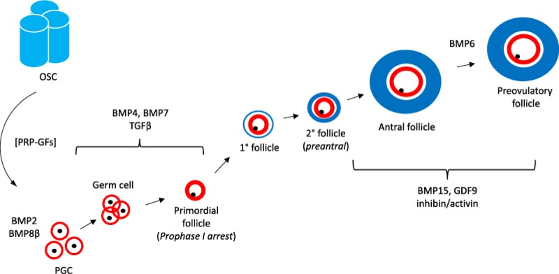
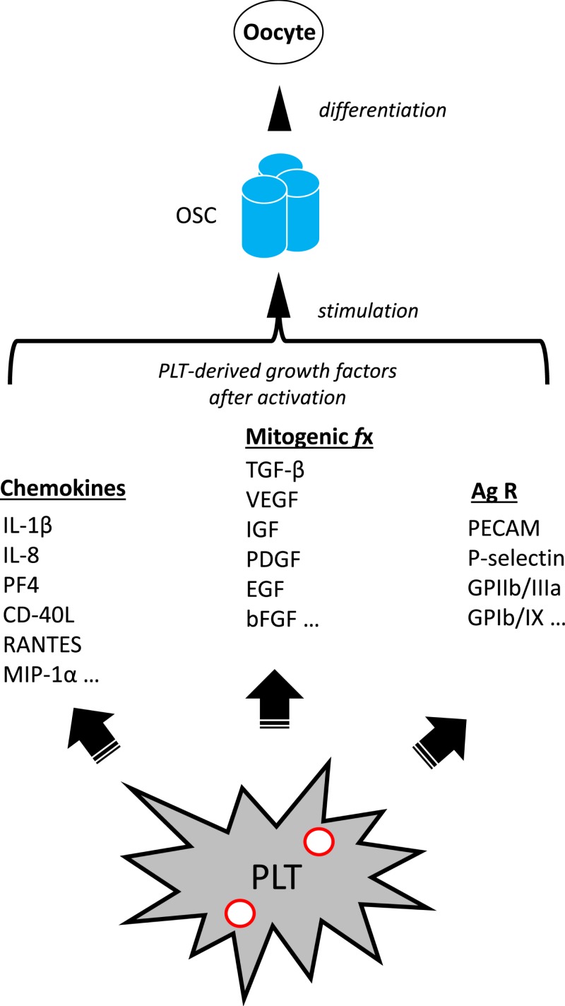
In clinical infertility practice, one intractable problem is low (or absent) ovarian reserve which in turn reflects the natural oocyte depletion associated with advancing maternal age. The number of available eggs has been generally thought to be finite and strictly limited, an entrenched and largely unchallenged tenet dating back more than 50 years. In the past decade, it has been suggested that renewable ovarian germline stem cells (GSCs) exist in adults, and that such cells may be utilized as an oocyte source for women seeking to extend fertility. Currently, the issue of whether mammalian females possess such a population of renewable GSCs remains unsettled. The topic is complex and even agreement on a definitive approach to verify the process of 'ovarian rescue' or 're-potentiation' has been elusive. Similarities have been noted between wound healing and ovarian tissue repair following capsule rupture at ovulation. In addition, molecular signaling events which might be necessary to reverse the effects of reproductive ageing seem congruent with changes occurring in tissue injury responses elsewhere. Recently, clinical experience with such a technique based on autologous activated platelet-rich plasma (PRP) treatment of the adult human ovary has been reported. This review summarizes the present state of understanding of the interaction of platelet-derived growth factors with adult ovarian tissue, and the outcome of human reproductive potential following PRP treatment.

Keywords: aging; fertility; menopause; ovary; reproduction.


Conflict of interest statement

E.S.S. holds a provisional U.S. patent for process and treatment using ovarian PRP.

Figures

Figure 1
Figure 1. Recruitment and growth of oocytes, from PGC stage through mature follicle, illustrating various growth factors mediating development
Known components of this sequence include granulosa precursors (red), theca compartment (blue), and germ cells (black). Upstream contributions by ovarian stem cells (OSC) may be possible under conditions enabled by growth factors released by platelet-rich plasma (PRP-GFs). Other relevant regulators are BMP2, BMP6, and BMP8β, which are involved in cytokine–cytokine receptor interactions; and Transforming growth factor β (TGF-β), which activates various substrates and regulatory proteins inducing transcription of genes for differentiation, chemotaxis, and proliferation. Later direction is under control of BMP15, a paracrine signal exclusively expressed in ovarian tissue which is involved in oocyte and follicular growth, as well as GDF9, a down-regulator of inhibin-A and promoter of further follicular maturation.
Figure 2
Figure 2. Proposed mechanism of action for alteration of adult ovarian function by application of activated PRP
Autologous activated PRP sample generates an enriched platelet (PLT) substrate collected by peripheral venipuncture. PLT combination with calcium gluconate achieves activation of α granules [red circles], which subsequently initiates release of at least three classes of molecular mediators. These include chemokines such as Interleukin-1β (IL-1β), a central inflammatory mediator involved in cell proliferation, differentiation, and apoptosis; Interleukin-8 (IL-8, also known as neutrophil chemotactic factor) which coordinates migration toward sites of injury or infection and is a promoter of angiogenesis and improved tissue perfusion; Platelet factor 4 (PF4), a versatile chemotactic protein with high affinity for heparin, involved in platelet aggregation and selective antimicrobial activity; Ligand of CD40 (CD-40L), a potent inducer of inflammatory processes by enhancing interactions among platelets, leukocytes, and endothelium; a protein known as Regulated after Activation of Normal T-cell Expressed and Secreted (RANTES), itself a useful marker for PLT activation which strongly attracts monocytes; Macrophage inflammatory protein 1-α (MIP-1α), which conditionally triggers migration and signaling cascades to mediate cell survival and proliferation; Platelet-associated cellular mitogens include TGF-β, which activates different downstream substrates and regulatory proteins inducing transcription of multiple target genes for differentiation, chemotaxis, proliferation and activation of immune system cells; Vascular endothelial growth factor (VEGF), a signal protein stimulating blood vessel formation; Insulin like growth factors (IGFs) a group of proteins with close homology to insulin required for cell stimulation and communication with the local environment; Platelet derived growth factor (PDGF), critical in growth of blood vessels from extant nearby capillaries, mitogenesis and proliferation of mesenchymal cells including fibroblasts, osteoblasts, tenocytes, vascular SMCs and mesenchymal stem cells; epidermal growth factor (EGF), a central element in cellular proliferation, differentiation, and survival; Basic fibroblast growth factor (bFGF), a mediator with broad mitogenic and cell survival activities, and is involved in a variety of biological processes, including embryonic development, cell growth, morphogenesis, tissue repair, tumor growth and invasion function. Platelet expressed antigens include Platelet endothelial cell adhesion molecule (PECAM), which plays a key role in removing aged neutrophils from circulation; P-selectin which contributes to initial recruitment of leukocytes to injury sites during inflammation; Glycoprotein IIb/IIIa, part of the integrin complex found on platelets aiding in platelet activation; and Glycoprotein Ib and IX (GPIb/IX) which binds von Willebrand factor, allowing platelet adhesion and platelet plug formation at sites of vascular injury. PRP is placed inside the adult ovary (by direct ultrasound-guided needle injection) thus permitting these signaling elements access to ovarian stem cells (OSCs) as discussed by Johnson et al. (2004). PLT-derived moieties then trigger or enable differentiation of these OSCs. Subsequently, reduced serum FSH and/or higher post-treatment levels of serum AMH have been observed clinically, consistent with improved or ‘re-potentiated’ ovarian function.

References

    1. Sills E.S., Alper M.M. and Walsh A.P. (2009) Ovarian reserve screening in infertility: practical applications and theoretical directions for research. Eur. J. Obstet. Gynecol. Reprod. Biol. 146, 30–36 10.1016/j.ejogrb.2009.05.008 - DOI - PubMed
    1. Hsueh A.J., Kawamura K., Cheng Y. and Fauser B.C. (2015) Intraovarian control of early folliculogenesis. Endocr. Rev. 36, 1–24 10.1210/er.2014-1020 - DOI - PMC - PubMed
    1. Robin C., Ottersbach K., de Bruijn M., Ma X., van der Horn K. and Dzierzak E. (2003) Developmental origins of hematopoietic stem cells. Oncol. Res. 13, 315–321 10.3727/096504003108748519 - DOI - PubMed
    1. Hummitzsch K., Anderson R.A., Wilhelm D., Wu J., Telfer E.E., Russell D.L.. et al. (2015) Stem cells, progenitor cells, and lineage decisions in the ovary. Endocr. Rev. 36, 65–91 10.1210/er.2014-1079 - DOI - PMC - PubMed
    1. Guigon C.J. and Cohen-Tannoudji M. (2011) Reconsidering the roles of female germ cells in ovarian development and folliculogenesis. Biol. Aujourdhui 205, 223–233 10.1051/jbio/2011022 - DOI - PubMed