. 2019 Apr 9:2019:4353791.
doi: 10.1155/2019/4353791. eCollection 2019.

Efficiently Capturing Mitochondria-Targeted Constituents with Hepatoprotective Activity from Medicinal Herbs

Affiliations

Efficiently Capturing Mitochondria-Targeted Constituents with Hepatoprotective Activity from Medicinal Herbs

Xing-Xin Yang et al. Oxid Med Cell Longev. .

Abstract

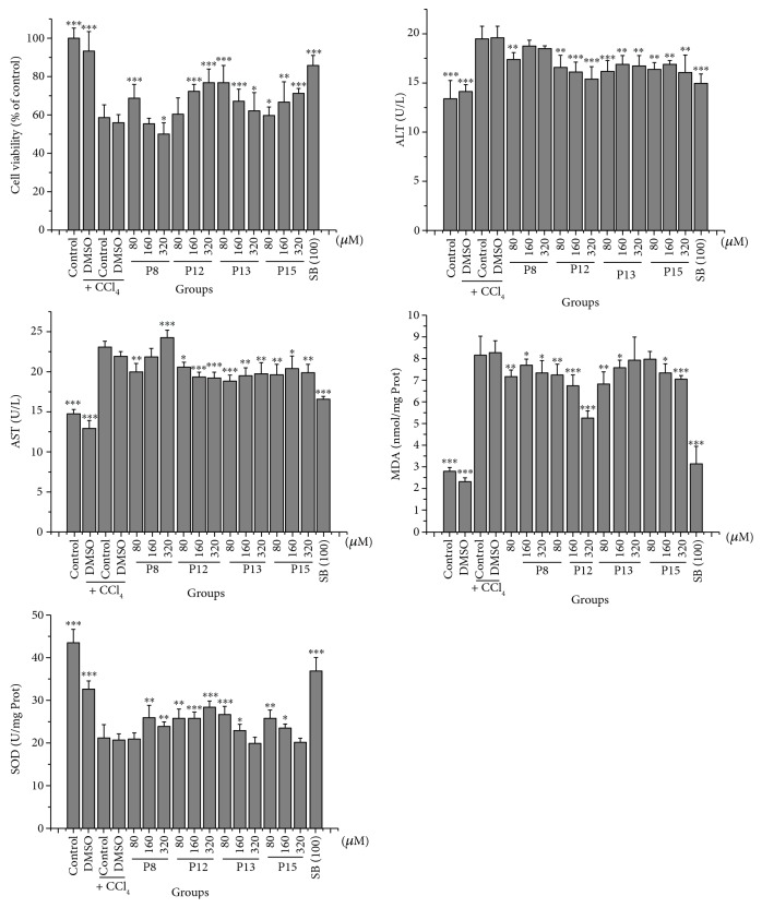
Targeting mitochondria as a hepatic-protective strategy has gained attention, because of their important roles in energy production, adjustment of apoptosis, and generation of reactive oxygen species. To promote the discovery of natural mitochondria-targeted hepatic-protectants, we established a hepatocellular mitochondria-based capturing method by coupling affinity ultrafiltration with liquid chromatography/mass spectrometry (LC/MS), which is suitable for identifying mitochondrial ligands from medicinal herbs (MHs). After evaluating the feasibility of the method, it was applied for capturing mitochondria-targeting constituents from Peucedani Radix extract. A total of 10 active compounds were identified by LC/MS, all of which were newly identified mitochondrial ligands. The mitochondria-remedying activity of 4 of the 10 hits was confirmed by pharmacological tests in vitro. Additionally, the hepatic-protective abilities of 4 hits were verified in both carbon tetrachloride-damaged liver L02 cells and mice. These results indicated that the method could be used for identifying hepatic mitochondria-targeting constituents in MHs, which might be beneficial for hepatic-protective development.


Figures

Figure 1
Figure 1
Overview of the analytical procedure of the search for mitochondria-targeted constituents from complex samples by CM-HMC.
Figure 2
Figure 2
Evaluation of the structural integrity and bio-functions of HM. (a) Transmission electron micrograph displaying typical mitochondrial morphology (×40,000). (b) Changes in the ΔΨm of HM after CCCP treatment. ΔΨm was determined as the difference in Rh123 uptake by HM and CCCP-treated HM and expressed in units of fluorescence intensity. HM: hepatic mitochondria. Data were obtained from 3 independent experiments and expressed as the mean ± SD. P < 0.05 compared with HM.
Figure 3
Figure 3
Analysis of four standard solutions by CM-HMC. HPLC chromatograms of ultrafiltrates comprised of standards released from active HM (black line) or denatured HM (red line), which served as a control, are presented. Enhancement of the peak area of the standards compared with the control indicates specific binding with HM.
Figure 4
Figure 4
Determination of a mixed standard solution using CM-HMC. (a) HPLC chromatograms of a mixed standard solution are shown for ultrafiltrates derived from active HM (black line) and denatured HM (red line), which served as a control. Peaks R2 and R3 exhibited remarkable area enhancement compared with the control. However, the area of the R1 peak was equal to that of the control. (b) HPLC chromatogram of a mixed standard solution, which was directly assayed by LC/MS. GL: glucuronide; AC: amoxicillin; DZ: daidzin; and SB: silybin.
Figure 5
Figure 5
Screening of the PR extract for HM-targeted constituents by CM-HMC. Compared with the control, which was comprised of denatured HM (red line), HPLC chromatograms of screened PR extract (A and B; (a) 0–48 min; (b) 48–85 min) presented fifteen peaks (P1–P15) that were significantly enhanced due to specific binding with HM (black line).
Figure 6
Figure 6
Chemical structures of the ten bioactive compounds that were identified from the PR extract.
Figure 7
Figure 7
Effects of hits on the mPTP opening (a), ∆Ψm (b), Na+-K+-ATPase activity (c), and Ca2+-Mg2+-ATPase activity (d) in isolated HM. Data were obtained from 5 independent measurements and are expressed as mean ± SD. The statistical significance of differences between groups was assayed by one-way analysis of variance (ANOVA) using Dunnett's method. P < 0.05, ∗∗ P < 0.01, and ∗∗∗ P < 0.001 compared with the model control group (a, c, and d) or between the control and PH treatment (B), under identical conditions. ## P < 0.01 and ### P < 0.001 compared with the black group (PH treatment) measured under identical conditions. P8: praeruptorin A; P12: praeruptorin B; P13: praeruptorin D; P15: praeruptorin E; CsA: cyclosporin A; SB: silybin; PH: pioglitazone hydrochloride.
Figure 8
Figure 8
Effects of hit compounds on the hepatocytes of CCl4-induced injury. Data were obtained from 5 independent determinations and are expressed as mean ± SD. The statistical significance of differences between groups was evaluated by one-way analysis of variance (ANOVA) using Dunnett's method. P < 0.05, ∗∗ P < 0.01, and∗∗∗ P < 0.001 compared with the model control group under identical conditions. P8: praeruptorin A; P12: praeruptorin B; P13: praeruptorin D; P15: praeruptorin E; CsA: cyclosporin A; SB: silybin.
Figure 9
Figure 9
Effects of the hit compound on the mice of CCl4-induced injury. ALT and AST in serum (a and b), liver index, SOD, GSH, and MDA in liver tissue (c–f), and Na+-K+-ATPase, Ca2+-Mg2+-ATPase, mPTP, MDA, and ∆Ψm in HM (g–k) were measured. Data were obtained from 5 independent experiments and are expressed as mean ± SD. The statistical significance of differences between groups was evaluated by one-way analysis of variance (ANOVA) using Dunnett's method. P < 0.05, ∗∗ P < 0.01, and ∗∗∗ P < 0.001 compared with the model control group under identical conditions. P12: praeruptorin B; SB: silybin.
Figure 10
Figure 10
Effect of the hit compound on the hepatic histomorphology injury by CCl4. As shown in (a), normal liver displayed a typical hepatolobular architecture, comprised of a clear central vein with radiating cords of hepatocytes separated by sinusoids. Hepatic cells were polygonal in shape, with distinctive nuclei and a uniform cytoplasm. Few binucleated cells were present, and the cytoplasm was regularly distributed. As shown in (b), hepatic injuries induced by CCl4 in mice were demonstrated by marked vacuolization of hepatocytes, necrosis around the central vein, sinusoidal dilation and congestion, infiltration of cells, loss of cellular boundaries and ballooning degeneration, and loss of architecture and were significantly different from those observed in the normal control group. However, treatment with silybin (c) and P12 (praeruptorin B) at doses of 8, 16, and 32 mg/kg for 7 days are shown in (d), (e), and (f), respectively and significantly attenuated liver injuries as shown by the absence of focal or bridging necrosis or mild hepatitis. The CCl4-induced histopathological changes improved by treatment with P12, and the recovery was significant in the low-dose group (8 mg/kg), which was comparable to that of silybin.

Similar articles

Cited by

References

    1. Friedman S.-L. Mechanisms of hepatic fibrogenesis. Gastroenterology. 2008;134(6):1655–1669. doi: 10.1053/j.gastro.2008.03.003. - DOI - PMC - PubMed
    1. Zhao Q., Peng Y., Huang K., et al. Salvianolate protects hepatocytes from oxidative stress by attenuating mitochondrial injury. Evidence-Based Complementary and Alternative Medicine. 2016;2016:10. doi: 10.1155/2016/5408705.5408705 - DOI - PMC - PubMed
    1. Pathania D., Millard M., Neamati N. Opportunities in discovery and delivery of anticancer drugs targeting mitochondria and cancer cell metabolism. Advanced Drug Delivery Reviews. 2009;61(14):1250–1275. doi: 10.1016/j.addr.2009.05.010. - DOI - PubMed
    1. Au J. S., Navarro V. J., Rossi S. Review article: drug-induced liver injury-its pathophysiology and evolving diagnostic tools. Alimentary Pharmacology & Therapeutics. 2011;34(1):11–20. doi: 10.1111/j.1365-2036.2011.04674.x. - DOI - PubMed
    1. McGill M.-R., Sharpe M.-R., Williams C.-D., Taha M., Curry S.-C., Jaeschke H. The mechanism underlying acetaminophen -induced hepatotoxicity in humans and mice involves mitochondrial damage and nuclear DNA fragmentation. Journal of Clinical Investigation. 2012;122(4):1574–1583. doi: 10.1172/JCI59755. - DOI - PMC - PubMed

MeSH terms

Substances

LinkOut - more resources