Case Reports
. 2019 Apr 4:2019:1492965.
doi: 10.1155/2019/1492965. eCollection 2019.

Jejunoileal GIST: A Rare Case of Transient Intussusception and Gastrointestinal Bleeding

Affiliations
Case Reports

Jejunoileal GIST: A Rare Case of Transient Intussusception and Gastrointestinal Bleeding

Sara Catarino Santos et al. Case Rep Surg. .

Abstract

Gastrointestinal stromal tumors (GIST) comprised 0,2% of all GI tumors. They are typically asymptomatic, but can manifest with nonspecific GI symptoms, GI bleeding, or intussusception. The authors report a case of a 55-year-old female patient with hematochezia and a palpable mass on the left lower quadrant. Ultrasound revealed possible intussusception. However, CT scan did not show any signs of lesions or intussusception. On reevaluation, the mass was no longer palpable. The patient had recurrent episodes of hematochezia with need of transfusional support. CT enterography revealed a 20-24 mm jejunoileal lesion. A laparotomy was undertaken with small bowel resection containing the lesion. Histological examination confirmed GIST. GIST presentation as transient intussusception and intermittent GI bleeding is rare. This case report emphasizes the rarity of jejunoileal GIST, its clinical details, diagnostic study, and treatment.


Figures

Figure 1
Figure 1
Abdominal ultrasound (possible intussusception).
Figure 2
Figure 2
CT scan (no signs of lesions or intussusception).
Figure 3
Figure 3
Lower endoscopy (hematic residues with no lesions detectable).
Figure 4
Figure 4
CT enterography—axial CT image with 20-24 mm jejunoileal lesion compatible with GIST.
Figure 5
Figure 5
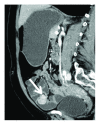
CT enterography—sagittal CT image with lesion compatible with GIST.
Figure 6
Figure 6
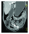
CT enterography—coronal CT image lesion compatible with GIST.
Figure 7
Figure 7
Surgery—jejunoileal lesion compatible with GIST with the endophytic and exophytic component.

References

    1. Sankey R. E., Maatouk M., Mahmood A., Raja M. Case Report: Jejunal gastrointestinal stromal tumour, a rare tumour, with a challenging diagnosis and a successful treatment. Journal of Surgical Case Reports. 2015;2015(5, article rjv050) doi: 10.1093/jscr/rjv050. - DOI - PMC - PubMed
    1. Joensuu H. Gastrointestinal stromal tumor (GIST) Annals of Oncology. 2006;17(Supplement 10):x280–x286. doi: 10.1093/annonc/mdl274. - DOI - PubMed
    1. Bresci G. Occult and obscure gastrointestinal bleeding: causes and diagnostic approach in 2009. World Journal of Gastrointestinal Endoscopy. 2009;1(1):3–6. doi: 10.4253/wjge.v1.i1.3. - DOI - PMC - PubMed
    1. Yamamoto M., Yamamoto K., Taketomi H., Yamamoto F., Yamamoto H. Obscure gastrointestinal bleeding due to a small intestinal gastrointestinal stromal tumor in a young adult. Case Reports in Gastroenterology. 2016;10(3):668–673. doi: 10.1159/000452207. - DOI - PMC - PubMed
    1. Marinis A., Yiallourou A., Samanides L., et al. Intussusception of the bowel in adults: a review. World Journal of Gastroenterology. 2009;15(4):407–411. doi: 10.3748/wjg.15.407. - DOI - PMC - PubMed

Publication types

LinkOut - more resources