. 2020 Mar 1;86(3):417-428.
doi: 10.1093/neuros/nyz128.

Brainstem Functional Connectivity Disturbances in Epilepsy may Recover After Successful Surgery

Affiliations

Brainstem Functional Connectivity Disturbances in Epilepsy may Recover After Successful Surgery

Hernán F J González et al. Neurosurgery. .

Abstract

Background: Focal seizures in temporal lobe epilepsy (TLE) are associated with widespread brain network perturbations and neurocognitive problems.

Objective: To determine whether brainstem connectivity disturbances improve with successful epilepsy surgery, as recent work has demonstrated decreased brainstem connectivity in TLE that is related to disease severity and neurocognitive profile.

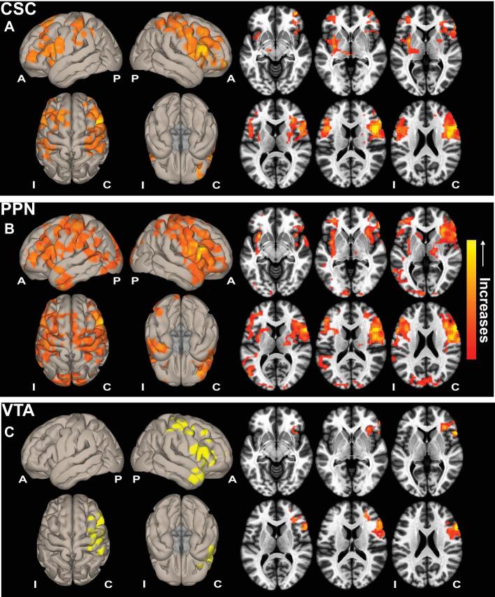
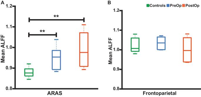
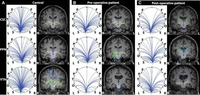
Methods: We evaluated 15 adult TLE patients before and after (>1 yr; mean, 3.4 yr) surgery, and 15 matched control subjects using magnetic resonance imaging to measure functional and structural connectivity of ascending reticular activating system (ARAS) structures, including cuneiform/subcuneiform nuclei (CSC), pedunculopontine nucleus (PPN), and ventral tegmental area (VTA).

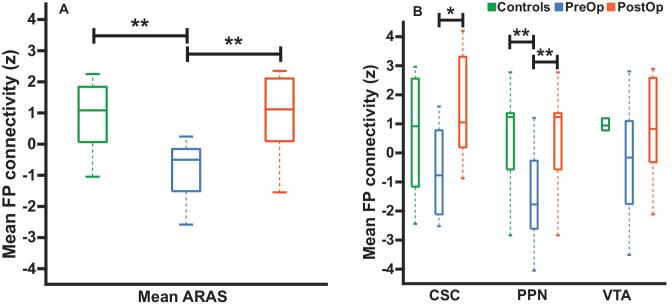
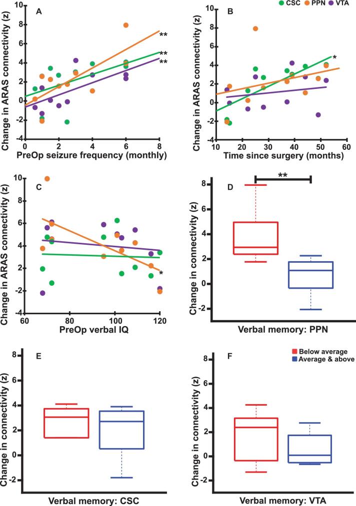
Results: TLE patients who achieved long-term postoperative seizure freedom (10 of 15) demonstrated increases in functional connectivity between ARAS structures and fronto-parietal-insular neocortex compared to preoperative baseline (P = .01, Kruskal-Wallis), with postoperative connectivity patterns resembling controls' connectivity. No functional connectivity changes were detected in 5 patients with persistent seizures after surgery (P = .9, Kruskal-Wallis). Among seizure-free postoperative patients, larger increases in CSC, PPN, and VTA functional connectivity were observed in individuals with more frequent seizures before surgery (P < .05 for each, Spearman's rho). Larger postoperative increases in PPN functional connectivity were seen in patients with lower baseline verbal IQ (P = .03, Spearman's rho) or verbal memory (P = .04, Mann-Whitney U). No changes in ARAS structural connectivity were detected after successful surgery.

Conclusion: ARAS functional connectivity disturbances are present in TLE but may recover after successful epilepsy surgery. Larger increases in postoperative connectivity may be seen in individuals with more severe disease at baseline.

Keywords: Brainstem; Epilepsy surgery; Functional connectivity; Postoperative; Temporal lobe epilepsy.


Figures

Figure 1.
Figure 1.
ARAS functional connectivity increases in seizure-free TLE patients after surgery. Cortical surface (left) and axial slice (right) views are shown, demonstrating functional connectivity increases in patients with TLE who achieved seizure freedom after surgery, seeded from CSC A, PPN B, and VTA C. Data represent seed-to-voxel functional connectivity (bivariate correlation) maps comparing postoperative vs preoperative fMRI (paired t-test, cluster threshold level P < .05, FDR correction) generated using the CONN toolbox (https://www.nitrc.org/projects/conn/). Positive contrasts are shown, and no connectivity decreases were observed on negative contrasts. Images are oriented across all patients with respect to the epileptogenic side. N = 10 patients before surgery and > 1 yr after surgery. A, anterior; ARAS, ascending reticular activating system; C, contralateral; CSC, cuneiform/subcuneiform nuclei; FDR, false discovery rate; I, ipsilateral; P, posterior; PPN, pedunculopontine nucleus; VTA, ventral tegmental area.
Figure 2.
Figure 2.
ARAS-frontoparietal functional connectivity in TLE patients before and after surgery and controls. A, Mean functional connectivity between ARAS and frontoparietal and insular neocortex is reduced in preoperative patients with TLE compared to controls. However, connectivity in the same TLE patients is increased > 1 yr after surgery, resembling connectivity in controls. B, Examining ARAS regions individually, increases in frontoparietal connectivity are seen after surgery in CSC and PPN, but not VTA. n = 10 patients before surgery and > 1 yr after surgery, who ultimately achieved seizure freedom vs 10 matched controls. *P = .05, Kruskal–Wallis with post hoc Dunn; **P value range = .01-.04, Kruskal–Wallis with post hoc Dunn. Central bar shows median, bottom and top edges of box indicate 25th and 75th percentiles, and whiskers indicate data extremes. ARAS, ascending reticular activating system; CSC, cuneiform/subcuneiform nuclei; FP, frontoparietal; PostOp, postoperative patients; PPN, pedunculopontine nucleus; PreOp, preoperative patients; VTA, ventral tegmental area.
Figure 3.
Figure 3.
ALFF in ARAS, but not frontoparietal neocortex, differs between patients and controls. A, Differences in mean ALFF values in ARAS between control subjects and both preoperative and postoperative patients. No ARAS ALFF differences are noted between preoperative and postoperative patients. B, No differences in mean ALFF values in bilateral frontoparietal/insular neocortical regions are observed between controls, preoperative patients, or postoperative patients. N = 10 patients before surgery and > 1 yr after surgery, who ultimately achieved seizure freedom vs 10 matched controls. **P < .05, Kruskal–Wallis with post hoc Dunn. Central bar shows median, bottom, and top edges of box indicate 25th and 75th percentiles, and whiskers indicate data extremes. ALFF, amplitude of low-frequency fluctuations, ARAS, ascending reticular activating system; CSC, cuneiform/subcuneiform nuclei; PostOp, postoperative patients; PPN, pedunculopontine nucleus; PreOp, preoperative patients; VTA, ventral tegmental area.
Figure 4.
Figure 4.
Relationships between ARAS postoperative functional connectivity changes and disease measures in seizure-free patients. A, Larger increases in functional connectivity between each ARAS structure (CSC, PPN, and VTA) and frontoparietal neocortex are associated with higher preoperative focal impaired consciousness seizure frequency. B, Larger increases in functional connectivity between CSC and frontoparietal neocortex are associated with longer time between surgery and the postoperative scan, whereas no similar relationship is observed with changes in PPN or VTA connectivity. C, Patients with lower preoperative verbal IQ before surgery demonstrate a larger postoperative increase in functional connectivity between PPN and frontoparietal neocortex, although no similar relationship is noted for CSC or VTA. D-F, Patients with worse preoperative verbal memory performance show a larger increase in PPN postoperatively D, but no such relationship is noted for CSC E or VTA F. N = 10 patients, who ultimately achieved seizure freedom after surgery. *P value range = .02-.03, uncorrected, **P value range = .03-.04 after Bonferroni-Holm correction for Spearman's rho A-C or Mann–Whitney U-test D-F. Central bar shows median, bottom and top edges of box indicate 25th and 75th percentiles, and whiskers indicate data extremes. ARAS, ascending reticular activating system; CSC, cuneiform/subcuneiform nuclei; PPN, pedunculopontine nucleus; PreOp, preoperative patients; VTA, ventral tegmental area.
Figure 5.
Figure 5.
Example ARAS structural connectivity. Diffusion tractography seeded from CSC (top row), PPN (middle row), and VTA (bottom row) in an example matched control A, preoperative patient B, and the same patient postoperative C for each region. Figures are generated using the BrainSuite Diffusion Pipeline (BDP; http://brainsuite.org). On the left in each column A-C are circle graphs that summarize projections seeded from ARAS regions to cortical and subcortical regions in BrainSuite SVReg Atlas. On the right in each column A-C are estimated diffusion tensors overlaid onto T1-weighted coronal anatomical images using a rigid mutual information-based registration. Overall, for the 3 ARAS seed regions, the most tracts are seen in the controls A compared to patients B and C. Additionally, visually, there are no differences in estimated tracts between preoperative patients B and postoperative patients C. BrainSuite settings: 1 seed per voxel, step-size = 0.25 mm, maximum steps = 500, angle-threshold = 10°, fractional anisotropy threshold = 0.05, orientation distribution function sampling = 20, and generalized fraction anisotropy/lambda 2 threshold = 0.01. CSC, cuneiform/subcuneiform nuclei; F, frontal; L, left; O, occipital; P, parietal; PPN, pedunculopontine nucleus; R, right; S, subcortical; T, temporal; VTA, ventral tegmental area.
Figure 6.
Figure 6.
Model for subcortical-cortical connectivity disturbances and recovery in TLE. A, At wakeful baseline, neocortical activation is maintained via direct and indirect excitatory projections from subcortical activating structures, including ARAS, intralaminar thalamus, and basal forebrain. B, During the transition to the ictal period, seizure activity begins in the mesial temporal lobe and may remain confined there without disturbing cortical activity, generating a small consciousness-sparing focal seizure, or aura. C, When seizure activity spreads to involve subcortical activating structures, the normal excitatory input from the subcortical regions to the neocortex is perturbed, and the neocortex defaults to a sleep-like inhibited state, resulting in a consciousness-impairing focal seizure. D, Over time, recurrent consciousness-impairing focal seizures may lead to progressive dysfunction of subcortical activating structures and aberrant connectivity between these regions and the neocortex, leading to a chronic state of reduced neocortical activation and impaired neurocognition. E, Seizure freedom after successful epilepsy surgery may allow recovery of certain subcortical-cortical functional connectivity pertubations. Adapted from Blumenfeld and Taylor, with permission and courtesy of Hal Blumenfeld.

References

    1. Engel J. What can we do for people with drug-resistant epilepsy? Neurology. 2016;87(23):2483-2489. - PMC - PubMed
    1. Choi H, Sell RL, Lenert L et al. .. Epilepsy surgery for pharmacoresistant temporal lobe epilepsy. JAMA. 2008;300(21):2497-2505. - PubMed
    1. Engel J Jr, McDermott MP, Wiebe S et al. .. Early surgical therapy for drug-resistant temporal lobe epilepsy. JAMA. 2012;307(9):922-930. - PMC - PubMed
    1. Tellez-Zenteno JF, Hernandez-Ronquillo L.. A review of the epidemiology of temporal lobe epilepsy. Epilepsy Res Treat. 2012;2012:630853. - PMC - PubMed
    1. Witt JA, Helmstaedter C.. Cognition in epilepsy. Curr Opin Neurol. 2017;30(2):174-179. - PubMed

Publication types