Review
. 2019 Aug;92(1100):20190058.
doi: 10.1259/bjr.20190058. Epub 2019 May 16.

Ultrasound of the pediatric chest

Affiliations
Review

Ultrasound of the pediatric chest

Priscilla Joshi et al. Br J Radiol. 2019 Aug.

Abstract

Cross-sectional imaging modalities like MRI and CT provide images of the chest which are easily understood by clinicians. However, these modalities may not always be available and are expensive. Lung ultrasonography (US) has therefore become an important tool in the hands of clinicians as an extension of the clinical exam, which has been underutilized by the radiologists. Reinforcement of the ALARA principle along with the dictum of "Image gently" have resulted in increased use of modalities which do not require radiation. Hence, ultrasound, which was earlier being used mainly to confirm the presence of pleural effusion as well as evaluate it and differentiate solid from cystic masses, is now being used to evaluate the lung as well. This review highlights the utility of ultrasound of the paediatric chest. It also describes the normal and abnormal appearances of the paediatric lung on ultrasound as well as the advantages and limitations of this modality.


Figures

Figure 1.
Figure 1.
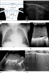
(A) Diagrammatic representation of acoustic windows used for evaluating the chest include- (1) Suprasternal notch (2) Parasternal region (3) Intercostal spaces (4) Trans diaphragmatic approach and (5) Sub costal/ Sub xiphoid approach. (B) Line diagram of normal appearance of the lung on a transcostal longitudinal US scan showing the chest wall, ribs, pleura, and A-lines (C) Corresponding transcostal longitudinal US image of normal lung with linear transducer showing the ribs with distal shadowing (R), intercostal spaces (IS), pleural line (arrow) and multiple horizontal echogenic lines, equidistant from each other suggestive of A lines (arrow head). (D) M mode ultrasound in normal lung showing the seashore sign. The cursor is placed on the pleura while sampling. The normal lung movement termed as “lung sliding” generates a “twinkling” or grainy appearance seen distal to the pleural line, distributed uniformly. The chest wall above the pleura shows no significant movement. This appearance mimics the sea and shore hence the term “seashore” sign. (E) Lung point in pneumothorax. Small pneumothorax showing separation of the pleura cranially and the normal pleura apposing caudally. (E inset) Diagrammatic representation of M-mode ultrasound in pneumothorax depicting the Bar code or stratosphere sign due to lack of movement wihtin the lung because of the presence of air. (F) Line diagram depicting small pneumothorax causing divergent visceral and parietal pleura at a point called the lung point.
Figure 2.
Figure 2.
RDS and TTN. (A & B) Line diagram and corresponding longitudinal US image depicting B-lines (Black arrow) and comet tail artefacts (arrow head/ short arrow). B- lines are lines vertical to the pleural surface, extending to the edge of the screen, deleting “A” lines. They are due to fluid rich interstitium and need to be differentiated from "comet tail" artefacts, which as the name suggests are artefacts which are vertically oriented ill defined lines perpendicular to the pleura. They are short, not reaching upto the edge of the screen, not erasing A lines and do not move with lung sliding. (C) Chest Radiograph supine view in a child showing extensive bilateral uniformly distributed fine granular opacities suggestive of respiratory distress syndrome (surfactant deficiency disease). (D) Subcostal US scan showing absence of mirror image of liver in the lung. Densely packed B-lines are seen instead, consistent with respiratory distress syndrome (RDS). (E) Subcostal scan (US) depicting mirror image artefact of the liver in the lung indicating normal aerated right lung base. Note presence of few “comet tail” artefacts (arrow), which are vertically oriented short lines which need to be differentiated from B lines. (F) Longitudinal US image of the chest demonstrating double lung point seen in Transient Tachypnoea of Newborn; SLF – Superior lung field, ILF – Inferior Lung field. (Image Courtesy - Dr Pradeep Suryawanshi, HOD Neonatology, Bharati Hospital, Pune)
Figure 3.
Figure 3.
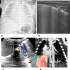
(A) Chest radiograph in a neonate with sudden drop in hemoglobin. Note the ground glass haziness over both lungs simulating RDS. (B) Longitudinal US image of the same neonate showing condensed B-lines similar to those seen in RDS. Diagnosis- Pulmonary haemorrhage. Note the pleural line is thick and irregular as also seen in RDS. (C) CPAM versus sequestration. Post natal colour Doppler Transverse image shows a branch from the aorta directly supplying the hypoechoic lesion in the left lung base. An antenatal Fetal MRI done at 38 weeks gestation revealed a wedge shaped hyperintense lesion near the left lower lobe (image not shown). (D) Coronal contrast enhanced CT image arterial phase confirmed the arterial supply from the aorta. (Inset) Operative photograph showing the wedge shaped lesion (arrow). Note the appearance of the normal lung in contrast.
Figure 4.
Figure 4.
Antenatally detected thoracic lesion (A) Antenatally detected cystic lesion left lung Postnatal chest radiograph in a 3 day old child revealed a homogenous opacity in the left hemithorax causing contralateral mediastinal shift. (B) US image showing the presence of a cystic lesion. Colour flow was seen in the mediastinal vessels medial to the cystic mass (dopper image not shown). (C) Axial contrast enhanced CT image revealed a cystic lesion in the left hemithorax. The child was operated on. Histopathological examination – CPAM.
Figure 5.
Figure 5.
Consolidation and atelectasis (A to D) 14 year old child with breathlessness and cough. CXR (not shown) revealed homogenous opacification of the right hemithorax with blunting of the right CP angle. Air bronchogram was noted suggestive of consolidation. (A) Longitudinal US image shows normal “A” lines replaced by consolidated lung. Associated small rim of pleural effusion is seen adjacent to the diaphragm. (B) Echogenic lines showing a branching pattern are seen within the consolidation due to the air within the bronchioles. This is termed “hepatisation” as the appearance mimics the appearance of the liver. No air bronchogram is seen distally, due to fluid filled bronchioles. (C) On colour doppler, branching pattern of the vessels is seen within the consolidation. This helps to differentiate consolidation from a mass. (D) Axial post contrast CT image confirming the ultrasound findings. No mass was seen. In retrospect the CT was probably not needed.
Figure 6.
Figure 6.
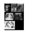
Three year old female child with pneumonia. (A) Transverse US image shows consolidation with air bronchograms. Overlying pleural effusion is seen with echoes within suggestive of an empyema. (B and C) Follow up longitudinal ultrasound images showing areas of breakdown within the consolidation bilaterally not appreciable on the chest radiograph taken at the same time. (D) Chest radiograph one month later revealed well defined cystic areas in the right mid and lower zones and ? left lower zone. (E) Longitudinal ultrasound image left lung base showing a hypoechoic lesion within the consolidation suggestive of an abscess. (F) Coronal CT lung window image reveals multiple thick walled cystic areas which had air fluid levels within them bilaterally suggestive of abscesses. Diagnosis – Necrotizing pneumonia.
Figure 7.
Figure 7.
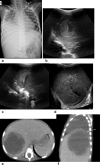
Subdiaphragmatic pathology involving the chest. 4 year old child presenting with fever, chills, dry cough and breathlessness. (A) Frontal chest radiograph revealed right lower zone opacity wth blunting of the CP angle suggestive of pleural effusion with associated consolidation. (B-D) US images showing consolidation of the right lower lobe and part of the upper lobe. Small pleural effusion with moving echoes within was also seen. A large well defined hypoechoic subdiaphragmatic intrahepatic lesion was seen suggestive of an abscess ? amoebic. Suspicious diaphragmatic discontinuity seen posteriorly ? abscess ruptured into the thorax. (E&F) Axial and sagittal post contrast CT images reveal a well defined round thick walled lesion with irregular enhancing internal margins suggestive of an abscess.The right hemidiaphragm shows discontinuity posteriorly (arrow). The child did not respond to antibiotics and had to be put on Metrogyl confirming the amoebic aetiology of the abscess.
Figure 8.
Figure 8.
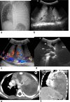
(A) Frontal chest radiograph of a three year old child with dyspnea showing homogenous opacification of the left hemithorax with contralateral mediastinal shift. No air bronchograms seen. (B-C) Longitudial US images left hemithorax reveal a large predominantly solid mass left hemithorax showing absence of air bronchograms with abnormal vascularity within. A small pleural effusion is also seen. (D) Transverse US image shows the mass in left hemithorax causing contralateral mediastinal shift. (E & F) Contrast enhanced axial and coronal reformatted CT image shows a large poorly enhancing heterogenous mass in the left hemithorax surrounding a collapsed left upper lobe. Diagnosis - Pleuropulmonary blastoma.
Figure 9.
Figure 9.
Eleven year old child with breathlessness. (A) Chest radiograph showing a homogenous opacity in the left hemithorax with no air bronchograms. Left CP angle is obliterated. Contralateral mediastinal shift is seen. (B) US image show a large pleural effusion with echoes within it as well as septae. As the patient was afebrile, and an intrathoracic mass was seen, this was although to represent a haemothorax rather than an empyema. (C and D) US images showing a large hyperechoic predominantly solid mass with few small cystic areas within it was also seen with no air bronchograms. Abnormal vessels were seen within the mass. (E) Axial post contrast CT image showing a large enhancing mass with few poorly enhancing areas suggestive of necrosis, bulging through the intercostal spaces. (F) Histopathological examination report - Malignant round cell tumour of the chest wall - PNET.
Figure 10.
Figure 10.
Thymus (A and B) A 3 day old male child born at term who cried weakly after physical stimulation, bag and mask ventilation given subsequently developed pneumomediastinum. (A) Frontal chest radiograph showing an opacity extending on either side-of the mediastinum touching the chest wall laterally, showing well defined margins. Pneumomediastinum was seen. (B) US axial image showing normal stippled appearance of the thymus confirming the opacity, is the thymus. (C & D) six month old child with fever for a week. (C) Digital chest radiograph showing superior mediastinal widening more to the left of the midline. (D) US image showing the opacity is due to the thymus which shows a classical stippled appearance (arrow). Chemical shift imaging (not included) showed signal loss on the out of Phase images confirming benign thymic enlargment.
Figure 11.
Figure 11.
Six month old child, chest radiograph (not shown) revealed a well defined opacity in the posterior mediastinum on the right. (A) Ultrasound done revealed a solid oval mass with few foci of calcification and vascularity within it. (B) T2 weighted coronal MR image revealed the mass had no intraspinal extension and confirmed the solid nature. US guided biopsy done. Histopathological examination – Ganglioneuroma.
Figure 12.
Figure 12.
A 7-year-old female child with cough, expectoration, fever and a left chest wall swelling for 10 days. (A)Frontal chest radiograph showing a well defined opacity in the left mid zone also involving the upper zone. No obvious rib destruction was seen. (B and C) US images confirm the consolidation. A large complex collection was seen overlying it anteriorly extending into the chest wall showing a heterogenous appearance. (D) Axial contrast enhanced CT image of the thorax at the level of the lesion confirmed the ultrasound findings. Diagnosis - Empyema necessitans.

References

    1. Pathan A . Sonography of the lungs and pleura . JLUMHS 2007. .
    1. Soni NJ , Franco R , Velez MI , Schnobrich D , Dancel R , Restrepo MI , et al. . Ultrasound in the diagnosis and management of pleural effusions . Journal of Hospital Medicine 2015. ; 10 : 811 – 6 . doi: 10.1002/jhm.2434 - DOI - PMC - PubMed
    1. Chira RI , Chira A , Mircea PA , Valean S . Mediastinal masses—transthoracic ultrasonography aspects . Medicine 2017. ; 96 : e9082 . doi: 10.1097/MD.0000000000009082 - DOI - PMC - PubMed
    1. Baert AL . Pediatric chest imaging: chest imaging in infants and children . Springer Science & Business Media 2010. ;.
    1. Enriquez G , Aso C , Serres X . Chest ultrasound (US) . : InPediatric Chest Imaging . Berlin, Heidelberg: : The British Institute of Radiology. ; 2008. . ( 1–35).