

Mueller-matrix-based polarization imaging and quantitative assessment of optically anisotropic polycrystalline networks
- PMID: 31095594
- PMCID: PMC6522018
- DOI: 10.1371/journal.pone.0214494
Mueller-matrix-based polarization imaging and quantitative assessment of optically anisotropic polycrystalline networks
Abstract
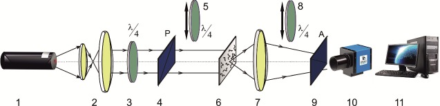
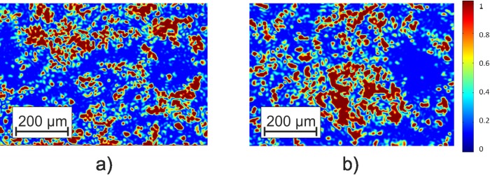
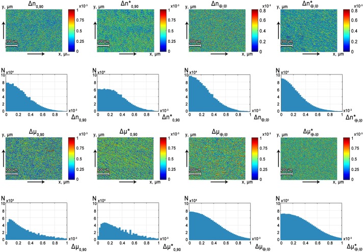
We introduce a Mueller-matrix imaging polarization-based approach for the quantitative digital screening of the polycrystalline structure of fibrillary-based biological tissues in vitro. The morphometric evaluation of histological sections of myocardium was performed utilizing the high-order statistical moments calculated based on the spatial distribution of linear and circular birefringence and dichroism obtained experimentally. We demonstrate that spatial distributions of phase of light and optical anisotropy of scattering inherent to fibrillar networks of myocardium at different necrotic stages can be effectively used as a quantitative marker of stages of myosin fibril degradation. Processing the images of phase of light scattered in biological tissues with high order statistical analysis provides a functional tool for the quantitative characterization of necrotic conditions of the myocardium.
Conflict of interest statement
The authors have declared that no competing interests exist.
Figures
References
-
- Tuchin V, Wang L, Zimnjakov D. Optical polarization in biomedical applications. New York, USA: Springer; 2006.
-
- Ghosh N, Wood M, Vitkin A. Polarized light assessment of complex turbid media such as biological tissues via Mueller matrix decomposition In: Tuchin V, editors. Handbook of Photonics for Biomedical Science. London: CRC Press, Taylor & Francis Group; 2010. pp. 253–282.
-
- Jacques S. Polarized light imaging of biological tissues In: Boas D, Pitris C, Ramanujam N, editors. Handbook of Biomedical Optics. Boca Raton, London, New York: CRC Press; 2011. pp. 649–669.
-
- Layden D, Ghosh N, Vitkin A. Quantitative polarimetry for tissue characterization and diagnosis In: Wang R, Tuchin V, editors. Advanced Biophotonics: Tissue Optical Sectioning. Boca Raton, London, New York: CRC Press, Taylor & Francis Group; 2013. pp. 73–108.
Publication types
MeSH terms
Substances
LinkOut - more resources
Full Text Sources
